Özet
Sakral yetmezlik kırığı, zayıflamış kemiğe normal biyomekanik kuvvetlerin uygulanmasıyla gelişir; osteoporoz sık bir zemin olup bel, sakral veya pelvik ağrıya yol açabilir. BT, sakral aladaki sklerozu, kırık çizgisini ve kortikal kesintiyi göstererek morfolojiyi tanımlar; erken kemik iliği ödemi için MR daha duyarlıdır. Bilateral vertikal alar hatlara transvers sakral bileşen eşlik ettiğinde H biçimli kırık örüntüsü görülebilir ve bu örüntü spinopelvik ayrışmayla ilişkili olabilir. Sakral yetmezlik kırığını metastazla karıştırmak yanlış tanıya ve gereksiz ek inceleme ya da biyopsiye yol açabilir; kırığın gözden kaçması tanıyı geciktirebilir.
Faz ve pencere
- Kemik algoritması tanısal
- Kontrastsız ince kesit pelvis BT'de sakral alada sakroiliak ekleme paralel dikey sklerotik bant, trabeküler düzensizlik, kortikal basamaklanma veya düşük dansiteli kırık çizgisi aranır. BT, sakral kırıkların tanı ve morfolojik karakterizasyonunda kullanılır. BT bulgusu negatif olsa bile klinik kuşku sürüyorsa kemik iliği ödemini gösterebilen MR değerlendirmesi gerekir.
Önerilen pencereler G genişlik, M merkez; değerler Hounsfield birimi (HU)
- KemikG 1800 / M +400
BT bulguları
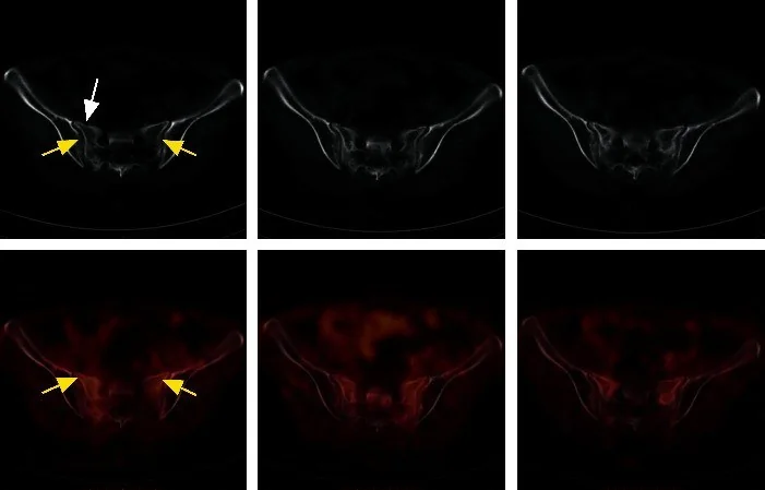
- Sakral ala içindeki dikey sklerotik bant sakroiliak eklem yakınlarında yerleşir.
- Kırık çizgisi çoğu kez sagittal yönlüdür; ince kortikal kesinti veya trabeküler çizgi aksiyel BT'de seçilebilir.
- Bilateral alar hatlar arasında sakral cisimden geçen yatay bileşen eklenirse koronal/sagittal rekonstrüksiyonda H ya da U benzeri konfigürasyon ortaya çıkar.
- Sakral alada skleroz, kırık çizgisi, kortikal kesinti veya miks osteolitik değişiklikler görülebilir; bu nedenle değerlendirmede tek başına skleroz bulgusuna bağlı kalınmamalıdır.
- Sakral kırıklarda yatay kırık örüntüsü görülebilir.
- BT'de foraminal veya kanal daralması ya da retropulse kemik fragmanı sinir basısı kuşkusunu artırabilir; nörolojik muayenenin yapılamadığı durumlarda BT, sinir basısı veya traksiyon yaralanması için dolaylı bir gösterge olabilir.
- Pubik ramus, parasimfiziyel bölge, asetabular çatı ve iliak kanattaki eşlik eden yetmezlik hatları aynı incelemede aranır.
Ölçütler ve sınıflamalar
- Osteoporotik sakral kırıkta BT duyarlılığı (bildirilen aralık)
- Tek merkezli, 44 hastalık bir çalışmada BT'nin MR'a göre klinik açıdan anlamlı pelvis kırıklarını (OF Pelvis III–V) saptama duyarlılığı %86,9 (%95 GA 71,9–95,5) bulunmuştur; bu değer sakral yetmezlik kırıkları için evrensel duyarlılık değildir.
- Denis anatomik zonu
- Denis sınıflaması, sakral kırıklar için kullanılan sınıflama sistemlerinden biridir. Bu zonlama kırık hattının anatomik yerini tanımlar; tek başına deplasman veya spinopelvik stabiliteyi sınıflamaz.
- AO Spine sakral sınıflaması
- C0, deplase olmayan U tipi sakral kırığı tanımlar; yalnız H/U görünümünden alt tip çıkarmak yerine kırık geometrisini ve deplasmanı birlikte değerlendir.
Normalde
Normal sakral alada lineer skleroz, kortikal kesinti ve kırık çizgisi gibi yetmezlik kırığı bulguları izlenmez. Tek başına skleroz yerine, sakral alada kırık çizgisi, kortikal kesinti ve miks osteolitik değişiklik gibi eşlik edebilecek BT bulguları da değerlendirilmelidir.
Normal BT ile kıyasla
Aynı bölgenin, kaynak veri setinde patoloji içermediği belirtilen bir BT incelemesini kesit kesit kaydırın; organ sınırlarını açarak anatomiyi hasta görüntüsüyle karşılaştırın. Küçük rastlantısal bulgular tümüyle dışlanmamıştır.
Kaynak: TotalSegmentator v2.01 veri seti (Wasserthal ve ark., Radiology: Artificial Intelligence 2023), CC BY 4.0, vaka s1256; organ sınırları veri setinin otomatik segmentasyonundan, birleştirilerek sadeleştirildi. Görüntüler 2,5 mm kesit aralığına yeniden örneklendi.
Ayırıcı tanı
- Sakral metastaz
- fokal litik yıkım, kortikal destrüksiyon veya yumuşak doku kitlesi kırık hattı görünümünü taklit edebilir; kemik iliği sinyali için MR ve onkolojik bağlam yardımcıdır.
- Osteitis condensans ilii gibi mekanik lezyonlar sakroiliak eklemin ön-orta bölümünde görülebilir.
- Sakroiliak eklem dejenerasyonu ligament ossifikasyonu veya mekanik stresle ilişkili artritik değişiklikler yapabilir; lezyonların eklemdeki dağılımı ayırıcı tanıya yardımcı olur.
- Sakral osteomiyelit/septik siyovit
- inflamatuar siyoviti taklit eden değişiklikler yapar; enfeksiyon varlığında eklem çevresindeki inflamatuvar ve yapısal değişiklikler dikkat çekicidir.
Tuzaklar
- BT'de kırık çizgisinin görülmemesi yetmezlik kırığını dışlamaz; skleroz ve kortikal basamaklanmayı ara, kuşku sürüyorsa MR'ın ödem duyarlılığını kullan.
- Tek taraflı alar sklerozu metastaz diye etiketlemeden önce yumuşak doku kitlesi ve kortikal destrüksiyon ara; kırık boyunca lineer skleroz ve karşı aladaki eşlik eden hat fragiliteyi destekler.
- Bilateral vertikal alar hatlara eşlik edebilen yatay sakral bileşeni de değerlendir; bu bileşen H tipi kırık örüntüsünün parçası olabilir.
- H tipi, sakral yetmezlik kırıklarında tanımlanan morfolojik örüntülerden biridir.
- Sakral yetmezlik kırığında FDG tutulumu sakroiliak ekleme paralel hafif, diffüz veya yamalı olabilir; PET/BT değerlendirmesinde BT'deki kırık hattı ve sistemik tümör durumu da dikkate alınır.
Kendini dene
Osteoporozlu hastanın pelvis BT'sinde iki alada SI eklemlere paralel sklerotik hatlar ve sakral cisimde yatay çizgi var. BT'de bilateral dikey sakral kırık hatları ve bunları birleştiren yatay bileşen hangi morfolojik örüntüyü oluşturur?
Cevabı göster
H biçimli yetmezlik kırığı. Bilateral dikey alar çizgiler ve bunları bağlayan sakral transvers bileşen H biçimli yetmezlik örüntüsüdür. Metastazda fokal yıkım/kitle; osteitis condensans ilii'de iliak tarafta üçgensi skleroz; artrozda eklem kenarı osteofit ve kistleri beklenir.
Ağrısı ve fragilite riski yüksek hastada BT'de skleroz var, belirgin kırık çizgisi yok. Erken kemik iliği ödemi için en duyarlı yöntem hangisi?
Cevabı göster
MR. MR, sakral kemik iliği ödemini BT'den daha duyarlı gösterir ve erken yetmezlik kırığında yardımcıdır. BT çizgi, korteks ve deplasmanı karakterize eder; direkt grafi duyarsızdır; arteriyel faz kırık ödemini değerlendirmez.
İlgili konular
- Sakral kırık taklitçileri ve normal varyantlar
- AO Spine sakral kırık sınıflaması
- Sakral foraminal/kanal fragmanı
Kaynaklar
Bu sayfadaki 44 cümle ve 2 quiz sorusu aşağıdaki kaynaklardan alıntılarla otomatik olarak karşılaştırıldı; 19 cümle bu karşılaştırmada düzeltildi (2026-10-09).
- Short-Course Radiotherapy in Locally Advanced Rectal Cancerpmc.ncbi.nlm.nih.gov
- Risk Factors for Sacral Insufficiency Fracture After Pelvic Irradiation for Cervical Cancerpmc.ncbi.nlm.nih.gov
- Sacral Insufficiency Fracture Following L4–S1 Circumferential Fusion in Corticosteroid-associated Bone Fragility: A Case Report and Biomechanical Considerationspmc.ncbi.nlm.nih.gov
- Delayed Sacral Insufficiency Fracture Following Oncologic Resection of a Chondrosarcoma: Emphasizing the Role of Biomechanical Risk Assessmentpmc.ncbi.nlm.nih.gov
- Biological activation of prolonged delayed fracture healing following abaloparatide under stable circular external fixation: successful limb salvage of a Gustilo type III-C pilon fracture in severe immune thrombocytopenia—a case reportpmc.ncbi.nlm.nih.gov
- Sacral Fractures and Sacroplasty.pubmed.ncbi.nlm.nih.gov
- Regional migratory osteoporosis and transient osteoporosis of the hip: are they all the same?pubmed.ncbi.nlm.nih.gov
- Sacral insufficiency fracture after partial sacrectomy.pubmed.ncbi.nlm.nih.gov
- Treatment for sacral insufficiency fractures: A systematic review.pubmed.ncbi.nlm.nih.gov
- Interventional Procedures in Metabolic Bone Diseases.pubmed.ncbi.nlm.nih.gov
- Sacral and Pelvic Insufficiency Fractures Following Adult Spinal Deformity Surgery: A Case Report and Systematic Literature Review.pubmed.ncbi.nlm.nih.gov
- Postpartum sacral insufficiency fractures.pubmed.ncbi.nlm.nih.gov
- Sacroiliitis or insufficiency fracture?pubmed.ncbi.nlm.nih.gov
- [Sacral insufficiency fractures].pubmed.ncbi.nlm.nih.gov
- Sacral Insufficiency Fracture on 68Ga-DOTATATE PET/CT.pubmed.ncbi.nlm.nih.gov
- Risk Factors for Sacral Insufficiency Fractures in Cervical Cancer After Whole Pelvic Radiation Therapy.pubmed.ncbi.nlm.nih.gov
- Short-Course Radiotherapy in Locally Advanced Rectal Cancer.pubmed.ncbi.nlm.nih.gov
- [Balloonsacroplasty: C-arm or CT controlled application? : Experience with 46 patients].pubmed.ncbi.nlm.nih.gov
- Reliability and effectiveness of percutaneous sacroplasty in sacral insufficiency fractures.pubmed.ncbi.nlm.nih.gov
- Acute management of spinal fractures: What you need to know.pubmed.ncbi.nlm.nih.gov
- Adult-onset hypophosphatemic osteomalacia as a cause of widespread musculoskeletal pain: A retrospective case series of single center experience.pubmed.ncbi.nlm.nih.gov
- Bridging the Gap: Pregnancy-And Lactation-Associated Osteoporosis.pubmed.ncbi.nlm.nih.gov
- Beyond Chordoma: A Comprehensive Review of Sacral Lesions.pubmed.ncbi.nlm.nih.gov
- Clinical and 18F-FDG PET/CT Imaging Characteristics of Post-radiotherapy Sacral Insufficiency Fractures in Cervical Cancer Patients.pubmed.ncbi.nlm.nih.gov
- Knowledge gaps in high-energy sacral fracture treatment and management.pubmed.ncbi.nlm.nih.gov
- Sacral and Pelvic Fractures: Historical Systems and Advancements with the AO Spine Classification.pubmed.ncbi.nlm.nih.gov
- Comparative biomechanical evaluation of the U-shaped lumbopelvic stabilization technique in treating unstable sacral fractures.pubmed.ncbi.nlm.nih.gov
- Recognizing Sacral Insufficiency Fractures Hidden in Plain Sight: An Illustrative Case Report.pubmed.ncbi.nlm.nih.gov
- Glossary of terms for musculoskeletal radiology.pubmed.ncbi.nlm.nih.gov
- Preliminary Retrospective Analysis of X-Ray, CT, and MRI Imaging Features of Brucellar Spondylodiscitis Diagnosis.pubmed.ncbi.nlm.nih.gov
- Imaging of Sacroiliac Pain: The Current State-of-the-Art.pubmed.ncbi.nlm.nih.gov
- Reliability of Fossae Lumbales Laterales and Pelvic Incidence for Estimating Transsacral Corridors Assessed Using Reconstruction Computed Tomography.pubmed.ncbi.nlm.nih.gov
- Evaluating the necessity of edema-sensitive imaging in assessing fragility fractures of the pelvis: a retrospective cohort study utilizing the OF Pelvis Classification and Score.pubmed.ncbi.nlm.nih.gov
- Multiple Occult Fractures at Hemodialysis Initiation in a Chronic Kidney Disease Patient: Diagnostic Value of MRI and Potential Role of Zinc Deficiency.pubmed.ncbi.nlm.nih.gov
- Fragility Fractures of the Pelvis-Current Understanding and Open Questions.pubmed.ncbi.nlm.nih.gov
- Perfecting the pour: A novel co-axial technique with sequential injections for optimising cement delivery during sacroplasty.pubmed.ncbi.nlm.nih.gov
- Vertebral body osteonecrosis: Evolving imaging features over time.pubmed.ncbi.nlm.nih.gov
- Anatomy of the intraosseous arterial microvasculature of the adult lumbar vertebral body and its role in fracture healing: a literature review and historical perspective.pubmed.ncbi.nlm.nih.gov
- Chronic pelvic insufficiency fractures and their treatment.pubmed.ncbi.nlm.nih.gov
- Surgical management strategies for unstable sacral fractures: Case series and surgical guideline.pubmed.ncbi.nlm.nih.gov
- Bone tumor mimickers: A pictorial essay.pubmed.ncbi.nlm.nih.gov
- Long-term safety and effectiveness of the "OptEase" vena cava filter.pubmed.ncbi.nlm.nih.gov
- Radiculopathy due to cement extravasation after sacroplasty: A case report.pubmed.ncbi.nlm.nih.gov
- Biomechanical stability evaluation of isolated straddle fracture using finite element analysis.pubmed.ncbi.nlm.nih.gov
- Fragility Fractures of the Pelvis With Acetabular and Femoral Neck Fractures in Elderly Patients.pubmed.ncbi.nlm.nih.gov
- Retrospective comparative study between conservative management and surgical fixation of pelvic insufficiency fractures.pubmed.ncbi.nlm.nih.gov
- CT of Sacral Fractures: Classification Systems and Management.pubmed.ncbi.nlm.nih.gov
- Sacral Fractures: A Review.pubmed.ncbi.nlm.nih.gov
- <sup>18</sup>F‑FDG PET/CT in metastatic chordoma: a retrospective analysis of imaging features and potential clinical relevance.pubmed.ncbi.nlm.nih.gov
- Axial Spondyloarthritis: Mimics and Pitfalls of Imaging Assessment.pubmed.ncbi.nlm.nih.gov
- Sacroiliitis mimics: a case report and review of the literature.pubmed.ncbi.nlm.nih.gov
- Early diagnosis and targeted intervention based on the pathogenesis of rapidly progressive osteoarthritis of the hip.pubmed.ncbi.nlm.nih.gov
- Advances in mechanical assessments of in vivo human lumbar spine tissues with noninvasive imaging techniques.pubmed.ncbi.nlm.nih.gov
- When it is not sacroiliitis.pubmed.ncbi.nlm.nih.gov
- Pictorial Review of Paediatric Limp.pubmed.ncbi.nlm.nih.gov
- Explainable AI for classifying vertebral fracture histology in digital spine pathology.pubmed.ncbi.nlm.nih.gov
- Diagnostics of Sacroiliac Joint Differentials to Axial Spondyloarthritis Changes by Magnetic Resonance Imaging.pubmed.ncbi.nlm.nih.gov
- Navigation-Guided Thoracolumbar Transpedicular Instrumentation Using the 7D Flash™ Navigation System (SeaSpine) in a Patient With L1 Vertebral Fracture and Spinal Deformity Secondary to Prior Instrumentation for Idiopathic Scoliosis: A Case Report.pubmed.ncbi.nlm.nih.gov
- Sacroplasty for Sacral Insufficiency Fractures: Narrative Literature Review on Patient Selection, Technical Approaches, and Outcomes.pubmed.ncbi.nlm.nih.gov
- Multilevel Lumbar CT Attenuation Beyond L1: Comparison with QCT-Derived Volumetric Bone Mineral Density and Prevalent Fragility Fracture Status in a Diagnostic Referral Cohort.pubmed.ncbi.nlm.nih.gov
- Are sacral stress fractures in young women truly rare? a retrospective MRI analysis of 1970 patients.pubmed.ncbi.nlm.nih.gov
- CT-based three-dimensional fracture mapping reveals characteristic patterns in pelvic insufficiency fractures.pubmed.ncbi.nlm.nih.gov
- Risk Factors for Sacral Insufficiency Fracture After Pelvic Irradiation for Cervical Cancer.pubmed.ncbi.nlm.nih.gov
- Minimally Invasive Lumbopelvic and Transiliac-Transsacral Fixation for a Pathological Sacral Fracture Due to Metastatic Breast Cancer: A Case Report.pubmed.ncbi.nlm.nih.gov
- Neuronavigated placement of iliosacral screws in sacral fractures: an additional tool in the spine surgeon's arsenal.pubmed.ncbi.nlm.nih.gov
- A positioning method for the transiliac-transsacral screw bone channel based on a manual tracing of a transparent overlay of preoperative computed tomography images in patients with pelvic fracture and a unilateral trans-sacroiliac corridor.pubmed.ncbi.nlm.nih.gov
- Sacral U-Type Fractures: A Comparative Study of Treatment Approaches.pubmed.ncbi.nlm.nih.gov
- Preferential sacral fracture sites in fragility fractures of the pelvis type IVb and comparison of internal fixation methods: CT-based morphological mapping and finite element analysis.pubmed.ncbi.nlm.nih.gov
- Traumatic lumbosacral instability: part 1 - proposing a definition and identifying underlying injury patterns.pubmed.ncbi.nlm.nih.gov
- Management of C0 Sacral Fractures Based on the AO Spine Sacral Injury Classification: A Narrative Review.pubmed.ncbi.nlm.nih.gov
- A Review on the Usage of Bone Single-Photon Emission Computed Tomography/Computed Tomography in Detecting Skeletal Metastases in the Post-COVID-19 Era: Is it Time to Ditch Planar and Single-Photon Emission Computed Tomography only Gamma Camera Systems?pubmed.ncbi.nlm.nih.gov
- Multidisciplinary management of spinal metastases: what the radiologist needs to know.pubmed.ncbi.nlm.nih.gov
- Imaging and treatment of sacral insufficiency fractures.pubmed.ncbi.nlm.nih.gov
- Incidental findings in female pelvis MRI performed for gynaecological malignancies.pubmed.ncbi.nlm.nih.gov
- Rapidly destructive hip osteoarthritis: a diagnosis not to miss.pubmed.ncbi.nlm.nih.gov
- Sacroplasty for Zone III Sacral Insufficiency Fracture: A Case Report Utilizing a Midline Approach and Articulating Osteotome.pubmed.ncbi.nlm.nih.gov
- Anterior Transpsoas Approach for Removal of Cement Leakage after Sacroplasty.pubmed.ncbi.nlm.nih.gov
- 'XX'-Sacroplasty: A Novel Technique for Management of "H-Type" Sacral Insufficiency Fractures.pubmed.ncbi.nlm.nih.gov
- Analysis of Fracture Patterns and Characteristics in Sacral Insufficiency Fracture: Do Sacral Fractures Occur in Patients Who Had Previous Lumbosacral Fusion Insufficiency Fractures or Stress Fractures?pubmed.ncbi.nlm.nih.gov
- Missed Bilateral Atraumatic Sacral Ala Insufficiency Fractures With Normal Dual-Energy X-ray Absorptiometry (DEXA) Scan and Radiofrequency Echographic Multi-spectrometry (REMS)-Confirmed Osteoporosis.pubmed.ncbi.nlm.nih.gov
- Atraumatic sacral fracture in a postpartum patient in the late postoperative period following lumbar decompression for disc herniation.pubmed.ncbi.nlm.nih.gov
- Preoperative evaluation and orthopedic surgical strategies for tumor-induced osteomalacia.pubmed.ncbi.nlm.nih.gov
- Fragility fractures of the sacrum: how to identify and when to treat surgically?pubmed.ncbi.nlm.nih.gov
- Abusive spinal injury: imaging and updates.pubmed.ncbi.nlm.nih.gov
- Sacral insufficiency fractures: a case of mistaken identity.pubmed.ncbi.nlm.nih.gov
- Radiographically occult and subtle fractures: a pictorial review.pubmed.ncbi.nlm.nih.gov
- Open and Percutaneous Fixation of Traumatic Sacral Fracture-Dislocation with Spinopelvic Dissociation: Two Adolescent Cases and a Systematic Literature Review.pubmed.ncbi.nlm.nih.gov
- Transsacral Bar Fixation for Osteoporotic H-Type Sacral Fractures: A Viable Alternative to Spinopelvic Fixation.pubmed.ncbi.nlm.nih.gov
- Spinal lesions: a comprehensive radiologic overview.pubmed.ncbi.nlm.nih.gov
- Transverse Process Fractures in Athletes: Mechanisms, Management and Return-to-Play Considerations.pubmed.ncbi.nlm.nih.gov
- Main Diagnostic Pitfalls in Reading the Sacroiliac Joints on MRI.pubmed.ncbi.nlm.nih.gov
- Bone marrow oedema of the sacroiliac joint without spondyloarthritis: a retrospective study of 34 cases of osteitis condensans ilii.pubmed.ncbi.nlm.nih.gov
- The treatment landscape in axial spondyloarthritis-do we need new therapies?pubmed.ncbi.nlm.nih.gov
- Axial Skeleton Bone Marrow Changes in Inflammatory Rheumatologic Disorders.pubmed.ncbi.nlm.nih.gov
- A new technique for percutaneous screw fixation for treating FFP IIIa and IIIb fragility fractures of the pelvis.pubmed.ncbi.nlm.nih.gov
- A Case of U-shaped Sacral Fracture After Longstanding Spinopelvic Fixation Treated With Percutaneous Sacroiliac Joint Fusion and Iliosacral Osteosynthesis.pubmed.ncbi.nlm.nih.gov
- Sacral Insufficiency Fracture Following L4-S1 Circumferential Fusion in Corticosteroid-associated Bone Fragility: A Case Report and Biomechanical Considerations.pubmed.ncbi.nlm.nih.gov
- Traumatic lumbosacral instability: part 2-indications and techniques for surgical management.pubmed.ncbi.nlm.nih.gov
- A Primer on Anatomy, Biophysics, Pathology, Imaging and Treatment of the Intervertebral Disc and the Anterior Spinal Column: The Discogenic, Intervertebral, Spinal Column (DISC) ASPN Workgroup.pubmed.ncbi.nlm.nih.gov
- The Radiologists' Guide to Spinal Separation Surgery: What Does the Surgeon Want to Know?pubmed.ncbi.nlm.nih.gov
- Diagnostic Value of Opportunistic CT-Based Bone Density Assessment in Patients with and Without Sacral Insufficiency Fractures.pubmed.ncbi.nlm.nih.gov
- Imaging of Multiple Myeloma: Present and Future.pubmed.ncbi.nlm.nih.gov
- An alphanumeric classification of osteoporotic pelvic ring injuries.pubmed.ncbi.nlm.nih.gov
- Imaging Review of Pelvic Ring Fractures and Its Complications in High-Energy Trauma.pubmed.ncbi.nlm.nih.gov
- Magnetic resonance imaging of the spine.pubmed.ncbi.nlm.nih.gov
- Preoperative 3D-Planned S1 Corridors Transferred into 2D Fluoroscopy Allow for Safe Intraoperative Large-Diameter Implant Placement: Description of a Novel Sacroiliac Fixation Technique and Proof of Concept in 137 Implantations.pubmed.ncbi.nlm.nih.gov
- Vertebral Augmentation Involving Vertebroplasty or Kyphoplasty for Cancer-Related Vertebral Compression Fractures: A Systematic Review.pubmed.ncbi.nlm.nih.gov
- A Guide to Improving the Care of Patients with Fragility Fractures, Edition 2.pubmed.ncbi.nlm.nih.gov
- Polymicrobial infection presenting as non-clostridial gas gangrene in a patient with an open pelvic ring fracture accompanied by abdominal evisceration: a case report.pubmed.ncbi.nlm.nih.gov
- Total laparoscopic radical hysterectomy for cervical cancer in prolapsed uterus.pubmed.ncbi.nlm.nih.gov
- Parathyroid Hormone in the Management of Pelvic Fragility Fractures: A Systematic Review and Meta-Analysis.pubmed.ncbi.nlm.nih.gov
- Case Report: Is the isolated bone change in advanced colorectal cancer necessarily malignant metastasis?pubmed.ncbi.nlm.nih.gov
- Clinical and biomechanical outcomes of single-position anterior fixation in anterior lumbar interbody fusion.pubmed.ncbi.nlm.nih.gov
- Disease- and Treatment-related Complication on F-18-Fluorodeoxyglucose Positron Emission Tomography/Computed Tomography in Oncology Practice: A Pictorial Review.pubmed.ncbi.nlm.nih.gov
- Role of MR Imaging and FDG PET/CT in Selection and Follow-up of Patients Treated with Pelvic Exenteration for Gynecologic Malignancies.pubmed.ncbi.nlm.nih.gov
Eğitim amaçlıdır; hasta başında tanı ve tedavi kararının yerine geçmez, tıbbi tavsiye değildir. Metinler yapay zekâ desteğiyle yazıldı ve otomatik bir adımda kaynak alıntılarıyla karşılaştırıldı; bu bir tıbbi doğrulama değildir ve içerik uzman radyolog incelemesinden geçmedi. Klinik kararlarda güncel kılavuzları, kurum protokollerini ve radyoloji raporunu esas alın.