Özet
Yavaş büyüyen ve çoğu kez ağrısız yüzeyel kitlelerde lipom sık görülen benign tümörlerden biridir; derin lipomlar kas içinde veya kaslar arasında yerleşebilir ve komşu nörovasküler yapılara bası yapabilir. Kontrastsız BT, lezyondaki yağın yanı sıra yumuşak doku bileşenlerinin varlığını ve yaygınlığını gösterir. Kontrastsız BT'de homojen yağ atenüasyonlu kitle tipik lipomu destekler; görüntüleme bulguları atipik lipomatöz tümörü her zaman kesin olarak dışlamaz. Derin veya kuşkulu kitleyi lipom varsayarak değerlendirmek, sarkomun plansız eksizyonuna yol açabilir.
Okuma sırası
- Yüzeyel yağ ve derin fasya. Aksiyel kesitlerde lezyonun yüzeyel fasyaya göre konumunu belirleyerek yüzeyel dokuda mı, fasya altında mı yerleştiğini değerlendirin.
- Lipom içi yağ. Kitle atenüasyonunun makroskopik yağla uyumlu düşük yoğunlukta olup olmadığını ve bu görünümün lezyon boyunca homojen sürüp sürmediğini değerlendirin.
- İntralezyonel yapı. Lezyon içinde kalın veya düzensiz septaları ve yağ dışı nodüler ya da solid bileşenleri arayın.
- Kas planı ve nörovasküler demet. Lezyon kas içindeyse, tümör içindeki kas lifleri benignite lehine bir ipucu olabilir; ancak tek başına tanı koydurmaz.
Faz ve pencere
- Kontrastsız tanısal
- İyi sınırlı kitle çoğunlukla homojen makroskopik yağ atenüasyonundadır; yayımlanmış klasik aralık −120 ile −65 HU'dur. İnce septalar görülebilir; kalın veya düzensiz septalar tipik basit lipomdan ayrımı güçleştirebilir. Bu tanı için intravenöz kontrast gerekmez.
Önerilen pencereler: Yumuşak doku (G 400 / M 40).
BT bulguları
- Lipom, çevre kas yerine kendi içindeki homojen yağ alanında −120 ile −65 HU arasında düşük yoğunluk gösterir.
- Düzgün sınırlı kontur — kontrastsız BT'de lipom, çevresi belirgin ve homojen yağ dansitesinde bir kitle olarak görülür.
- İnce lineer septalar — yağ lobülleri arasında uzanır; tek başına malignite anlamına gelmez.
- Yağ dışı nodül yokluğu — homojen yağ yapısı içinde belirgin yumuşak doku yoğunluklu küresel odak izlenmez.
- Yüzeyel fasya ilişkisi — lipomlar en sık yüzeyel yumuşak dokularda görülür; derin lipomlar fasya altında, kas içinde veya kaslar arasında yerleşebilir.
- Basınç etkisi — komşu kas demetleri ve damar-sinir yapıları yer değiştirir; kemik korteksinde agresif yıkım beklenmez.
Ölçütler ve sınıflamalar
- Klasik lipom için bildirilen kontrastsız BT yağ atenüasyonu
- −120 ile −65 HU
Normalde
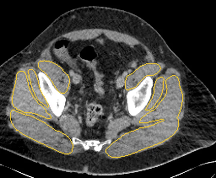
Subkutan yağ dokusu yüzeyel fasya üzerinde uzanırken, lipom benzeri kitleler bu plane'i iter veya kas içine sızmış olabilir. Aynı düzeyde kitlenin yağ dansitesini subkutan yağla, sınırını yüzeyel ve derin fasya ile, bası etkisini komşu kas ve nörovasküler demetle kıyaslayın.
Normal BT ile kıyasla
Aynı bölgenin, kaynak veri setinde patoloji içermediği belirtilen bir BT incelemesini kesit kesit kaydırın; organ sınırlarını açarak anatomiyi hasta görüntüsüyle karşılaştırın. Küçük rastlantısal bulgular tümüyle dışlanmamıştır.
Kaynak: TotalSegmentator v2.01 veri seti (Wasserthal ve ark., Radiology: Artificial Intelligence 2023), CC BY 4.0, vaka s0685; organ sınırları veri setinin otomatik segmentasyonundan, birleştirilerek sadeleştirildi. Görüntüler 2,5 mm kesit aralığına yeniden örneklendi.
Ayırıcı tanı
- Atipik lipomatöz tümör/iyi diferansiye liposarkom
- iyi diferansiye liposarkom bu adla da anılır; kalın veya düzensiz septalar şüphe uyandırır, ancak görüntüleme lipomdan ayrımı her zaman kesinleştirmez.
- İntramüsküler lipom
- yağ kütlesi kas içinde büyür ve çevre kas lifleri arasına şeritler halinde uzanabilir.
- Hibernom
- kahverengi yağ kaynağıdır; MR'de subkutan yağa izointense ama tam aynı değildir ve kontrast sonrası beklenenden fazla difüz güçlenir.
Tuzaklar
- Kontrastsız BT'de lipom, −120 ile −65 HU arasında homojen yağ atenüasyonunda, çevresi sınırlı bir kitle görünümündedir.
- Lipomlarda ince internal septalar görülebilir; ALT açısından kalın veya düzensiz septalar daha şüphelidir.
- Homojen yağ görünümü şüpheli klinik ve derin büyük kitlede ALT'yi bütünüyle dışlamaz; BT'nin karakterizasyon sınırını raporda belirtin.
- İntramüsküler lezyonda çevre kas lifleri tümör nodülü sanılabilir; şeritlerin kasla devamlılığını ardışık kesitlerde izleyin.
Kendini dene
Uyluk cilt altındaki düzgün sınırlı kitle, kontrastsız BT'de homojen yağ yoğunluğunda; yağ dışı nodül yok. En olası tanı nedir?
Cevabı göster
Lipom. Homojen makroskopik yağ ve düzgün sınırlı kontur tipik lipom lehinedir. Hibernomada MR sinyali subkutan yağla benzer fakat özdeş olmayabilir ve kontrast sonrası belirgin difüz güçlenme görülebilir; kalın veya düzensiz septalar ile nodüler bileşen ALT şüphesini artırır.
Yağ baskın derin uyluk kitlesinde düzensiz kalın septalar ve kontrastlı yumuşak doku odağı görülüyor. Hangi tanı öncelikle düşünülmeli?
Cevabı göster
Atipik lipomatöz tümör. Yağ içindeki kalın düzensiz septa ve kontrastlanan nodül ALT/iyi diferansiye liposarkom şüphesini artırır. Basit lipom kontrastsız BT'de sınırlı ve homojen yağ atenüasyonlu görünür; elastofibrom dorsi ise tipik olarak infraskapular bölgede, kasla benzer yoğunlukta ve yağ çizgilenmeleri içeren bir kitledir.
İlgili konular
Kaynaklar
Bu sayfadaki 36 cümle ve 2 quiz sorusu aşağıdaki kaynaklardan alıntılarla tek tek karşılaştırıldı; 19 cümle bu karşılaştırmada düzeltildi (2026-10-09).
- Imaging review of lipomatous musculoskeletal lesionspmc.ncbi.nlm.nih.gov
- Use of radiologic imaging to differentiate lipoma from atypical lipomatous tumor/well-differentiated liposarcoma: Systematic reviewpmc.ncbi.nlm.nih.gov
- Lipoma arborescens.pubmed.ncbi.nlm.nih.gov
- [Cardiac tumors].pubmed.ncbi.nlm.nih.gov
- Lipomatous Soft-tissue Tumors.pubmed.ncbi.nlm.nih.gov
- Colonic Lipoma Causing Bowel Intussusception: An Up-to-Date Systematic Review.pubmed.ncbi.nlm.nih.gov
- Complex forms of spinal dysraphism.pubmed.ncbi.nlm.nih.gov
- T2 black lesions on routine knee MRI: differential considerations.pubmed.ncbi.nlm.nih.gov
- Approach to a patient with monoarticular disease.pubmed.ncbi.nlm.nih.gov
- Imaging of benign soft tissue tumors.pubmed.ncbi.nlm.nih.gov
- [Diagnosis and therapy of benign intraarticular tumors].pubmed.ncbi.nlm.nih.gov
- Incidental findings on brain and spine imaging in children.pubmed.ncbi.nlm.nih.gov
- Nonschwannoma tumors of the cerebellopontine angle.pubmed.ncbi.nlm.nih.gov
- Lipomatous tumours of the head and neck: a spectrum of biological behaviour.pubmed.ncbi.nlm.nih.gov
- Endobronchial Lipoma: Case Report and Literature Review.pubmed.ncbi.nlm.nih.gov
- An Unusual Lipoma at the Lateral Margin of the Tongue: A Case Report.pubmed.ncbi.nlm.nih.gov
- A Soft Tissue Osteochondrolipoma of the Lateral Ankle: A Case Report and Clinical Insight.pubmed.ncbi.nlm.nih.gov
- Giant nonencapsulated pubic lipoma: a case report and literature review on differential diagnoses of atypical lipomas.pubmed.ncbi.nlm.nih.gov
- Endobronchial lipoma presenting as a fat-attenuation lesion causing left upper lobar bronchial obstruction: A case report.pubmed.ncbi.nlm.nih.gov
- Ancillary Diagnostic Utility of Glycoprotein Nonmetastatic Melanoma Protein B (GPNMB) in Diagnostically Challenging Hepatic Angiomyolipoma.pubmed.ncbi.nlm.nih.gov
- The space of Retzius: anatomy, imaging features, and patterns of disease spread.pubmed.ncbi.nlm.nih.gov
- Nasopharyngeal Lipoma Anterior to the Eustachian Tube Orifice: A Case Report.pubmed.ncbi.nlm.nih.gov
- Falciform Ligament Lipoma: A Rare Cause of Chronic Epigastric Pain.pubmed.ncbi.nlm.nih.gov
- Gastrocnemius-soleus intramuscular lipoma misinterpreted as hemangioma: a rare case report.pubmed.ncbi.nlm.nih.gov
- Large lipoma of the finger masquerading as a giant cell tumor: A diagnostic pitfall in an uncommon site.pubmed.ncbi.nlm.nih.gov
- A case of fat-forming solitary fibrous tumor that is prone to be confused with liposarcoma.pubmed.ncbi.nlm.nih.gov
- Endobronchial Lipoma: A Rare Benign Tumor Managed by Minimally Invasive Bronchoscopic Excision.pubmed.ncbi.nlm.nih.gov
- Incidental pulmonary findings on CT in daily practice: the nodule and the interstitial lung abnormalities - what's old, what's new.pubmed.ncbi.nlm.nih.gov
- Aesthetic Thermotherapy-Induced Panniculitis in a Patient with Psoriasis: A Case Report.pubmed.ncbi.nlm.nih.gov
- MRI findings for differentiating benign and malignant soft tissue tumors: a systematic review-part 2: key imaging findings.pubmed.ncbi.nlm.nih.gov
- Common cystic pancreatic neoplasms: A comprehensive magnetic resonance imaging-based review of typical and atypical imaging features.pubmed.ncbi.nlm.nih.gov
- Report-Documented Frequency and Spectrum of Incidental Findings on Emergency Abdominopelvic Computed Tomography: A Retrospective Clinical Data Study from Saudi Arabia.pubmed.ncbi.nlm.nih.gov
- Classifying diagnostic pitfalls in juxtapleural CT lesions: A root cause.pubmed.ncbi.nlm.nih.gov
- Gastric bronchogenic cyst pooled case analysis: a case report and systematic review.pubmed.ncbi.nlm.nih.gov
- Endoscopic Management of Acute Gastrointestinal Bleeding From a Gastric Lipoma: A Case Report, Literature Review, and Treatment Recommendation With Reference to ESGE Guidelines.pubmed.ncbi.nlm.nih.gov
- Postoperative Computed Tomographic Assessment of the Complete Resection of an Infiltrative Lipoma Compressing the Spinal Cord in a Dog.pubmed.ncbi.nlm.nih.gov
- Sclerosing Atypical Lipomatous Tumor with Myxomatous Features Presenting in the Plantar Heel: A Case Report.pubmed.ncbi.nlm.nih.gov
- Subcutaneous Sacrococcygeal Lipoma in a 7 Months Old Female Mimicking Myelomeningocele and Sacrococcygeal Teratoma, A Case Report.pubmed.ncbi.nlm.nih.gov
- Management of a large angiomyofibroblastoma of the vulva: a case report describing radical excision and reconstruction.pubmed.ncbi.nlm.nih.gov
- Case Report: Laparoscopic repair of indirect inguinal hernia combined with ipsilateral giant subcutaneous inguinoperineal lipoma.pubmed.ncbi.nlm.nih.gov
- Imaging findings of intra-articular tumor/tumor-like lesions based on pathologic correlation.pubmed.ncbi.nlm.nih.gov
- CT and MRI Key Features of Benign Tumors and Tumor-like Lesions of the Tongue: A Pictorial Review.pubmed.ncbi.nlm.nih.gov
- Tumors and pseudotumors of the soft tissues: Imaging semiology and strategy.pubmed.ncbi.nlm.nih.gov
- Glossary of terms for musculoskeletal radiology.pubmed.ncbi.nlm.nih.gov
- Abdominal Wall Endometriosis Following Cesarean Section: A Retrospective Analysis.pubmed.ncbi.nlm.nih.gov
- Fat-containing soft-tissue tumors: Imaging findings and pathologic correlation.pubmed.ncbi.nlm.nih.gov
- Temporal Lipoma in the Interfascial Fat Pad: A Rare Finding.pubmed.ncbi.nlm.nih.gov
- Surgical and anatomical nuances in trigeminal neuralgia due to cerebellopontine angle lipoma: illustrative case.pubmed.ncbi.nlm.nih.gov
- When Fat Takes Over: A Progressively Enlarging Giant Hand Lipoma.pubmed.ncbi.nlm.nih.gov
- Rare co-occurrence of parosteal lipoma and growing osteochondroma: A case report.pubmed.ncbi.nlm.nih.gov
- Surgical removal of giant adrenal lipoma with gallstones in limited resources areas: A case report.pubmed.ncbi.nlm.nih.gov
- An Orbicularis Oculi-Related Band as a Potential Modifier of Tear-Trough Visibility: A Hypothesis-Generating Observation of a Layered Boundary in a Patient.pubmed.ncbi.nlm.nih.gov
- Clinical and imaging features of tufted angioma in children.pubmed.ncbi.nlm.nih.gov
- Key Points of Endoscopic Minimally Invasive Surgery for Gynecomastia Based on Refined Anatomy: A Case Series.pubmed.ncbi.nlm.nih.gov
- A retrospective analysis of initial ultrasonographic misdiagnosis of fibrous hamartoma of infancy at the ultrasound center of a tertiary hospital in China.pubmed.ncbi.nlm.nih.gov
- A Rare Case Report on Dedifferentiated Liposarcoma of the Thigh Mimicking an Abscess With Vascular Features: A Diagnostic Challenge With Fatal Outcome.pubmed.ncbi.nlm.nih.gov
- Giant retroperitoneal dedifferentiated liposarcoma with intraperitoneal extension: A case report.pubmed.ncbi.nlm.nih.gov
- Well-Differentiated Liposarcoma of the Spermatic Cord Involving Testicular Parenchyma.pubmed.ncbi.nlm.nih.gov
- Adenocarcinoma Developing Overlying a Colonic Lipoma: A Case Report.pubmed.ncbi.nlm.nih.gov
- Colonic lipoma presenting as colo-colic intussusception in an adult.pubmed.ncbi.nlm.nih.gov
- Imaging features of soft tissue hibernoma: a review of 73 cases.pubmed.ncbi.nlm.nih.gov
- Lateral Abdominal Wall Hibernoma: A Rare Case From the Maldives.pubmed.ncbi.nlm.nih.gov
- Rare Cause for a Fluorodeoxyglucose (18F) avid Paravertebral Mass: Mediastinal Hibernoma.pubmed.ncbi.nlm.nih.gov
- Spontaneous transomental internal hernia causing small bowel obstruction in an elderly patient with a virgin abdomen: a case report.pubmed.ncbi.nlm.nih.gov
- Wünderlich syndrome mimicking acute cholecystitis: a case report.pubmed.ncbi.nlm.nih.gov
- Traumatic hip dislocation in a child with an acetabular fleck sign indicating posterior osteochondral avulsion: a case report.pubmed.ncbi.nlm.nih.gov
- Society for Cardiovascular Magnetic Resonance 2024 Cases of SCMR case series.pubmed.ncbi.nlm.nih.gov
- Application of postmortem imaging modalities in cases of sudden death due to cardiovascular diseases-current achievements and limitations from a pathology perspective : Endorsed by the Association for European Cardiovascular Pathology and by the International Society of Forensic Radiology and Imaging.pubmed.ncbi.nlm.nih.gov
- Giant Pancreatic Lipoma of the Uncinate Process: A Case Report.pubmed.ncbi.nlm.nih.gov
- A Diagnostic Limitation of Carnett's Maneuver: A Case of Deep-Seated Abdominal Wall Angiolipoma Mimicking Visceral Pain.pubmed.ncbi.nlm.nih.gov
- Retroperitoneal Dumbbell-Shaped Lipoma: Laparoscopy-Assisted Hybrid Surgical Approach.pubmed.ncbi.nlm.nih.gov
- A rare case of intramuscular granular cell tumor in the right thigh: case report and literature review.pubmed.ncbi.nlm.nih.gov
- Intermuscular Adipose Tissue as a Multimodality Quantitative Imaging Biomarker: Evidence Across Metabolic, Cardiovascular and Oncologic Diseases.pubmed.ncbi.nlm.nih.gov
- Anatomy and Pathology of Anterior and Lateral Hip Compartments Bursae: An Ultrasound Structured Approach in Sports Medicine.pubmed.ncbi.nlm.nih.gov
- Ultrasonography of the Fasciae and Common Pathologies: The Game Changer.pubmed.ncbi.nlm.nih.gov
- CT colonography: revisited after 30 years.pubmed.ncbi.nlm.nih.gov
- Imaging features of right atrial lipoma on multimodality imaging: A case report.pubmed.ncbi.nlm.nih.gov
- Morphological Variability of the Thigh Muscle Traps in an Ultrasound That Awaits Clinicians.pubmed.ncbi.nlm.nih.gov
- Skeletal Muscle Disease: Imaging Findings Simplified.pubmed.ncbi.nlm.nih.gov
- Increasing differential diagnosis between lipoma and liposarcoma through radiomics: a narrative review.pubmed.ncbi.nlm.nih.gov
- Giant Asymptomatic Intermuscular Lipoma of Right Thigh: Case Report and Review of Literature.pubmed.ncbi.nlm.nih.gov
- Incidental nondysraphic cervical intradural extramedullary lipoma at C1-C2: CT and MRI findings.pubmed.ncbi.nlm.nih.gov
- Giant lipoma with an unusual anatomical location successfully managed in a resource-limited setting: a case report.pubmed.ncbi.nlm.nih.gov
- Hibernoma: A case report of a rare soft lipoid tissue in the neck.pubmed.ncbi.nlm.nih.gov
- The Imaging of Primary Fallopian Tube Carcinoma: A Literature Review.pubmed.ncbi.nlm.nih.gov
- Multimodal Imaging of Osteosarcoma: From First Diagnosis to Radiomics.pubmed.ncbi.nlm.nih.gov
- Imaging of Osteosarcoma: Presenting Findings, Metastatic Patterns, and Features Related to Prognosis.pubmed.ncbi.nlm.nih.gov
- Mesenchymal Tumors of the Liver: An Update Review.pubmed.ncbi.nlm.nih.gov
- Rapidly Growing Chest Wall Lipoma: Case Report and Calculating Volume Doubling Time.pubmed.ncbi.nlm.nih.gov
- Bilateral Peri-Scapular and Gluteal Elastofibromas: A Report of an Incidental Finding During Oncologic Follow-Up Imaging.pubmed.ncbi.nlm.nih.gov
- Practical approach to ultrasound of soft tissue tumors and the added value of MRI: how I do it.pubmed.ncbi.nlm.nih.gov
- Elastofibroma dorsi: clinicopathological analysis of 76 cases.pubmed.ncbi.nlm.nih.gov
- Laryngeal Fibrolipoma: A Case Report.pubmed.ncbi.nlm.nih.gov
- Giant omental lipoma, a rare etiology of right-iliac fossa pain in adult: A surgical case report.pubmed.ncbi.nlm.nih.gov
- High-Frequency and Ultra-High-Frequency Ultrasound in Dermatologic Diseases and Aesthetic Medicine.pubmed.ncbi.nlm.nih.gov
- Right Ventricular Lipoma With an Aneurysm-Like Appearance: A Case Report of Successful Surgical Resection.pubmed.ncbi.nlm.nih.gov
- Small but lethal: cystic tumour of the atrioventricular node-a case report.pubmed.ncbi.nlm.nih.gov
- Epicardial and pericardial adipose tissue: anatomy, physiology, imaging, segmentation, and treatment effects.pubmed.ncbi.nlm.nih.gov
- Visceral Fat Volume and Inguinal Hernia After Robot-Assisted Radical Prostatectomy: A Three-Dimensional CT Analysis.pubmed.ncbi.nlm.nih.gov
- Giant disfiguring occipital lipoma: successful surgical management of a late-presenting case in a resource-limited setting in Somaliland.pubmed.ncbi.nlm.nih.gov
- Pediatric omental torsion: A rare radiologic diagnostic challenge.pubmed.ncbi.nlm.nih.gov
- Intraosseous lipoma of the sphenoid bone causing optic nerve compression: A case report.pubmed.ncbi.nlm.nih.gov
- Comparative Evaluation of Endoscopic Ultrasonography and Multi-Slice Spiral CT in Diagnosing Gastrointestinal Stromal Tumors: A Retrospective Study.pubmed.ncbi.nlm.nih.gov
- A Retrospective Analysis on the Effects and Complications of Endoscope-Assisted Transoral Approach and Lateral Cervical Approach in the Resection of Parapharyngeal Space Tumors.pubmed.ncbi.nlm.nih.gov
- Infraorbital myxofibrosarcoma: Imaging features and surgical management of a rare facial case.pubmed.ncbi.nlm.nih.gov
- De Garengeot's hernia: A rare presentation of acute appendicitis and challenging preoperative diagnosis.pubmed.ncbi.nlm.nih.gov
- International Society of Paediatric Surgical Oncology (IPSO) Surgical Practice Guidelines.pubmed.ncbi.nlm.nih.gov
- Does laparoscopic surgery decrease the risk of atrial fibrillation after foregut surgery?pubmed.ncbi.nlm.nih.gov
- The Enigmatic Mass Over the Clavicle: A Case Report Unveiling a Rapidly Growing Spindle Cell Lipoma.pubmed.ncbi.nlm.nih.gov
- Fascia as a Functional System in Health and Disease: From Fundamental Biology to Assessment and Targeted Interventions.pubmed.ncbi.nlm.nih.gov
- Leiomyosarcomas of the Great Saphenous Vein: Diagnostic and Therapeutic Strategies to Prevent Unplanned Excisions and Improve Oncologic, Functional, and Psychological Outcomes.pubmed.ncbi.nlm.nih.gov
- Perigastric appendagitis mimicking biliary colic: Recognition of the central dot sign on CT.pubmed.ncbi.nlm.nih.gov
Eğitim amaçlıdır; hasta başında tanı ve tedavi kararının yerine geçmez. İçerik yapay zekâ destekli bir süreçle güncel literatüre göre cümle cümle kaynaklarla karşılaştırıldı, ancak uzman radyolog incelemesinden geçmedi. Klinik kararlarda güncel kılavuzları, kurum protokollerini ve radyoloji raporunu esas alın.