Özet
Atipik lipomatöz tümör (ALT), çoğunlukla ekstremitelerin derin yumuşak dokusunda gelişen, lokal nüks potansiyeli taşıyan yağ hücreli neoplazidir; anatomik bölgeye göre iyi diferansiye liposarkom adı da kullanılır. BT, kitlenin yağ oranını, septalarını, yağ dışı nodüllerini ve çevre yapılarla ilişkisini gösterir; kontrastlanan solid odaklar kuşkuyu artırabilir. Bu bulgular olasılık bildirir; kesin tanı yalnız BT görünümünden kurulamaz ve MR/uzman değerlendirmesi gerekebilir. Kalın septalı veya nodüler alanı gözden kaçırmak, biyopsi hedefini ve sarkom değerlendirmesini geciktirebilir.
Faz ve pencere
- Kontrastsız tanısal
- Makroskopik yağ içeren kitlenin lobülasyonu, yağ dışı septaları ve nodülleri baz görüntüde ayırt edilir; homojen yağdan daha yüksek atenüasyonlu solid odak aranır. Aynı kesitte derin fasya ve kas kompartımanlarının aşılması değerlendirilir.
- Kontrastlı tanısal
- Kontrastlı seride kalın/düzensiz septaların veya yağ dışı nodülün kontrastlanması araştırılır; kontrast tutan solid bileşen şüpheli bölgeyi belirginleştirir. MR görüntüleme, lipom ile ALT/WDL ayrımında BT'den üstün bildirilmiştir; MR kontrendike olduğunda BT kullanılabilir.
Önerilen pencereler: Yumuşak doku (G 400 / M 40).
BT bulguları
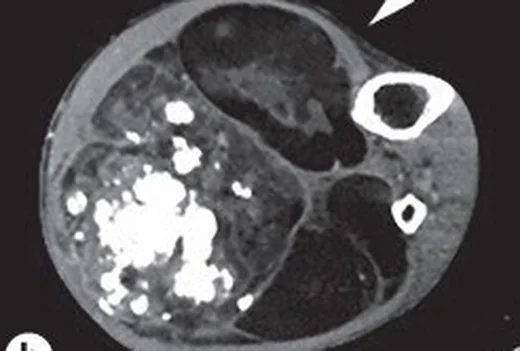
- Makroskopik yağ ağırlıklı kitle — tipik olarak subkutan yağla eşit veya düşük atenüasyonda görünür; ancak kalın septalar ve nodüler yumuşak doku bileşenleri nedeniyle heterojenlik ve lokal olarak daha yüksek yoğunluk gösterilebilir.
- Kalın septa — özellikle 2 mm'den kalın, düzensiz veya kontrastlanan fibröz bantlar benign lipoma göre ALT olasılığını yükseltir.
- Yağ dışı nodül — yağ zemininde yuvarlak ya da lobüle yumuşak doku bileşeni görülmesi şüphe uyandırır.
- Lobüle ve geniş kitle — büyük hacim, derin fasya altı konum ve kas kompartımanı boyunca uzanım ek kuşku işaretleridir.
- Septal/nodüler kontrastlanma — kontrastlı görüntüde vasküler yumuşak doku bileşenini makroskopik yağdan ayırır.
- Kas liflerinin kitlenin içinde tipik şeritler halinde korunmaması — intramüsküler lipomun araya giren lif görünümüne karşı uyarıcı olabilir.
- Komşu yapı etkisi — nörovasküler demetlerde kütle etkisi, kas planlarının kaybolması veya kemik korteksinde bası skulptrasyonu/infiltrasyonu izlenebilir.
Ölçütler ve sınıflamalar
- ALT şüphesini artıran kalın septa
- >2 mm
- Lipomatöz tümörde liposarkom olasılığını artıran büyük boyut (tek başına tanı koydurmaz)
- ≥10 cm
Normalde
Kas kompartımanlarında intermuscular adipose tissue, kas grupları arasında veya kas fasyasının altında yer alır. Lipomatöz kitleyi değerlendirirken septaların kalın veya düzensiz olmasına, nodül varlığına ve lezyon içinde kas liflerinin bulunup bulunmamasına bakın.
Normal BT ile kıyasla
Aynı bölgenin, kaynak veri setinde patoloji içermediği belirtilen bir BT incelemesini kesit kesit kaydırın; organ sınırlarını açarak anatomiyi hasta görüntüsüyle karşılaştırın. Küçük rastlantısal bulgular tümüyle dışlanmamıştır.
Kaynak: TotalSegmentator v2.01 veri seti (Wasserthal ve ark., Radiology: Artificial Intelligence 2023), CC BY 4.0, vaka s0685; organ sınırları veri setinin otomatik segmentasyonundan, birleştirilerek sadeleştirildi. Görüntüler 2,5 mm kesit aralığına yeniden örneklendi.
Ayırıcı tanı
- Konvansiyonel lipom
- genellikle homojen makroskopik yağdan oluşur; belirgin kalın septa veya nodüler nonadipoz bileşen beklenmez.
- İntramüsküler lipom
- yağ kas lifleri arasına şeritler halinde girer; bu lifler düzenli, lineer ve lezyon boyunca devamlı olabilir.
- Dediferansiye liposarkom
- yağlı kitle içinde belirgin, daha büyük ve solid yumuşak doku kitlesi veya agresif invazyon görülür.
- Hibernom
- yağ atenüasyonu klasik lipomdan yüksek, iç yapı heterojen ve vaskülarite belirgin olabilir.
- Elastofibrom
- tipik skapula altı yerleşimde fibröz yumuşak doku ile normal yağ bantlarının dönüşümlü dizilimi görülür.
Tuzaklar
- 2 mm üzerindeki septa ALT lehine olabilir ancak benign lipomda da kalın septa bildirilmiştir; bu eşik tek başına tanı ölçütü değildir.
- Büyük boyut olasılığı yükseltir fakat küçük ALT bulunabilir; yalnız çap üzerinden benign/malign sınıflaması yapmayın.
- Kontrastsız incelemede damar veya septa nodül gibi görünebilir; kontrast sonrası kesitlerde aynı odağın davranışını karşılaştırın.
- Lezyon yalnız yağdan oluşuyor gibi görünse de BT ALT'yi bütünüyle dışlamaz; derin, büyüyen veya klinikçe kuşkulu kitleyi kesin benign diye etiketlemeyin.
Kendini dene
Derin uyluk kitlesi çoğunlukla yağ yoğunluğunda; içinde kalın düzensiz septa ve kontrastlanan yumuşak doku odağı var. En olası tanı nedir?
Cevabı göster
Atipik lipomatöz tümör. Kalın septa ve kontrastlanan nonadipoz nodül ALT şüphesini artırır. Basit lipomlar genellikle homojen makroskopik yağ yapısındadır; hibernomlar ise belirgin vasküler yapılar ve beklenenden fazla kontrast tutulumuyla öne çıkar.
Yağ içeren kitlede belirgin kontrastlanan solid nodül komşu kas planını siliyor. Hangi ayırıcı tanı özellikle dışlanmalıdır?
Cevabı göster
Dediferansiye liposarkom. Yağlı zemin içinde belirgin solid kitle ve infiltratif davranış dediferansiyasyonu düşündürür. Konvansiyonel lipom homojen yağlıdır; elastofibrom çoğunlukla skapula altındadır; kas dejenerasyonu kitleleşen nodül oluşturmaz.
İlgili konular
Kaynaklar
Bu sayfadaki 41 cümle ve 2 quiz sorusu aşağıdaki kaynaklardan alıntılarla otomatik olarak karşılaştırıldı; 5 cümle bu karşılaştırmada düzeltildi (2026-10-09).
- Use of radiologic imaging to differentiate lipoma from atypical lipomatous tumor/well-differentiated liposarcoma: Systematic reviewpmc.ncbi.nlm.nih.gov
- Imaging review of lipomatous musculoskeletal lesionspmc.ncbi.nlm.nih.gov
- Primary endobronchial liposarcoma successfully resected via bronchoscopy: A rare case report with genetic analysis.pubmed.ncbi.nlm.nih.gov
- [A CASE OF WELL-DIFFERENTIATED LIPOSARCOMA IN THE RIGHT SCROTUM, DIAGNOSED AFTER LOCAL TUMOR EXCISION].pubmed.ncbi.nlm.nih.gov
- Spindle Cell Lipoma with Ossification Mimicking Atypical Lipomatous Tumor/Well-Differentiated Liposarcoma: A Case Report.pubmed.ncbi.nlm.nih.gov
- Case Report: Primary low-grade dedifferentiated liposarcoma of the urinary bladder with molecular confirmation.pubmed.ncbi.nlm.nih.gov
- Orbital liposarcoma.pubmed.ncbi.nlm.nih.gov
- Lipoblastoma of the extremities.pubmed.ncbi.nlm.nih.gov
- COMPUTED TOMOGRAPHIC FINDINGS IN 24 DOGS WITH LIPOSARCOMA.pubmed.ncbi.nlm.nih.gov
- Radiomic features of primary retroperitoneal sarcomas: a prognostic study.pubmed.ncbi.nlm.nih.gov
- Pleural lipoma: a non-surgical lesion?pubmed.ncbi.nlm.nih.gov
- Pancreatic liposarcoma: a case report.pubmed.ncbi.nlm.nih.gov
- Myxoid Liposarcoma Originating In The Anterior Mediastinum.pubmed.ncbi.nlm.nih.gov
- High-Grade Myxoid Liposarcoma (Round Cell Variant) in a Dog.pubmed.ncbi.nlm.nih.gov
- Pancreatic ductal adenocarcinoma arising from the pancreatic parenchyma compressed by a huge pancreatic lipoma: a case report.pubmed.ncbi.nlm.nih.gov
- Liposarcoma of the spermatic cord masquerading as an inguinal hernia.pubmed.ncbi.nlm.nih.gov
- Molecular mechanisms underlying liposarcoma subtype transition and proteasome-associated targeted interventions.pubmed.ncbi.nlm.nih.gov
- Unusual Retroperitoneal Soft Tissue Sarcomas: Giant Masqueraders.pubmed.ncbi.nlm.nih.gov
- Large lipoma of the finger masquerading as a giant cell tumor: A diagnostic pitfall in an uncommon site.pubmed.ncbi.nlm.nih.gov
- Intramuscular lipoma: a review of the literature.pubmed.ncbi.nlm.nih.gov
- A Rare Case Report on Dedifferentiated Liposarcoma of the Thigh Mimicking an Abscess With Vascular Features: A Diagnostic Challenge With Fatal Outcome.pubmed.ncbi.nlm.nih.gov
- Nasopharyngeal Lipoma Anterior to the Eustachian Tube Orifice: A Case Report.pubmed.ncbi.nlm.nih.gov
- Giant Retroperitoneal Liposarcoma Without Organ Invasion: A Rare Case Report.pubmed.ncbi.nlm.nih.gov
- MRI findings for differentiating benign and malignant soft tissue tumors: a systematic review-part 2: key imaging findings.pubmed.ncbi.nlm.nih.gov
- Indications and clinical outcomes in patients with soft-tissue sarcoma attached to the bone receiving tumorendoprosthetic replacement after tumor resection.pubmed.ncbi.nlm.nih.gov
- Diagnosis and Therapy of Soft Tissue Sarcomas: Spanish Group for Research in Sarcomas (GEIS) Guidelines.pubmed.ncbi.nlm.nih.gov
- CT and MRI features of tumors and tumor-like lesions in the abdominal wall.pubmed.ncbi.nlm.nih.gov
- Forefoot plantar multilobular noninfiltrating angiolipoma: a case report and review of the literature.pubmed.ncbi.nlm.nih.gov
- Diagnostic performance and dose reduction of virtual noncontrast imaging in pediatric computed tomography urography.pubmed.ncbi.nlm.nih.gov
- Diagnosis and Management of Soft-Tissue Sarcomas: Current Insights.pubmed.ncbi.nlm.nih.gov
- Percutaneous Management of Desmoid Tumors.pubmed.ncbi.nlm.nih.gov
- Fat-containing soft-tissue tumors: Imaging findings and pathologic correlation.pubmed.ncbi.nlm.nih.gov
- Imaging and Clinical Findings in a Series of Six Cases of Rare Primary Mediastinal Liposarcoma.pubmed.ncbi.nlm.nih.gov
- Soft tissue sarcomas of the proximal adductor area of the thigh: Indications, results and complications at medium follow-up in a series of 43 surgically treated patients.pubmed.ncbi.nlm.nih.gov
- Radiotherapy for soft tissue sarcoma of the proximal lower extremity.pubmed.ncbi.nlm.nih.gov
- Lipoid Pneumonia: HRCT and MRI Spectrum, Diagnostic Pitfalls, and Imaging-Based Diagnostic Workflow.pubmed.ncbi.nlm.nih.gov
- Imaging review of lipomatous musculoskeletal lesions.pubmed.ncbi.nlm.nih.gov
- Does composite bone resection for soft-tissue sarcoma with cortical contact result in better local control and survival compared to sub-periosteal dissection? : a comparative retrospective cohort study.pubmed.ncbi.nlm.nih.gov
- Practical approach to ultrasound of soft tissue tumors and the added value of MRI: how I do it.pubmed.ncbi.nlm.nih.gov
- Lump on back: a rare case of parosteal lipoma of scapula.pubmed.ncbi.nlm.nih.gov
- Imaging of acute musculoskeletal infections in children and their differential diagnoses.pubmed.ncbi.nlm.nih.gov
- Intermuscular Adipose Tissue as a Multimodality Quantitative Imaging Biomarker: Evidence Across Metabolic, Cardiovascular and Oncologic Diseases.pubmed.ncbi.nlm.nih.gov
- Imaging approach to retroperitoneal vascular tumors: a narrative review.pubmed.ncbi.nlm.nih.gov
- Sequential EBUS-Guided Fine Needle Aspiration and Transbronchial Cryobiopsy for the Diagnosis of a Giant Mediastinal Lipoma: A Novel Intraoperative Pivot Strategy.pubmed.ncbi.nlm.nih.gov
- Common cystic pancreatic neoplasms: A comprehensive magnetic resonance imaging-based review of typical and atypical imaging features.pubmed.ncbi.nlm.nih.gov
- Giant retroperitoneal dedifferentiated liposarcoma with intraperitoneal extension: A case report.pubmed.ncbi.nlm.nih.gov
- Malignant phyllodes tumor of the breast with heterologous osteosarcomatous and pleomorphic liposarcomatous differentiation: a rare case report and literature review.pubmed.ncbi.nlm.nih.gov
- Myxoid liposarcoma of sigmoid mesocolon: a case report and review of the literature.pubmed.ncbi.nlm.nih.gov
- Imaging and clinicopathological features of a high-SUV hibernoma mimicking liposarcoma on PET/CT, MRI, and CT: A case report.pubmed.ncbi.nlm.nih.gov
- A pictorial essay of thoracic wall diseases: multiple pathologies in the same anatomical site.pubmed.ncbi.nlm.nih.gov
- Elastofibroma dorsi: case report and review of the literature.pubmed.ncbi.nlm.nih.gov
- Elastofibroma dorsi: clinicopathological analysis of 76 cases.pubmed.ncbi.nlm.nih.gov
- Developing a diagnostic model to differentiate the well-differentiated lipomatous tumors based on clinicopathological characteristics.pubmed.ncbi.nlm.nih.gov
- Bayesian additive regression trees for machine learning to classify benign vs atypical lipomatous tumors on MRI.pubmed.ncbi.nlm.nih.gov
- Use of radiologic imaging to differentiate lipoma from atypical lipomatous tumor/well-differentiated liposarcoma: Systematic review.pubmed.ncbi.nlm.nih.gov
- Atypical Spindle Cell/Pleomorphic Lipomatous Tumor with Atypical Imaging Features.pubmed.ncbi.nlm.nih.gov
- Well-Differentiated Liposarcoma of the Spermatic Cord Involving Testicular Parenchyma.pubmed.ncbi.nlm.nih.gov
- Gynecologic imaging errors and pitfalls: how to avoid misdiagnosis on pelvic MRI.pubmed.ncbi.nlm.nih.gov
- Benign focal liver lesions: The role of magnetic resonance imaging.pubmed.ncbi.nlm.nih.gov
- Understanding a mass in the paraspinal region: an anatomical approach.pubmed.ncbi.nlm.nih.gov
- Combined Extra- and Intrathoracic (Dumbbell) Lipoma Crossing the Third Intercostal Space.pubmed.ncbi.nlm.nih.gov
- State of the art of 18 F-FDG PET/CT application in inflammation and infection: a guide for image acquisition and interpretation.pubmed.ncbi.nlm.nih.gov
- Pictorial Review of Soft Tissue Lesions with Calcification.pubmed.ncbi.nlm.nih.gov
- Surface bone sarcomas: an update on current clinicopathological diagnosis and treatment.pubmed.ncbi.nlm.nih.gov
- Pleomorphic liposarcoma of bone: a rare primary malignant bone tumour.pubmed.ncbi.nlm.nih.gov
- Sarcoma or haematoma? If only it was that simple! Part 2.pubmed.ncbi.nlm.nih.gov
- CT and MRI Key Features of Benign Tumors and Tumor-like Lesions of the Tongue: A Pictorial Review.pubmed.ncbi.nlm.nih.gov
- A diagnostic approach to the mediastinal masses.pubmed.ncbi.nlm.nih.gov
- When Fat Takes Over: A Progressively Enlarging Giant Hand Lipoma.pubmed.ncbi.nlm.nih.gov
- Case Report: Laparoscopic repair of indirect inguinal hernia combined with ipsilateral giant subcutaneous inguinoperineal lipoma.pubmed.ncbi.nlm.nih.gov
- Differentiation of Atypical Lipomatous Tumors from Lipomas: Our Experience with Visual Analysis of Conventional Magnetic Resonance Imaging.pubmed.ncbi.nlm.nih.gov
- MRI characteristics of appendicular soft tissue lymphoma: a retrospective analysis.pubmed.ncbi.nlm.nih.gov
- Minimal One-Quarter Incision and Four-Step (MOQIF) Excision Method for Subcutaneous Lipoma.pubmed.ncbi.nlm.nih.gov
- Multiparametric US and MRI Features of Femoral Myxoid Liposarcoma-Case Report and Literature Review.pubmed.ncbi.nlm.nih.gov
- Adult retroperitoneal Ganglioneuroma: Case report of an incidental mass treated with robotic excision.pubmed.ncbi.nlm.nih.gov
- Abdominal Wall in Cross-Sectional Imaging as an Essential Knowledge for Radiologists: A Pictorial Review.pubmed.ncbi.nlm.nih.gov
- FocusedON-BC: A Robust Deep Learning Framework for Automated Body Composition Assessment.pubmed.ncbi.nlm.nih.gov
- Methodological standards for body composition assessment-an expert-endorsed guide for research and clinical applications: bioimpedance, dual-energy X-ray absorptiometry, computerized tomography, and ultrasound methods.pubmed.ncbi.nlm.nih.gov
- Intramuscular Fat as a Biomarker of Muscle Health: Strengths, Limitations and Open Challenges.pubmed.ncbi.nlm.nih.gov
- An Unusual Lipoma at the Lateral Margin of the Tongue: A Case Report.pubmed.ncbi.nlm.nih.gov
- Madelung's disease involving the scrotum: a case report and diagnostic pitfalls with liposarcoma.pubmed.ncbi.nlm.nih.gov
- Fascia Lata and Tensor Fasciae Latae in Lower-Limb Reconstruction.pubmed.ncbi.nlm.nih.gov
- Soft Tissue Masses of the Hand.pubmed.ncbi.nlm.nih.gov
- Diffuse lipofibromatosis with hand bone involvement: expanding the clinical spectrum: A case report and literature review.pubmed.ncbi.nlm.nih.gov
- Recognizing the Benign Behind Worrisome Histology: A Case Report of Proliferative Fasciitis.pubmed.ncbi.nlm.nih.gov
- The space of Retzius: anatomy, imaging features, and patterns of disease spread.pubmed.ncbi.nlm.nih.gov
- Navigating pelvic anatomy for exenteration: a clinical guide for radiologists.pubmed.ncbi.nlm.nih.gov
- Tumors and pseudotumors of the soft tissues: Imaging semiology and strategy.pubmed.ncbi.nlm.nih.gov
Eğitim amaçlıdır; hasta başında tanı ve tedavi kararının yerine geçmez, tıbbi tavsiye değildir. Metinler yapay zekâ desteğiyle yazıldı ve otomatik bir adımda kaynak alıntılarıyla karşılaştırıldı; bu bir tıbbi doğrulama değildir ve içerik uzman radyolog incelemesinden geçmedi. Klinik kararlarda güncel kılavuzları, kurum protokollerini ve radyoloji raporunu esas alın.