Özet
Koroner köken ve seyir anomalileri, bir damarın olağan aort sinüsünden çıkmaması veya başlangıçtan itibaren farklı bir rota izlemesiyle oluşan konjenital anatomik farklılıklardır. Karşı sinüsten çıkan damarda interarteriyel ya da aort duvarı içi segment, dar yarık biçimli ostium ve keskin çıkış açısı özellikle dikkatle tarif edilir. EKG eşzamanlı koroner BT anjiyografi, ostiumu ve proksimal damarı aort ile pulmoner arter ilişkisi içinde yüksek uzaysal çözünürlükle gösterir. Anormal başlangıç veya risk taşıyan proksimal segment gözden kaçarsa klinik olarak önemli koroner anatomi raporlanmayabilir.
Faz ve pencere
- BTA tanısal
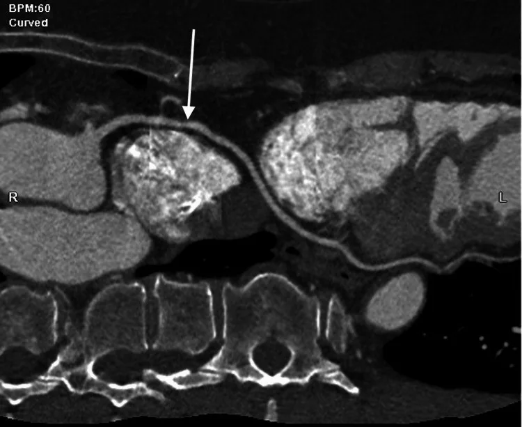
- EKG eşzamanlı ince kesit koroner BT anjiyografide aort kökü ve pulmoner trunkusun yeterli opasifikasyonunu kullanarak her ostiumdan proksimal koroner yatağa ilerle. Ostiumun sinüsünü, çıkış açısını, aort duvarı içindeki segmenti ve damarın büyük damarların önünden, arasından ya da arkasından geçtiği rotayı merkez hat ve oblik rekonstrüksiyonlarda belirle.
Önerilen pencereler: Anjiyo (G 600 / M 150), Mediasten (G 350 / M 50).
BT bulguları
- Ostiumun kökeni — sağ, sol veya nonkoroner sinüs; karşı sinüs, yüksek aort ya da pulmoner arter çıkışını adlandır.
- Ostium biçimi ve çıkış açısı — yarık görünüm, dar giriş veya aort duvarına teğet başlangıcı koronal-oblik ve kısa eksen kesitlerinde incele.
- İnterarteriyel rota — başlangıç segmentinin aorta ile pulmoner trunkus arasındaki koridordan geçtiğini damar boyunca doğrula.
- İntramural segment — koroner lümenin bir bölümünün aort duvarı içinde ve aort lümenine komşu seyrettiğini göster.
- Retroaortik rota — damar aort kökünün arkasından dolaşıp karşı tarafın koroner dağılımına yönelir.
- Prepulmonik rota — koroner dal pulmoner trunkusun önünden kalbin ön yüzüne doğru geçer.
- Transseptal/subpulmonik rota — damar miyokard içinden, septum boyunca veya pulmoner kökün altından seyreder.
- Retrokardiyak rota — anormal koroner dal aort kökünün arkasından geçer; bu anatomik varyant güncel sınıflamalarda retroaortik rota olarak adlandırılır ve genellikle hemodinamik önemi yoktur.
- Proksimal lümen biçimi — eliptikleşme ya da odaksal daralma varsa damarı merkez hattına dik kısa eksen kesitlerinde doğrula.
Ölçütler ve sınıflamalar
- Karşı sinüsten köken alan koroner arterin proksimal seyri
- Tek bir evrensel sınıflama yoktur. Anormal kökenli koroner arterin proksimal seyri interarteriyel, prepulmonik, retroaortik veya transseptal olarak adlandırılır. Aort duvarı içindeki intramural segment ayrı bir anatomik özellik ve olası risk morfolojisi olarak ayrıca belirtilir.
Normalde
Normal başlangıçta sağ koroner arter sağ koroner sinüsten, sol ana koroner arter sol koroner sinüsten çıkar; proksimal segmentler aort duvarı içinde uzun bir rota izlemez. Kıyaslamada sinüsleri ve ostiumları tanımladıktan sonra her damarı merkez hattı boyunca takip et; böylece duvar içi bölüm ile damarların önünden, arasından veya arkasından geçen rotayı ayırabilirsin.
Normal BT ile kıyasla
Aynı bölgenin, kaynak veri setinde patoloji içermediği belirtilen bir BT incelemesini kesit kesit kaydırın; organ sınırlarını açarak anatomiyi hasta görüntüsüyle karşılaştırın. Küçük rastlantısal bulgular tümüyle dışlanmamıştır.
Kaynak: TotalSegmentator v2.01 veri seti (Wasserthal ve ark., Radiology: Artificial Intelligence 2023), CC BY 4.0, vaka s0977; organ sınırları veri setinin otomatik segmentasyonundan, birleştirilerek sadeleştirildi. Görüntüler 2,5 mm kesit aralığına yeniden örneklendi.
Ayırıcı tanı
- Koroner arter fistülü
- geniş veya kıvrımlı besleyici damar kalp boşluğuna ya da büyük damara sonlanır; anormal köken-seyir lezyonu değildir.
- Miyokard köprüsü
- ostium olağan sinüste kalırken epikardiyal arterin bir segmenti miyokard içinden geçer.
- Sol ana koroner arter atrezisi, koroner arter anomalileri arasında yer alır.
- Pulmoner arterden koroner çıkış
- ostium aort sinüsü yerine pulmoner arter duvarından kaynaklanır.
- Cerrahi reimplantasyon
- koroner ağız doğumsal varyant gibi görünebilir; ameliyat hattı, greftler ve eski görüntüler kaynak yerini açıklar.
Tuzaklar
- Bir damarın tek kesitte aorta ile pulmoner arter arasında görünmesi rotayı kanıtlamaz; ostiumdan distal segmente kesintisiz merkez hat oluştur.
- Aort duvarı içindeki bölümle iki büyük damar arasındaki rota farklı özelliklerdir; aynı koroner arterde art arda bulunabileceklerini raporda ayrı yaz.
- Karşı sinüsten çıkan tüm arterler aynı anatomiye sahip değildir; pulmoner kök önü ve aort arkası rotalarını damarlar arası geçişle karıştırma.
Kendini dene
Bir yenidoğanda sol koroner sistemin pulmoner arterden kaynaklandığı görülüyor. Bu anatomik sendromun adı hangisidir?
Cevabı göster
Bland-White-Garland sendromu. Sol koroner arterin pulmoner arterden çıkışı ALCAPA sendromu olarak bilinir ve tedavi edilmezse yaşamı tehdit eder. ARCAPA sağ koroner arterin pulmoner kökenden çıkışıdır; diğer seçenekler koroner köken anomalisi değildir.
Aort kökünden ortak tek ostiumla başlayan ve iki ana koroner dala ayrılan konjenital varyant nasıl adlandırılır?
Cevabı göster
Tek koroner arter. Tek aortik ostiumdan çıkan ortak koroner gövde, distal dallanma gösterse de tek koroner arter varyantıdır. Fistülde anormal boşalma bağlantısı, köprüde kas içi segment, atrezide ise lümen yokluğu veya kesintisi söz konusudur.
İlgili konular
Kaynaklar
Bu sayfadaki 42 cümle ve 2 quiz sorusu aşağıdaki kaynaklardan alıntılarla tek tek karşılaştırıldı; 5 cümle bu karşılaştırmada düzeltildi (2026-10-08).
- Coronary Artery Anomalies: A Computed Tomography Angiography Pictorial Reviewpmc.ncbi.nlm.nih.gov
- [ANOCOR registry].pubmed.ncbi.nlm.nih.gov
- Coronary anomalies: what the radiologist should know.pubmed.ncbi.nlm.nih.gov
- Coronary CTA assessment of coronary anomalies.pubmed.ncbi.nlm.nih.gov
- Coronary artery anomalies evaluation with cardiac computed tomography: A review.pubmed.ncbi.nlm.nih.gov
- Coronary artery abnormalities and sudden cardiac death.pubmed.ncbi.nlm.nih.gov
- Overview of coronary artery variants, aberrations and anomalies.pubmed.ncbi.nlm.nih.gov
- Anomalies of coronary artery origin: Evaluation on multidetector CT angiography.pubmed.ncbi.nlm.nih.gov
- One Ostium to Rule Them All: Rare Case of a Single Coronary Artery.pubmed.ncbi.nlm.nih.gov
- Automated Quantitative Stress Perfusion Cardiac Magnetic Resonance in Pediatric Patients.pubmed.ncbi.nlm.nih.gov
- Single coronary artery: spectrum of imaging findings with multidetector CT.pubmed.ncbi.nlm.nih.gov
- Anomalous origination of a coronary artery from the opposite sinus.pubmed.ncbi.nlm.nih.gov
- Anomalous origin of the right coronary artery.pubmed.ncbi.nlm.nih.gov
- The first multicentre study on coronary anomalies in the Netherlands: MuSCAT.pubmed.ncbi.nlm.nih.gov
- Coronary Arterial Anomalies in Adults with Congenital Heart Disease: From Detection to Management Strategies.pubmed.ncbi.nlm.nih.gov
- Circumaortic Configuration of the Left Coronary Arteries.pubmed.ncbi.nlm.nih.gov
- Single Coronary Artery in the Context of Vascular Disease: Anatomy, Development, Multimodality Imaging, and Clinical Interpretation.pubmed.ncbi.nlm.nih.gov
- Classification, diagnosis and clinical strategy of congenital coronary artery disease in children.pubmed.ncbi.nlm.nih.gov
- Anomalous origin of left main coronary artery from right coronary artery in a patient presenting with inferior wall myocardial infarction: a case report and literature review.pubmed.ncbi.nlm.nih.gov
- Coronary artery anomalies overview: The normal and the abnormal.pubmed.ncbi.nlm.nih.gov
- Bringing paediatric coronary artery abnormalities into view: the role of virtual reality.pubmed.ncbi.nlm.nih.gov
- Evaluation of High-Risk Anatomical Features in Single and Opposite-Sinus Origin Coronary Artery Anomalies Using Coronary Computed Tomography Angiography.pubmed.ncbi.nlm.nih.gov
- The Initial Diagnostic Approach to Adult Congenital Heart Disease: A Practical Imaging Pathway for General Cardiologists and Cardiac Imagers.pubmed.ncbi.nlm.nih.gov
- Incidental noninvasive diagnosis of isolated persistent left superior vena cava in asymptomatic adults: A case report.pubmed.ncbi.nlm.nih.gov
- High-risk anatomical features: a new perspective on refined echocardiographic diagnosis of anomalous coronary artery origin from the opposite sinus.pubmed.ncbi.nlm.nih.gov
- Cardiovascular Computed Tomography Angiographic Assessment of Simple Cardiac Shunts in Adults.pubmed.ncbi.nlm.nih.gov
- Complex adult congenital heart disease on cross-sectional imaging: an introductory overview.pubmed.ncbi.nlm.nih.gov
- Coronary Artery Origin Anomalies on Coronary CT Angiography: A Single-Center Tertiary-Care Cohort.pubmed.ncbi.nlm.nih.gov
- Image and Clinical Characteristics of the Right Coronary Artery Originating From the Left Coronary Sinus: A Database Review.pubmed.ncbi.nlm.nih.gov
- Aberrant Right Coronary Artery from the Left Coronary Sinus Presenting with Inferior Wall Myocardial Infarction.pubmed.ncbi.nlm.nih.gov
- Risk stratification with exercise N(13)-ammonia PET in adults with anomalous right coronary arteries.pubmed.ncbi.nlm.nih.gov
- Long intramural left coronary artery confined to the left sinus of Valsalva with diastolic deformation: a case report.pubmed.ncbi.nlm.nih.gov
- Histological assessment of the intramural segment in adults with an anomalous aortic origin of a coronary artery with an interarterial course.pubmed.ncbi.nlm.nih.gov
- Coronary Artery Anomalies: A Computed Tomography Angiography Pictorial Review.pubmed.ncbi.nlm.nih.gov
- Exercise stress cardiovascular magnetic resonance imaging is feasible in adolescents and young adults with anomalous coronary arteries.pubmed.ncbi.nlm.nih.gov
- Clinical and CT Features, Clinical Management, and Decision on Sport Eligibility of Professional Athletes with Congenital Coronary Anomalies: A Case Series Study.pubmed.ncbi.nlm.nih.gov
- Coronary Artery Anomalies Revisited: Description of the Types, Pathophysiology and Treatment Options Based on Latest Guidelines.pubmed.ncbi.nlm.nih.gov
- Ablation in Adult Congenital Heart Disease Using a Dual-Energy, Lattice-Tip, Large-Footprint, Map-and-Ablate Catheter.pubmed.ncbi.nlm.nih.gov
- Parent Artery Disease-Related Stroke: What Is the Impact on Endovascular Treatment? A Narrative Review.pubmed.ncbi.nlm.nih.gov
- Exploring the Association Between Heart Rate Variability and Intracranial Atherosclerosis in Middle-Aged or over Community-Dwelling Adults.pubmed.ncbi.nlm.nih.gov
- 2024 Consensus Statement on Coronary Stenosis and Plaque Evaluation in CT Angiography From the Asian Society of Cardiovascular Imaging-Practical Tutorial (ASCI-PT).pubmed.ncbi.nlm.nih.gov
- Application of postmortem imaging modalities in cases of sudden death due to cardiovascular diseases-current achievements and limitations from a pathology perspective : Endorsed by the Association for European Cardiovascular Pathology and by the International Society of Forensic Radiology and Imaging.pubmed.ncbi.nlm.nih.gov
- Coronary artery fistulas: a systematic review and pooled analysis of anatomy, risk stratification and contemporary management.pubmed.ncbi.nlm.nih.gov
- Assessment of Coronary Arteries during the Prenatal and Postnatal Periods using Two- and Three-dimensional Ultrasound.pubmed.ncbi.nlm.nih.gov
- CCTA of Pediatric Congenital Right Heart Obstructive Lesions: A Pictorial Review.pubmed.ncbi.nlm.nih.gov
- Chest Discomfort: Could Coronary Pathology Extend Beyond Atherosclerosis?pubmed.ncbi.nlm.nih.gov
- Case Report: Low-profile covered stents in CHD: the role of Gore Viabahn (VBX) stents.pubmed.ncbi.nlm.nih.gov
- Computational studies on visceral artery lesions and aortic pathologies involving visceral branches: a comprehensive review for the clinician.pubmed.ncbi.nlm.nih.gov
- Intrapleural left axillary artery bypass grafting compromising subsequent left internal thoracic artery harvesting: a case report.pubmed.ncbi.nlm.nih.gov
- Transcatheter Aortic Valve Replacement After David Procedure for Severe Aortic Regurgitation.pubmed.ncbi.nlm.nih.gov
- Towards Fixing Vessel Segmentation Breakage: A Systematic Review.pubmed.ncbi.nlm.nih.gov
- Comparative Efficacy of Integrated Stent-Graft versus Conventional Covered Stent-Graft Alone in Type B Aortic Dissection: A Retrospective Study Focusing on Aortic Remodeling and Branch Perfusion.pubmed.ncbi.nlm.nih.gov
- Cardiac Computed Tomography in Structural Heart Interventions: From Preprocedural Planning to Procedural Strategy.pubmed.ncbi.nlm.nih.gov
- Outcomes after surgical management of anomalous aortic origin of a coronary artery (AAOCA): an up-to-date systematic review and meta-analysis.pubmed.ncbi.nlm.nih.gov
- The Incidence, Impact, and Techniques of Commissural Alignment in Transcatheter Aortic Valve Implantation: A Review.pubmed.ncbi.nlm.nih.gov
- Usefulness of Routine Coronary CT Angiography in Patients with Transposition of the Great Arteries After an Arterial Switch Operation.pubmed.ncbi.nlm.nih.gov
- Detectability of intracranial vessel wall atherosclerosis using black-blood spectral CT: a phantom and clinical study.pubmed.ncbi.nlm.nih.gov
- Mechanisms of aortic dissection: From pathological changes to experimental and <i>in silico</i> models.pubmed.ncbi.nlm.nih.gov
- State-of-the-art CT and MR imaging and assessment of atherosclerotic carotid artery disease: the reporting-a consensus document by the European Society of Cardiovascular Radiology (ESCR).pubmed.ncbi.nlm.nih.gov
- Intracardiac Echocardiography during Invasive Electrophysiological Procedures: a Scientific Statement of the European Heart Rhythm Association of the ESC, and the European Association of Percutaneous Cardiovascular Interventions of the ESC.pubmed.ncbi.nlm.nih.gov
- From Modified Haller Index to a Novel Patented Anatomical Measurement Device: Engineering Development, Validation, and Clinical Applications of Non-Invasive Thoracic Morphometry.pubmed.ncbi.nlm.nih.gov
- Bicuspid Aortic Valve Disease-Associated Aortopathy in Pediatric Subjects-From Traditional Assessment to Current Advances and Future Perspectives: A Narrative Review.pubmed.ncbi.nlm.nih.gov
- Case Report: A rare case of coronary artery anomaly (right coronary artery-pulmonary artery fistula with diffuse dilatation).pubmed.ncbi.nlm.nih.gov
- CT Angiography for Aortic Arch Anomalies: Prevalence, Diagnostic Efficacy, and Illustrative Findings.pubmed.ncbi.nlm.nih.gov
- MDCT Imaging of Non-Traumatic Thoracic Aortic Emergencies and Its Impact on Diagnosis and Management-A Reappraisal.pubmed.ncbi.nlm.nih.gov
- Cardioplegia strategies in ALCAPA surgery: A narrative review on practical comparison of techniques.pubmed.ncbi.nlm.nih.gov
- Congenital Aortic-Left Atrial Tunnel with Coarctation and Anomalous Left Coronary Artery from the Pulmonary Artery: A First-of-its-kind Case Report.pubmed.ncbi.nlm.nih.gov
- Anomalous Origin of Left Anterior Descending and Left Circumflex Arteries From the Proximal Right Coronary Arteries Individually With an Absent Left Main Coronary Artery: A Case Report.pubmed.ncbi.nlm.nih.gov
- Anomalous aortic origin of a coronary artery in 16,680 patients evaluated by cardiac computed tomography.pubmed.ncbi.nlm.nih.gov
- Imaging considerations in tetralogy of Fallot: A comprehensive review.pubmed.ncbi.nlm.nih.gov
- A Case Series of Right Coronary Artery Anomalies With a Malignant Course.pubmed.ncbi.nlm.nih.gov
- First- and Second-Trimester Cardiovascular Anomalies in Trisomy 21 Fetuses: Anatomy, Embryology, Genetics and Imaging.pubmed.ncbi.nlm.nih.gov
- Contemporary sequential segmental approach to congenital heart disease using four-dimensional magnetic resonance imaging with ferumoxytol: an illustrated editorial.pubmed.ncbi.nlm.nih.gov
- Materials Advances in Devices for Heart Disease Interventions.pubmed.ncbi.nlm.nih.gov
- 2025 Heart Disease and Stroke Statistics: A Report of US and Global Data From the American Heart Association.pubmed.ncbi.nlm.nih.gov
- Recurrent Syncope and Sudden Cardiac Death due to Anomalous Origin of the Right Coronary Artery From the Pulmonary Artery (ARCAPA): A Case Report.pubmed.ncbi.nlm.nih.gov
- Detection of anomalous aortic origin of a coronary artery (AAOCA) by echocardiogram: When does computed tomographic angiography add value?pubmed.ncbi.nlm.nih.gov
- Anomalous left coronary artery from the pulmonary artery (ALCAPA) in adult patients - a multimodality imaging approach.pubmed.ncbi.nlm.nih.gov
- Congenital anomalies of coronary arteries: Diagnosis with 64 slice multidetector CT.pubmed.ncbi.nlm.nih.gov
- Right coronary artery originated from left coronary sinus associated with children hypertrophic cardiomyopathy: report of two cases and literature review.pubmed.ncbi.nlm.nih.gov
- Anomalous Left Coronary Artery Originating From the Right Coronary Sinus With a Septal Curse in a Patient With Myocardial Infarction Due to Right Coronary Lesion: Innocent Until Proven Otherwise.pubmed.ncbi.nlm.nih.gov
- Management of Pediatric Pulmonary Vein Stenosis.pubmed.ncbi.nlm.nih.gov
- Left Main Coronary Artery Hypoplasia in Elderly.pubmed.ncbi.nlm.nih.gov
- Anomalous single coronary artery on low dose MDCT.pubmed.ncbi.nlm.nih.gov
- Voxel-scale diffusion tensor phenomapping of the healthy and pressure-overloaded human heart.pubmed.ncbi.nlm.nih.gov
- Cardiac phase variability in pulmonary root geometry: implications for electrophysiological mapping and ablation.pubmed.ncbi.nlm.nih.gov
- Hemodynamic Impact of the Aberrant Subclavian Artery: A CFD Investigation.pubmed.ncbi.nlm.nih.gov
- The lone lifeline-a rare case report of a single right coronary circulation with hypoplastic left coronary system.pubmed.ncbi.nlm.nih.gov
- Stress CT myocardial perfusion imaging after arterial switch operation for transposition of the great arteries.pubmed.ncbi.nlm.nih.gov
- A novel multimodal imaging approach for working diagnosis of acute myocardial infarction with non-obstructive coronary arteries: a promising diagnostic strategy.pubmed.ncbi.nlm.nih.gov
- Coronary artery anomalies: what are they? when to suspect? how to treat?-a narrative review.pubmed.ncbi.nlm.nih.gov
- Spontaneous Coronary Artery Dissection in Complex Congenital Coronary Anatomy.pubmed.ncbi.nlm.nih.gov
- Duodenal Obstruction and Grade III Acute Cholangitis Following Pancreaticoduodenal Artery Aneurysm Rupture During Conservative Management of Acute Type B Aortic Dissection: A Case Report.pubmed.ncbi.nlm.nih.gov
Eğitim amaçlıdır; hasta başında tanı ve tedavi kararının yerine geçmez. İçerik yapay zekâ destekli bir süreçle güncel literatüre göre cümle cümle kaynaklarla karşılaştırıldı, ancak uzman radyolog incelemesinden geçmedi. Klinik kararlarda güncel kılavuzları, kurum protokollerini ve radyoloji raporunu esas alın.