Özet
Künt göğüs travması veya yüksek enerjili yavaşlama sonrası koroner arter diseksiyonu gelişebilir. Koroner arter diseksiyonu künt göğüs travmasının seyrek ve yaşamı tehdit edebilen bir komplikasyonudur; eşlik eden yaralanmalar koroner hasar veya infarktüs tanısının gözden kaçmasına yol açabilir. Koroner BT anjiyografi, travmatik koroner yaralanma şüphesinde koroner anatomiyi değerlendirmeye yardımcı olabilir; metal artefaktı tanısal görüntü kalitesini sınırlayabilir. Travma BT'sinde koroner arterlerin değerlendirilmesi teknik olarak kısıtlanabilir; metal artefaktı tanısal görüntü kalitesini düşürebilir.
Faz ve pencere
- BTA tanısal
- Travmatik koroner yaralanma şüphesinde koroner BT anjiyografi koroner anatomiyi değerlendirmeye yardımcı olabilir; metal artefaktı görüntü kalitesini sınırlayabilir.
- Arteriyel
- Travma BT'sinde perikardiyal hematom ve odaksal kontrast ekstravazasyonu görülebilir; koroner BT anjiyografi, şüpheli travmatik koroner yaralanmada koroner anatomiyi değerlendirmeye yardımcı olur.
Önerilen pencereler: Anjiyo (G 600 / M 150), Mediasten (G 350 / M 50), Yumuşak doku (G 400 / M 40).
BT bulguları
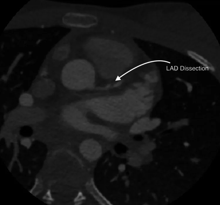
- Koroner arter diseksiyonu, künt göğüs travması sonrası gelişebilen yapısal bir koroner yaralanmadır.
- Fokal düzensiz daralma, ani kalibrasyon kaybı ya da kontrast dolmayan segment tromboz/oklüzyon lehinedir.
- Damar dışına kontrast geçişi, koroner yaralanma ve aktif kanama olasılığını düşündürür.
- LAD yaralanmasında anterior duvar ve septum miyokart kontrastlanması özellikle gözden geçirilir; LAD apeksi besliyorsa apeks de değerlendirilir.
- Miyokart kontrastlanma azalması koroner tıkanıklıkla ilişkili hipoperfüzyonda görülebilir; travma sonrası kontüzyon da fokal düşük atenüasyon oluşturabildiğinden BT bulgusu tek başına nedeni ayırt ettirmez.
- Hemoperikard veya perikardiyal hematom koroner duvar rüptürüne eşlik edebilir.
- Kalp hareketi ve yetersiz koroner opasifikasyon lümen konturunu bozarak yalancı darlık veya değerlendirme kısıtlılığı oluşturabilir.
- Koroner yaralanma travma sonrası başlangıçta belirgin olmayabilir; yeni iskemi bulguları, eşlik eden koroner hasar olasılığının yeniden değerlendirilmesini gerektirebilir.
Normalde
Şüpheli koroner lümen bulgusunu miyokart perfüzyonuyla birlikte değerlendirin; LAD dolum kusuruna sol ventrikül hipoperfüzyonu eşlik edebilir.
Normal BT ile kıyasla
Aynı bölgenin, kaynak veri setinde patoloji içermediği belirtilen bir BT incelemesini kesit kesit kaydırın; organ sınırlarını açarak anatomiyi hasta görüntüsüyle karşılaştırın. Küçük rastlantısal bulgular tümüyle dışlanmamıştır.
Kaynak: TotalSegmentator v2.01 veri seti (Wasserthal ve ark., Radiology: Artificial Intelligence 2023), CC BY 4.0, vaka s0977; organ sınırları veri setinin otomatik segmentasyonundan, birleştirilerek sadeleştirildi. Görüntüler 2,5 mm kesit aralığına yeniden örneklendi.
Ayırıcı tanı
- Koroner ateroskleroz
- kalsifiye veya yoğun nonkalsifiye plak lümen daralmasına yol açabilir; plak morfolojisi diseksiyondan farklıdır.
- Koroner vazospazm
- travma sonrası koroner daralma ve iskemiye eşlik edebilir; tek başına lümen daralması diseksiyon tanısı koydurmaz.
- Spontan koroner arter diseksiyonu
- travma öyküsü yerine spontan iskemi bağlamı öne çıkar; BT morfolojisi örtüşebilir.
- Hareket artefaktı
- kalp hareketi koroner sınırları bulanıklaştırabilir ve komşu kesitlerde yinelenmeyen yalancı çift kontur oluşturabilir.
- Miyokart kontüzyonu
- miyokart hipoenhansmanı görülebilir; bu bulgu tek başına koroner lümen hasarını kanıtlamaz.
Tuzaklar
- Kalp hareketi koroner boyunca basamaklanma yaratır; olası flebin aynı düzlemde ardışık kesitlerde sürüp sürmediğini ve ortogonal rekonstrüksiyonları kontrol edin.
- Koroner ostium çevresindeki hareket artefaktı lümen konturunu bulanıklaştırabilir; ostiumu ardışık kesit ve farklı düzlemde yeniden izleyin.
- Rutin travma BT’sinde, özellikle metal artefaktı koroner görünürlüğü bozuyorsa, damar değerlendirmesi kısıtlanabilir; şüpheli bölgede çok düzlemli rekonstrüksiyonlar değerlendirmeye yardımcı olur.
Kendini dene
Travmatik koroner BT anjiyografide sağ koroner arterin akut marjinal dalından kontrast ekstravazasyonu ve perikardiyal hematom görülüyor. En olası tanı hangisidir?
Cevabı göster
Travmatik koroner arter yaralanması. Koroner daldan kontrast ekstravazasyonu ve çevresindeki perikardiyal hematom travmatik koroner arter yaralanmasını destekler.
Travmatik koroner BT anjiyografide damar görünürlüğünü sınırlayabilecek bulgu hangisidir?
Cevabı göster
Metal artefaktı. Metal artefaktı koroner BT anjiyografide tanısal görüntü kalitesini sınırlayabilir.
İlgili konular
Kaynaklar
Bu sayfadaki 33 cümle ve 2 quiz sorusu aşağıdaki kaynaklardan alıntılarla otomatik olarak karşılaştırıldı; 25 cümle bu karşılaştırmada düzeltildi (2026-10-09).
- ACR Appropriateness Criteria® Blunt Chest Trauma-Suspected Cardiac Injurypubmed.ncbi.nlm.nih.gov
- Imaging traumatic cardiac injury: A mechanism-based diagnostic approachpubmed.ncbi.nlm.nih.gov
- Traumatic Coronary Artery Dissection Diagnosed Immediately with Cardiac CTemjreviews.com
- Early detection of myocardial infarction following blunt chest trauma by computed tomography: a case reportlink.springer.com
- Imaging in blunt cardiac injury: Computed tomographic findings in cardiac contusion and associated injuriespubmed.ncbi.nlm.nih.gov
- Cardiac Injuries: A Review of Multidetector Computed Tomography Findingspubmed.ncbi.nlm.nih.gov
- Spontaneous Coronary Artery Dissection: Current State of the Science: A Scientific Statement From the American Heart Associationpmc.ncbi.nlm.nih.gov
- Coronary Artery Anatomyaats.org
- Coronary artery rupture in blunt thoracic trauma: a case report and review of literaturepubmed.ncbi.nlm.nih.gov
- Imaging Features of Complications after Coronary Interventions and Surgical Procedurespubs.rsna.org
- CT imaging of blunt chest traumalink.springer.com
- Left Ventricular Thrombosis Secondary to Severe Myocardial Contusion Without Coronary Artery Injury Following Blunt Injury: A Case Reportpmc.ncbi.nlm.nih.gov
- Lawn Mower Versus Left Ventricular Assist Devicepmc.ncbi.nlm.nih.gov
- Delayed revascularization following complete transection of left anterior descending artery after a stab woundpmc.ncbi.nlm.nih.gov
- Predictors of Intracranial Hemorrhage on Initial Head Computed Tomography After Mild Traumatic Brain Injurypmc.ncbi.nlm.nih.gov
- Fatal pedestrian injury secondary to rupture of a right coronary artery aneurysm: A case reportpmc.ncbi.nlm.nih.gov
- Lawn Mower Versus Left Ventricular Assist Device: A Case of Traumatic Coronary Injury.pubmed.ncbi.nlm.nih.gov
- A case of acute and late coronary events after blunt chest trauma: Attention to the late onset angina.pubmed.ncbi.nlm.nih.gov
- Left Ventricular Thrombosis Secondary to Severe Myocardial Contusion Without Coronary Artery Injury Following Blunt Injury: A Case Report.pubmed.ncbi.nlm.nih.gov
- Clinical Applications of Dual-Energy CT.pubmed.ncbi.nlm.nih.gov
- Imaging of Cardiac Trauma.pubmed.ncbi.nlm.nih.gov
- Penetrating cardiac injury: a narrative review.pubmed.ncbi.nlm.nih.gov
- 36th International Symposium on Intensive Care and Emergency Medicine : Brussels, Belgium. 15-18 March 2016.pubmed.ncbi.nlm.nih.gov
- State of the Art Imaging Review of Blunt and Penetrating Cardiac Trauma.pubmed.ncbi.nlm.nih.gov
- The anesthetic management of cardiovascular trauma.pubmed.ncbi.nlm.nih.gov
- Post-traumatic left anterior descending artery dissection.pubmed.ncbi.nlm.nih.gov
- Traumatic coronary artery dissection: a case report and literature review.pubmed.ncbi.nlm.nih.gov
- Factors Favoring the Development of Chronic Subdural Hematoma After Traumatic Acute Subdural Hematoma in the Elderly.pubmed.ncbi.nlm.nih.gov
- Large Aneurysmal Rupture of Coronary-Pulmonary Artery Fistula After Buffalo Goring.pubmed.ncbi.nlm.nih.gov
- Acute Traumatic Subdural Hematoma and Anticoagulation Risk.pubmed.ncbi.nlm.nih.gov
- Left Main Coronary Dissection Masquerading in Pediatric Polytrauma: A Diagnostic Challenge.pubmed.ncbi.nlm.nih.gov
- Traumatic Failure of a Left Internal Mammary Artery Graft Following Coronary Artery Bypass Grafting: A Case Report.pubmed.ncbi.nlm.nih.gov
- Successful Outcome Following Repair of Stanford Type A Ascending Aortic Dissection and Sternal Fracture Following Blunt Trauma: A Report of a Rare Case.pubmed.ncbi.nlm.nih.gov
- Fatal pedestrian injury secondary to rupture of a right coronary artery aneurysm: A case report.pubmed.ncbi.nlm.nih.gov
- Multimodal Imaging Unmasks Occult Acute Anterior Myocardial Infarction After Blunt Chest Trauma in a Young Male Patient.pubmed.ncbi.nlm.nih.gov
- Trauma-triggered coronary embolism causing extensive anterolateral ST-segment elevation myocardial infarction in a young man with a mechanical aortic valve: a case report.pubmed.ncbi.nlm.nih.gov
- Aortic Injury and Hemopericardium: Key Findings and Evaluation.pubmed.ncbi.nlm.nih.gov
- Cinematic rendering of penetrating cardiac injury.pubmed.ncbi.nlm.nih.gov
- Traumatic injury of the left anterior descending coronary artery with fistula to the right ventricular outflow tract postgunshot wound.pubmed.ncbi.nlm.nih.gov
- Evaluation of Atherosclerotic Changes in the Superior Mesenteric Artery Using MDCT.pubmed.ncbi.nlm.nih.gov
- Computed Tomography-Verified Deep Learning Model for Rib Fracture Prediction and Complication Association from Chest Radiographs.pubmed.ncbi.nlm.nih.gov
- Understanding Photon-Counting CT: Physics, Detector Technology, and Image Reconstructions.pubmed.ncbi.nlm.nih.gov
- Myocardial Infarction with Nonobstructive Coronary Artery Disease-Definition, Etiopathogenesis, Diagnosis, and Management.pubmed.ncbi.nlm.nih.gov
- Uncommon Causes of Acute Abdominal Pain - A Pictorial Essay.pubmed.ncbi.nlm.nih.gov
- Commentary: an eye on PET quantification.pubmed.ncbi.nlm.nih.gov
- Radiation dose optimization in coronary CT angiography clinical application of absolute phase gating in patients with atrial fibrillation.pubmed.ncbi.nlm.nih.gov
- Poor Distal Left Atrial Appendage Opacification on Early-Phase Cardiac CTA and Association With Arterial Thromboembolism.pubmed.ncbi.nlm.nih.gov
- Low- versus high-concentration iodine contrast media for coronary computed tomography angiography and dynamic stress computed tomography perfusion: a study protocol for a prospective randomized trial.pubmed.ncbi.nlm.nih.gov
- Isolated abdominal aortic dissection: Key role of computed tomography in diagnosis and management, and the etiological dilemma (spontaneous vs iatrogenic)-case report.pubmed.ncbi.nlm.nih.gov
- Isolated Hepatic Artery Dissection in a Nonsurgical Patient: A Case Report and Literature Review.pubmed.ncbi.nlm.nih.gov
- Chest Discomfort: Could Coronary Pathology Extend Beyond Atherosclerosis?pubmed.ncbi.nlm.nih.gov
- Covered Stent Treatment of a Saphenous Vein Graft Pseudoaneurysm: A Rare Late Complication of CABG.pubmed.ncbi.nlm.nih.gov
- Presumed Primary Cardiac Angiosarcoma With Atrioventricular Groove Rupture and Right Coronary Artery Compromise: A Case Report and Literature Review.pubmed.ncbi.nlm.nih.gov
- Stanford Type B aortic dissection following transradial coronary angiography in a patient with an aberrant right subclavian artery: a case report.pubmed.ncbi.nlm.nih.gov
- Giant left main coronary artery aneurysm with fistulous drainage to the right atrium: a rare case report and surgical management approach.pubmed.ncbi.nlm.nih.gov
- The Contemporary Role of Intracoronary Physiological Assessment: Fractional Flow Reserve, Non-Hyperemic Pressure Ratios, Wireless Technologies, and Microcirculation.pubmed.ncbi.nlm.nih.gov
- Stress CT myocardial perfusion imaging after arterial switch operation for transposition of the great arteries.pubmed.ncbi.nlm.nih.gov
- Fractional Flow Reserve Measurement Using Dynamic CT Perfusion Imaging in Patients with Heavy Coronary Calcification and Stent.pubmed.ncbi.nlm.nih.gov
- Association Between Coronary Artery Calcium Score and Right Ventricular Dysfunction: Insights from Combined Echocardiographic and CT Assessment.pubmed.ncbi.nlm.nih.gov
- Non-pulmonary postoperative complications of cardiothoracic surgery.pubmed.ncbi.nlm.nih.gov
- Spontaneous Coronary Artery Dissection Unveiled: Pathophysiology, Imaging, and Evolving Management Strategies.pubmed.ncbi.nlm.nih.gov
- Comprehensive review of pulmonary embolism imaging: past, present and future innovations in computed tomography (CT) and other diagnostic techniques.pubmed.ncbi.nlm.nih.gov
- Society for Cardiovascular Magnetic Resonance 2024 Cases of SCMR case series.pubmed.ncbi.nlm.nih.gov
- Cardiovascular Computed Tomography Angiographic Assessment of Simple Cardiac Shunts in Adults.pubmed.ncbi.nlm.nih.gov
- Pathogenesis and Surgical Treatment of Dextro-Transposition of the Great Arteries (D-TGA): Part II.pubmed.ncbi.nlm.nih.gov
- The imaging spectrum of myocardial infarction and its associated complications: a contemporary pictorial review of computed tomography and magnetic resonance imaging.pubmed.ncbi.nlm.nih.gov
- Ischemic Heart Disease: Noninvasive Imaging Techniques and Findings.pubmed.ncbi.nlm.nih.gov
- Multimodality cardiac imaging in the 21st century: evolution, advances and future opportunities for innovation.pubmed.ncbi.nlm.nih.gov
- Assessment of cardiac allograft vasculopathy in heart transplant patients using multidimensional dynamic CTA and principal components analysis.pubmed.ncbi.nlm.nih.gov
- Coronary cross-sectional area stenosis severity determined using coronary CT highly correlated with coronary functional flow reserve: a pilot study.pubmed.ncbi.nlm.nih.gov
- Exploring double low-dose CT technology for achieving coronary mixed reality radiation dose reduction: a preliminary study in coronary artery disease patients.pubmed.ncbi.nlm.nih.gov
- Predictors of Intracranial Hemorrhage on Initial Head Computed Tomography After Mild Traumatic Brain Injury.pubmed.ncbi.nlm.nih.gov
- Coronary Intramural Hematoma in a Young Athlete After Direct Ball Impact to the Chest.pubmed.ncbi.nlm.nih.gov
- Best of JACC: Case Reports From ACC.23.pubmed.ncbi.nlm.nih.gov
- Conference Proceedings: Select Abstracts Presented at the 2025 Advocate Aurora Scientific Day.pubmed.ncbi.nlm.nih.gov
- Left Subclavian Artery Thrombosis Causing Myocardial Ischemia in Patients With Prior Coronary Artery Bypass Grafting.pubmed.ncbi.nlm.nih.gov
- ST-elevation myocardial infarction following road traffic accident in a young patient: a case report.pubmed.ncbi.nlm.nih.gov
- Limited intimal tear of the ascending aorta in a young patient: a case report.pubmed.ncbi.nlm.nih.gov
- Combined Bentall, Coronary Artery Bypass Grafting and Implantation of Ascyrus Medical Dissection Stent Landed Inside a Thoracic Endovascular Aortic Repair Stent.pubmed.ncbi.nlm.nih.gov
- Cardiac Injury Causing Traumatic Ventricular Septal Rupture With Right Ventricular Pseudoaneurysm.pubmed.ncbi.nlm.nih.gov
- Effect of Cangrelor on Infarct Size in ST-Segment-Elevation Myocardial Infarction Treated by Primary Percutaneous Coronary Intervention: A Randomized Controlled Trial (The PITRI Trial).pubmed.ncbi.nlm.nih.gov
- Multidetector CT Imaging Biomarkers as Predictors of Prognosis in Shock: Updates and Future Directions.pubmed.ncbi.nlm.nih.gov
- Multisystem Imaging Manifestations of COVID-19, Part 2: From Cardiac Complications to Pediatric Manifestations.pubmed.ncbi.nlm.nih.gov
- Case Report: Multimodal ultrasound diagnosis of traumatic right coronary sinus rupture combined with atrial septal tears.pubmed.ncbi.nlm.nih.gov
- From congenital variants to critical clues: A radiologic review of inferior vena cava pathologies.pubmed.ncbi.nlm.nih.gov
- Aorta unveiled: the crucial role of imaging in diagnosing and managing aortic disease-a review.pubmed.ncbi.nlm.nih.gov
- Case Report: Iatrogenic left main coronary artery dissection during premature ventricular contraction ablation managed with emergent coronary artery bypass grafting.pubmed.ncbi.nlm.nih.gov
- Minimally invasive hybrid retrieval of a fractured sion guidewire during transcatheter coronary intervention: a case report of multidisciplinary collaborative management.pubmed.ncbi.nlm.nih.gov
- Deep Learning-Based Image Reconstruction for CT Angiography of the Aorta.pubmed.ncbi.nlm.nih.gov
- Transcaval Access and Closure Best Practices.pubmed.ncbi.nlm.nih.gov
- Dynamic changes of facial skeletal fractures with time.pubmed.ncbi.nlm.nih.gov
- Spontaneous Coronary Artery Dissection and Takotsubo Syndrome: Converging Mechanisms, Diagnostic Pitfalls, and a Unified Clinical Algorithm.pubmed.ncbi.nlm.nih.gov
- Clinical Decision-Making and Imaging-Guided Follow-Up Strategies in Spontaneous Coronary Artery Dissection.pubmed.ncbi.nlm.nih.gov
- Artificial Intelligence Techniques in Cardiac Neuromodulation: Mechanisms, Applications, and Pathways to Clinical Translation.pubmed.ncbi.nlm.nih.gov
- Artificial intelligence applied to post-resuscitation ECGs for early prognostication after out-of-hospital cardiac arrest.pubmed.ncbi.nlm.nih.gov
- An Integrated Approach on the Diagnosis of Cerebral Veins and Dural Sinuses Thrombosis (a Narrative Review).pubmed.ncbi.nlm.nih.gov
- Multidetector computed tomography of chest trauma: indications, technique and interpretation.pubmed.ncbi.nlm.nih.gov
- An Overview of the Diagnostic and Treatment Strategies for Aortic Disease.pubmed.ncbi.nlm.nih.gov
- Idiopathic Ascending Aortic Thrombus With Limb Ischemia and Myocardial Infarction in a Young Woman.pubmed.ncbi.nlm.nih.gov
Eğitim amaçlıdır; hasta başında tanı ve tedavi kararının yerine geçmez, tıbbi tavsiye değildir. Metinler yapay zekâ desteğiyle yazıldı ve otomatik bir adımda kaynak alıntılarıyla karşılaştırıldı; bu bir tıbbi doğrulama değildir ve içerik uzman radyolog incelemesinden geçmedi. Klinik kararlarda güncel kılavuzları, kurum protokollerini ve radyoloji raporunu esas alın.