Özet
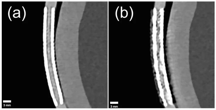
Koroner stent strutları yüksek atenüasyonlu oldukları için BT'de gerçek kalınlıklarından daha geniş görünebilir ve lümeni kısmen örtebilir. Stent blooming artefaktının başlıca nedenleri kısmi hacim etkisi, hareket artefaktları ve ışın sertleşmesidir. Koroner BT'de stentli segmentlerin değerlendirilmesi blooming artefaktları nedeniyle sınırlanabilir.
Faz ve pencere
- BTA tanısal
- Stent strutları gerçek boyutlarından kalın/yayvan görünerek çevre kontrast lümenine taşar; yüksek atenüasyon yanında koyu çizgilenme, lümen sınırında belirsizlik ve kısmi hacim etkisi görülebilir. Hareket kaynaklı bulanıklık kardiyak faza göre değişebilir; çok fazlı incelemede artefaktın fazlar arasında belirgin değişmesi gerçek sabit stenozdan çok hareket etkisini düşündürür.
Önerilen pencereler: Anjiyo (G 600 / M 150).
BT bulguları
- Metal blooming — blooming artefaktı stentli koroner segmentlerin BT ile değerlendirilmesini sınırlayabilir.
- Lümen örtülmesi — metal artefaktları stent içi lümenin BT'de değerlendirilmesini sınırlayabilir.
- Çizgilenme — parlak metal çevresinde koyu veya parlak çizgiler komşu dokulara uzanarak sınırları bozar.
- Kısmi hacim etkisi — metal ve kontrast aynı vokselde ortalanır, gerçek lümen çapı olduğundan küçük ölçülür.
- Hareket bulanıklığı — damar veya stent konturu çiftleşir, uzar ya da kardiyak faza göre farklı görünür.
- Yalancı lümen darlığı — CTA'da metalik stent materyalinin blooming artefaktı restenoz derecesinin olduğundan fazla ölçülmesine yol açabilir.
- Stent içi tanısal yetersizlik — strut gölgeleri lümeni bütünüyle kapatıyorsa bu bulgu stenoz derecesi ölçülemeyeceğini gösterir.
Normalde
Stentli segmentte strutların keskin ve lümen içinin görünür olması, görüntünün değerlendirilmesini kolaylaştırır. Şüpheli stent görünümünü aksiyel/kesitsel ve MPR görüntülerinde karşılaştırın; CPR görüntüsünü tek başına değerlendirmeyin.
Normal BT ile kıyasla
Aynı bölgenin, kaynak veri setinde patoloji içermediği belirtilen bir BT incelemesini kesit kesit kaydırın; organ sınırlarını açarak anatomiyi hasta görüntüsüyle karşılaştırın. Küçük rastlantısal bulgular tümüyle dışlanmamıştır.
Kaynak: TotalSegmentator v2.01 veri seti (Wasserthal ve ark., Radiology: Artificial Intelligence 2023), CC BY 4.0, vaka s0977; organ sınırları veri setinin otomatik segmentasyonundan, birleştirilerek sadeleştirildi. Görüntüler 2,5 mm kesit aralığına yeniden örneklendi.
Ayırıcı tanı
- Gerçek in-stent restenoz
- stentli segmentte stenoz değerlendirmesi, blooming artefaktları ve sınırlı uzaysal çözünürlük nedeniyle güçleşebilir.
- Stent trombozu
- stent içi lümenin değerlendirilmesi metal artefaktları nedeniyle sınırlanabilir.
- Kalsifiye koroner plak
- yoğun kalsiyum stent strut örgüsü yerine damar duvarında yerleşir ve benzer blooming ile yalancı darlık doğurabilir.
- Kardiyak hareket artefaktı
- bulanıklık faza ve damar yönüne bağlı değişir; stent strut geometrisi tek başına bu değişimi açıklamaz.
Tuzaklar
- Blooming kaynaklı yalancı darlık olasılığında CPR'yi tek başına değerlendirmeyin; aksiyel/kesitsel ve MPR görüntülerle karşılaştırın.
- Metal blooming değerlendirilirken kısmi hacim etkisi, hareket artefaktları ve ışın sertleşmesi başlıca nedenler arasında göz önünde bulundurulmalıdır.
- Stent strut kalınlığı veya çap için klinik evrensel eşik varmış gibi davranma; ölçüme dayanak yoksa yalnız görsel tanısal yeterliliği tarif et.
- Yüksek atenüasyonlu stentli segmentte lümen değerlendirmesinin blooming artefaktları nedeniyle sınırlanabileceğini göz önünde bulundurun.
- Stent lümenini yeniden değerlendirirken yüksek çözünürlüklü BT donanımı veya gelişmiş yüksek çözünürlüklü rekonstrüksiyon, kısmi hacim etkisini ve buna bağlı bloomingi azaltabilir.
Kendini dene
Koroner BT'de blooming artefaktının başlıca nedeni aşağıdakilerden hangisidir?
Cevabı göster
Kısmi hacim etkisi. Kardiyak BT'de blooming artefaktının başlıca nedeni kısmi hacim etkisi olarak bildirilmiştir.
Kardiyak BT'de blooming artefaktını azaltmak için derlemede hangi çözüm grupları önerilmiştir?
Cevabı göster
Yüksek çözünürlüklü BT donanımı ve rekonstrüksiyonu, çıkarma ve son işlem teknikleri. Derlemede çözüm grupları yüksek çözünürlüklü BT donanımı, yüksek çözünürlüklü BT rekonstrüksiyonu, çıkarma teknikleri ve son işlem teknikleri olarak sınıflandırılmış; derin öğrenme tekniklerine özellikle vurgu yapılmıştır.
İlgili konular
Kaynaklar
Bu sayfadaki 33 cümle ve 2 quiz sorusu aşağıdaki kaynaklardan alıntılarla otomatik olarak karşılaştırıldı; 14 cümle bu karşılaştırmada düzeltildi (2026-10-09).
- Coronary computed tomography angiography for clinical practicepmc.ncbi.nlm.nih.gov
- Photon-counting computed tomography in coronary artery bypass grafting planning and follow-up: a narrative review of a high-resolution adjunct to invasive angiography.pubmed.ncbi.nlm.nih.gov
- Advanced quantitative CT for coronary artery disease: integrating stenosis quantification, plaque quantification, and fractional flow reserve-CT.pubmed.ncbi.nlm.nih.gov
- Technical advances and clinical impact of photon-counting computed tomography angiography for peripheral artery disease.pubmed.ncbi.nlm.nih.gov
- Aortic Dissection Mimicry Under Extracorporeal Membrane Oxygenation (ECMO) After Cardiac Arrest: A Case Report of Emergency Imaging Dilemmas.pubmed.ncbi.nlm.nih.gov
- Hemodynamic Significance of APEXCT Coronary Angiography Combined With Dynamic CT Myocardial Perfusion Imaging in Assessing Restenosis After Coronary Stenting.pubmed.ncbi.nlm.nih.gov
- Intravascular ultrasound wall shear stress imaging in stented coronary arteries with ultrafast Doppler.pubmed.ncbi.nlm.nih.gov
- Beyond Angiography: Integrating Advanced Diagnostic Modalities in Coronary Stent Restenosis-a Systematic Review.pubmed.ncbi.nlm.nih.gov
- Clinical Applications of Fractional Flow Reserve Derived from Computed Tomography in Coronary Artery Disease.pubmed.ncbi.nlm.nih.gov
- Cardiac computed tomography: Current practice, guidelines, applications, and prospects.pubmed.ncbi.nlm.nih.gov
- Ultra-High-Resolution Photon-Counting CT Angiography for Noninvasive Detection of Neurovascular In-Stent Restenosis.pubmed.ncbi.nlm.nih.gov
- Left main coronary artery stenosis assessment on coronary CT angiography.pubmed.ncbi.nlm.nih.gov
- Understanding Photon-Counting CT: Physics, Detector Technology, and Image Reconstructions.pubmed.ncbi.nlm.nih.gov
- Image quality assessment of ECG-less coronary CT angiography: A comparative study with conventional ECG-gated CCTA.pubmed.ncbi.nlm.nih.gov
- Motion correction algorithm improves right coronary artery image quality at heart rates > 75 bpm in non-ECG-gated coronary CT angiography.pubmed.ncbi.nlm.nih.gov
- Cardiac CT blooming artifacts: clinical significance, root causes and potential solutions.pubmed.ncbi.nlm.nih.gov
- Pictorial review: a step-by-step guide to coronary CT angiography with photon-counting detector CT.pubmed.ncbi.nlm.nih.gov
- Coronary Computed Tomography Angiography for the Diagnosis and Revascularization Guidance of Coronary Bifurcation Lesions: A Contemporary Review.pubmed.ncbi.nlm.nih.gov
- Clinical benefits and current challenges of photon-counting detector CT in vascular imaging.pubmed.ncbi.nlm.nih.gov
- Machine Learning for Coronary Plaque Characterization: A Multimodal Review of OCT, IVUS, and CCTA.pubmed.ncbi.nlm.nih.gov
- Technical principles, benefits, challenges, and applications of photon counting computed tomography in coronary imaging: a narrative review.pubmed.ncbi.nlm.nih.gov
- Effect of quantum iterative reconstruction on coronary stent assessment in ultra-high resolution photon-counting detector CT: a phantom study.pubmed.ncbi.nlm.nih.gov
- Computed Tomography Imaging of Thoracic Aortic Surgery: Distinguishing Life-Saving Repairs from Life-Threatening Complications.pubmed.ncbi.nlm.nih.gov
- Society for Cardiovascular Magnetic Resonance 2024 Cases of SCMR case series.pubmed.ncbi.nlm.nih.gov
- Clinical Impact of CT-Based FFR in Everyday Cardiology: Bridging Computation and Decision-Making.pubmed.ncbi.nlm.nih.gov
- Case Report: Vascular abdominal pain in systemic sclerosis-chronic mesenteric ischemia from high-grade superior mesenteric artery stenosis with coexisting aortomesenteric narrowing.pubmed.ncbi.nlm.nih.gov
- Future Applications of Cardiothoracic CT.pubmed.ncbi.nlm.nih.gov
- Innovations in MRI and AI Integration for Vascular Plaque Evaluation and Overview of Deep Learning Techniques in Peripheral Vascular Disease.pubmed.ncbi.nlm.nih.gov
- Photon-Counting Computed Tomography for In-Stent Occlusion: Lumen Visualization and Myocardial Viability.pubmed.ncbi.nlm.nih.gov
- Coronary Computed Tomography Angiography as a Method for Diagnosing a Thrombotic Occlusion of a Giant Right Coronary Artery Aneurysm in a Patient with Difficulty in Visualizing the Right Coronary Artery During Invasive Coronary Angiography.pubmed.ncbi.nlm.nih.gov
- Real-Time Intravascular Ultrasound-Guided Percutaneous Coronary Intervention for Spontaneous Coronary Artery Dissection.pubmed.ncbi.nlm.nih.gov
- Mechanically governed fracture resistance of biodegradable Zn-Cu alloy stents: Role of ring-length optimized structural design.pubmed.ncbi.nlm.nih.gov
- Computational Analysis of Intravascular OCT Images for Future Clinical Support: A Comprehensive Review.pubmed.ncbi.nlm.nih.gov
- A novel iron bioresorbable scaffold: a potential strategy for pulmonary artery stenosis.pubmed.ncbi.nlm.nih.gov
- The Role of Coronary Imaging in Chronic Total Occlusions: Applications and Future Possibilities.pubmed.ncbi.nlm.nih.gov
- Artificial Intelligence-Assisted Intravascular Imaging in Percutaneous Coronary Intervention: Current Status and Advances.pubmed.ncbi.nlm.nih.gov
- Integrating Invasive and Noninvasive Imaging for Coronary Atherosclerosis: A Systematic Review With Pragmatic Algorithms From Anatomy to Physiology.pubmed.ncbi.nlm.nih.gov
- Spectral Computed Tomography Angiography in Visceral Artery Aneurysms: Technical Principles and Clinical Applications.pubmed.ncbi.nlm.nih.gov
- Single-Bolus Sequential Triple-Rule-Out CT Angiography: Image Quality and Radiation Dose on Wide-Area Detector and Dual-Source CT.pubmed.ncbi.nlm.nih.gov
- Radiation dose optimization in coronary CT angiography clinical application of absolute phase gating in patients with atrial fibrillation.pubmed.ncbi.nlm.nih.gov
- State-of-the-Art Review on Finite Element Analysis Applied to Ultrathin-Strut Drug-Eluting Stents: Examining the Present and Looking Ahead to the Future.pubmed.ncbi.nlm.nih.gov
- Additively manufactured reinforced polymeric stents for peripheral artery disease: design, manufacture and performance evaluation.pubmed.ncbi.nlm.nih.gov
- Intracoronary imaging for left main percutaneous coronary intervention: a clinical consensus statement of the European Association of Percutaneous Cardiovascular Interventions (EAPCI) of the ESC and the European Bifurcation Club (EBC).pubmed.ncbi.nlm.nih.gov
- Clinical OCT computation-modeled prognosis of coronary plaque progression with validation by fusogenic macrophage nano-targeting.pubmed.ncbi.nlm.nih.gov
- Progressive Stenosis of Thoraflex Hybrid Prosthesis after Total Arch Replacement Leading to a Fatal Outcome: A Case Report.pubmed.ncbi.nlm.nih.gov
- Recent Advances in 3D-Printed Bioresorbable Vascular Stents: Toward Patient-Specific, Novel-Structured, Functional, and Smart Devices.pubmed.ncbi.nlm.nih.gov
- Severe arterial hypertension, a silent complication of Williams-Beuren syndrome: A case report with literature review.pubmed.ncbi.nlm.nih.gov
- FFR-CT: Technical Advances and Implementation in Clinical Practice.pubmed.ncbi.nlm.nih.gov
- Dual complications of type I endoleak and aorto-enteric fistula post-EVAR: A case report and review of literature.pubmed.ncbi.nlm.nih.gov
- Evaluating flow modulating treatment response in intracranial aneurysms using black-blood MRI in vitro.pubmed.ncbi.nlm.nih.gov
- Wearable Flexible Sensors for Cardiovascular Disease Monitoring.pubmed.ncbi.nlm.nih.gov
- Impact of the confluence of cardiac motion and high spatial resolution on performance of ECG-gated imaging with an investigational photon-counting CT system: A phantom study.pubmed.ncbi.nlm.nih.gov
- CAD-RADS™ 2.0 - 2022 Coronary Artery Disease - Reporting and Data System An Expert Consensus Document of the Society of Cardiovascular Computed Tomography (SCCT), the American College of Cardiology (ACC), the American College of Radiology (ACR) and the North America Society of Cardiovascular Imaging (NASCI).pubmed.ncbi.nlm.nih.gov
- Non-contrast coronary magnetic resonance angiography: current frontiers and future horizons.pubmed.ncbi.nlm.nih.gov
- From intravascular imaging to adaptive vascular care: intelligent photonics and digital twins in panvascular disease.pubmed.ncbi.nlm.nih.gov
- Aortic pseudoaneurysm caused by Tempofilter II inferior vena cava filter strut perforation: a case report.pubmed.ncbi.nlm.nih.gov
- Chest Pain Evaluation: Diagnostic Testing.pubmed.ncbi.nlm.nih.gov
- Coronary calcium quantification using contrast-enhanced dual-energy computed tomography scans.pubmed.ncbi.nlm.nih.gov
- Clinical Utility and Limitations of Photon-Counting Detector CTA in the Follow-Up of Flow Diverter Stent.pubmed.ncbi.nlm.nih.gov
- Photon-counting detector CTA to assess intracranial stents and flow diverters: an in vivo study with ultrahigh-resolution spectral reconstructions.pubmed.ncbi.nlm.nih.gov
- Long-term safety and effectiveness of the "OptEase" vena cava filter.pubmed.ncbi.nlm.nih.gov
- Photon-Counting Computed Tomography (PCCT): Technical Background and Cardio-Vascular Applications.pubmed.ncbi.nlm.nih.gov
- Novel Imaging-Based Biomarkers for Identifying Carotid Plaque Vulnerability.pubmed.ncbi.nlm.nih.gov
- Diagnostic accuracy of low-radiation coronary computed tomography angiography with low tube voltage and knowledge-based model reconstruction.pubmed.ncbi.nlm.nih.gov
- Ultra-high spatial resolution at photon-counting computed tomography: technical insights and sustainable applications in cardiothoracic imaging.pubmed.ncbi.nlm.nih.gov
- The Shifting Boundary Between Invasive and Non-Invasive Angiographic Investigation in Contemporary Cardiology and Cardiac Surgery: An Up-to-Date Narrative Review.pubmed.ncbi.nlm.nih.gov
- Beyond Angiography: Cardiac CT for Planning Complex PCI in Calcified Coronary Lesions.pubmed.ncbi.nlm.nih.gov
- Improving Measurement Accuracy of the Degree of Carotid In-Stent Stenosis Using CT Angiography: A Comparison With DSA.pubmed.ncbi.nlm.nih.gov
- Coronary computed tomography angiography for clinical practice.pubmed.ncbi.nlm.nih.gov
- Cardiovascular Applications of Photon-Counting CT Technology: A Revolutionary New Diagnostic Step.pubmed.ncbi.nlm.nih.gov
- Photon-counting CT in cancer radiotherapy: technological advances and clinical benefits.pubmed.ncbi.nlm.nih.gov
- Multimodality Imaging Approach in Diagnosis and Follow-Up of Aortic Coarctation in Adulthood.pubmed.ncbi.nlm.nih.gov
- Commentary: an eye on PET quantification.pubmed.ncbi.nlm.nih.gov
- Design and fabrication of a 3D-printed oral stent for head and neck radiotherapy from routine diagnostic imaging.pubmed.ncbi.nlm.nih.gov
- 2.5-mm articulated endoluminal forceps using a compliant mechanism.pubmed.ncbi.nlm.nih.gov
- Computer Vision Techniques for Transcatheter Intervention.pubmed.ncbi.nlm.nih.gov
- Multilayer and crushed stent visualization using photon- counting detector computed tomography - a proof of concept.pubmed.ncbi.nlm.nih.gov
- Ultra-high resolution CT angiography for the assessment of intracranial stents and flow diverters using photon counting detector CT.pubmed.ncbi.nlm.nih.gov
- Emerging quantitative CCTA imaging biomarkers for cardiovascular risk stratification: a narrative review.pubmed.ncbi.nlm.nih.gov
- Comprehensive risk prediction of acute coronary syndrome by integrating coronary plaque morphology and hemodynamic characteristics.pubmed.ncbi.nlm.nih.gov
- Association of AI-assisted quantitative coronary plaque burden and CT-derived fractional flow reserve with major adverse cardiovascular events.pubmed.ncbi.nlm.nih.gov
- Radiation and Contrast Medium Dosage Optimization of Coronary CT Angiography While Combining Personalized Patient Protocol Technology and Automated Tube Voltage Selection Technology.pubmed.ncbi.nlm.nih.gov
- Spectral ultrahigh-resolution photon-counting CT for coronary stent imaging: evaluation in a dynamic phantom.pubmed.ncbi.nlm.nih.gov
- Development of Biodegradable Polymeric Stents for the Treatment of Cardiovascular Diseases.pubmed.ncbi.nlm.nih.gov
- AI-driven multimodal retinal imaging for early detection and risk stratification of vascular and neurodegenerative diseases.pubmed.ncbi.nlm.nih.gov
- Deep learning predicts stent implantation in borderline coronary lesions from angiography.pubmed.ncbi.nlm.nih.gov
- Case Report: Zero-contrast multivessel PCI with left main bifurcation guided by dual IVUS strategies using a coronary CTA roadmap.pubmed.ncbi.nlm.nih.gov
- Successful management of the recurrent acute in-stent thrombosis despite optimal medical therapy: a case report and review of the literature.pubmed.ncbi.nlm.nih.gov
Eğitim amaçlıdır; hasta başında tanı ve tedavi kararının yerine geçmez, tıbbi tavsiye değildir. Metinler yapay zekâ desteğiyle yazıldı ve otomatik bir adımda kaynak alıntılarıyla karşılaştırıldı; bu bir tıbbi doğrulama değildir ve içerik uzman radyolog incelemesinden geçmedi. Klinik kararlarda güncel kılavuzları, kurum protokollerini ve radyoloji raporunu esas alın.