Özet
Mantle hücreli lenfoma (MCL), B-hücreli bir non-Hodgkin lenfomadır; tanı anında sıklıkla ileri evre nodal hastalığa ek olarak dalak, kemik iliği ve gastrointestinal sistem tutulumu bulunabilir. Kontrastlı abdomen-pelvis BT, bağırsak duvarı kalınlaşmasını, polipoid lezyonları, lenfadenopatiyi ve hepatosplenomegaliyi değerlendirebilir. Mantle hücreli lenfomanın gastrointestinal tutulumu ileri evre hastalıkta sık görülür; gastrointestinal tutulum FDG PET/BT’de düşük avidite gösterebildiğinden gözden kaçabilir, bu nedenle PET/BT endoskopik değerlendirme ve biyopsinin yerini tutmaz ve evrelemede gastrointestinal sistemin değerlendirilmesi gerekir. Bağırsak lenfomasına obstrüksiyon, invajinasyon veya perforasyon gibi komplikasyonlar eşlik edebilir; BT bu bulguları değerlendirmeye yardımcı olur, ancak histolojik tanının yerine geçmez.
Okuma sırası
- Terminal ileum. Abdominal BT’de bağırsak veya kolondaki kitle ya da polipler görülebilir.
- İleoçekal valv. İleoçekal bölgedeki kitle veya polip BT’de görülebilir; MCL’nin kesin tanısı histolojik incelemeyle konur.
- İnce bağırsak ve kolon lümeni. Abdominal BT’de ince bağırsak veya kolondaki kitle ya da polipler görülebilir.
- Proksimal anslar ve mezenter. Abdominal BT bağırsak veya kolondaki kitle ya da polipleri gösterebilir ve komplike formları değerlendirmeye yardımcı olabilir; evreleme için tüm vücut BT veya PET kullanılabilir.
Faz ve pencere
- Portal tanısal
- İntravenöz kontrastlı portal venöz faz abdomen-pelvis BT’de bağırsak duvarı, mezenter ve solid organlar değerlendirilir; lenfoma infiltrasyonu duvar kalınlaşması, polipoid lezyon veya kitle şeklinde görülebilir. Küçük polipoid mukozal odaklar BT’de gözden kaçabilir; lümen açıklığı, duvar kalınlığı ve geçiş/dilatasyon bulguları birlikte değerlendirilir.
Önerilen pencereler G genişlik, M merkez; değerler Hounsfield birimi (HU)
- BatınG 400 / M +50
- Yumuşak dokuG 400 / M +40
- KaraciğerG 150 / M +30
BT bulguları
- İleri evre mantle hücreli lenfomada yaygın lenfadenopati görülebilir.
- Sistemik lenfoma yükünde hepatosplenomegali görülebilir; dalakta diffüz ya da fokal tutulum eşlik edebilir, ancak organ boyutu tek başına MCL tanısı koydurmaz.
- Gastrointestinal lenfomatozis polipozisi — ince bağırsak ve kolonda çok sayıda intraluminal polipoid yumuşak doku odağı görülebilir; küçük lezyonlar BT’de saptanamayabilir.
- Segmental bağırsak duvarı kalınlaşması — mide, distal ileum veya kolon duvarında düzgün ya da yaygın kalınlaşma lenfomatöz infiltrasyonu düşündürebilir.
- Lümen genişlemesi — bağırsak lenfomasında duvar kalınlaşmasına rağmen segment anevrizmatik biçimde genişleyebilir; geçiş noktası ve proksimal ans çapı ayrıca incelenir.
- Komplikasyon işaretleri — BT’de hedef işareti ileokolik invajinasyonu düşündürebilir; abdominal BT lenfomanın komplike formlarını değerlendirmeye yardımcı olabilir.
Ölçütler ve sınıflamalar
- Lugano anatomik evreleme
- Lugano evrelemesi lenfomanın evresini sınıflandırmak için kullanılır. MCL evrelemesinde PET/BT çoğunlukla kullanılır; kemik iliği ve gerektiğinde gastrointestinal doku incelemesi görüntülemeyi tamamlar, görüntüleme histopatolojik tanının yerine geçmez.
Normalde
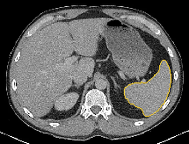
Dalak boyutunu değerlendirerek splenomegaliyi tanımlayın.
Normal BT ile kıyasla
Aynı bölgenin, kaynak veri setinde patoloji içermediği belirtilen bir BT incelemesini kesit kesit kaydırın; organ sınırlarını açarak anatomiyi hasta görüntüsüyle karşılaştırın. Küçük rastlantısal bulgular tümüyle dışlanmamıştır.
Kaynak: TotalSegmentator v2.01 veri seti (Wasserthal ve ark., Radiology: Artificial Intelligence 2023), CC BY 4.0, vaka s0119; organ sınırları veri setinin otomatik segmentasyonundan, birleştirilerek sadeleştirildi. Görüntüler 2,5 mm kesit aralığına yeniden örneklendi.
Ayırıcı tanı
- Foliküler lenfoma ve MCL ayrı lenfoma alt tipleridir; gastrointestinal lenfomatozis polipozisi MCL’de görülebilir, ancak bu bulgu tek başına alt tipi doğrulamaz.
- Gastrointestinal adenokarsinom
- fokal veya asimetrik duvar kalınlaşması ayırıcı tanıda değerlendirilir; görüntüleme örtüşebilir ve kesin tanı histolojiyle konur.
- Gastrointestinal stromal tümör
- bağırsak duvarı kaynaklı fokal kitle ayırıcı tanıdadır; çoklu lenfomatöz polipler MCL’yi düşündürür ancak histoloji gerekir.
- Lenfoid hiperplazi
- terminal ileumda mukozal nodülerlik yapabilir; yaygın lenfadenopati, dalak bulguları ve klinik bağlamla birlikte değerlendirilir.
- Peritoneal karsinomatozis
- peritoneal implantlar, asit ve serozal nodüller örtüşen görüntüleme bulgularıdır; primer tümör öyküsü ve gerektiğinde doku örneklemesiyle ayırıcı tanı yapılır.
Tuzaklar
- Gastrointestinal polipler MCL tutulumunda görülebilir; abdominal BT bağırsak veya kolondaki polipleri gösterebilir, endoskopi ise biyopsi alınmasına olanak verir.
- Bağırsak lenfomasında duvar kalınlaşması lümen genişlemesiyle birlikte olabilir ve proksimal obstrüksiyon görülmeyebilir; yine de geçiş noktası ve proksimal ansları birlikte değerlendir.
- Hepatosplenomegali lenfoma tutulumunu destekleyebilir ancak özgül değildir; tek başına organ hacmi MCL tanısı koydurmaz.
- Bağırsak polipoid lezyonları BT’de görülebilir; yalnız radyolojik değerlendirme MCL’yi diğer polipozislerden ayıramaz ve kesin tanı histolojiyle konur.
- Çoklu lenfomatöz polipozis MCL ile ilişkili olabilir, ancak tek başına alt tipi belirlemez; radyolojik değerlendirme kesin tanı koydurmaz.
Kendini dene
MCL’de yaygın lenfadenopati görülebilir; gastrointestinal tutulum çok sayıda polipoid odak şeklinde olabilir. Bu gastrointestinal polipozis paterniyle ilişkili lenfoma alt tipi hangisidir?
Cevabı göster
Mantle hücreli lenfoma. Lenfomatozis polipozis MCL ile ilişkilidir; kesin alt tip histopatolojik incelemeyle belirlenir.
MCL olgusunda ileal duvar kitlesiyle birlikte ileokolik invajinasyon saptanıyor. Görüntülemede tanımlanan bağırsak komplikasyonu hangisidir?
Cevabı göster
İleokolik invajinasyon. BT’deki hedef işareti ileokolik invajinasyonla uyumlu olabilir; MCL tanısı ise histopatolojik incelemeyle doğrulanır.
İlgili konular
Kaynaklar
Bu sayfadaki 37 cümle ve 2 quiz sorusu aşağıdaki kaynaklardan alıntılarla otomatik olarak karşılaştırıldı; 12 cümle bu karşılaştırmada düzeltildi (2026-10-09).
- Primary mantle-cell lymphoma of small intestine presenting with intussusceptions: A case report and review of the literaturepmc.ncbi.nlm.nih.gov
- [Lymphomas].pubmed.ncbi.nlm.nih.gov
- Surveillance scanning in lymphoma.pubmed.ncbi.nlm.nih.gov
- Diagnosis and staging in gastrointestinal lymphoma.pubmed.ncbi.nlm.nih.gov
- Mantle cell lymphoma: evolving management strategies.pubmed.ncbi.nlm.nih.gov
- Molecular Monitoring of Cell-Free Circulating Tumor DNA in Non-Hodgkin Lymphoma.pubmed.ncbi.nlm.nih.gov
- CXCR4-targeted theranostics in oncology.pubmed.ncbi.nlm.nih.gov
- Is Limited-Stage Mantle Cell Lymphoma Curable and How Is It Best Managed?pubmed.ncbi.nlm.nih.gov
- In situ mantle cell lymphoma in the nasopharynx.pubmed.ncbi.nlm.nih.gov
- Refining the Mantle Cell Lymphoma Paradigm: Impact of Novel Therapies on Current Practice.pubmed.ncbi.nlm.nih.gov
- 18 F-FDG PET or PET/CT in Mantle Cell Lymphoma.pubmed.ncbi.nlm.nih.gov
- The predictive value of interim and final [18F] fluorodeoxyglucose positron emission tomography after rituximab-chemotherapy in the treatment of non-Hodgkin's lymphoma: a meta-analysis.pubmed.ncbi.nlm.nih.gov
- [Mantle cell lymphoma presenting with spontaneous splenic rupture].pubmed.ncbi.nlm.nih.gov
- Mantle cell lymphoma involvement of the pleura and tuberculous pleurisy with pulmonary tuberculosis: a case report and literature review.pubmed.ncbi.nlm.nih.gov
- Mantle cell lymphoma with a unique pattern of CD5 expression: a case report with review of the literatures.pubmed.ncbi.nlm.nih.gov
- Acute abdominal presentation of mantle cell lymphoma mimicking acute appendicitis: a case report.pubmed.ncbi.nlm.nih.gov
- Leukemic Non-Nodal Mantle Cell Lymphoma Presenting with Traumatic Splenic Rupture.pubmed.ncbi.nlm.nih.gov
- Mantle Cell Lymphoma Mimicking Parotid Neoplasm: A Rare Case Report.pubmed.ncbi.nlm.nih.gov
- Primary duodenal T/histiocyte-rich large B-cell lymphoma complicated with obstructive jaundice: A case report and review of literature.pubmed.ncbi.nlm.nih.gov
- Disseminated Talaromyces marneffei infection presenting as multiple intestinal perforations and diffuse hepatic granulomatous inflammation in an infant with STAT3 mutation: a case report.pubmed.ncbi.nlm.nih.gov
- Mastocytosis-A Review of Disease Spectrum with Imaging Correlation.pubmed.ncbi.nlm.nih.gov
- Case Report: Two cases of primary pulmonary endobronchial extranodal marginal zone lymphoma of mucosa-associated lymphoid tissue localized in the trachea and bronchus.pubmed.ncbi.nlm.nih.gov
- Small Intestine Tumors: Diagnostic Role of Multiparametric Ultrasound.pubmed.ncbi.nlm.nih.gov
- Primary Gastrointestinal B-Cell Lymphomas: A Clinicopathological Review.pubmed.ncbi.nlm.nih.gov
- When Histology Is Unknown: Surgical Decision-Making in Small Bowel Tumors-From Preoperative Imaging to Intraoperative Findings: A Narrative Review.pubmed.ncbi.nlm.nih.gov
- Updates in Evidence-Based Decision Making for Upfront Surgical Interventions in Paediatric Lymphoma.pubmed.ncbi.nlm.nih.gov
- A Twisted Tale: Ileocolic Intussusception in an Adult: A Case Report.pubmed.ncbi.nlm.nih.gov
- Mesenteric lymph node cavitation syndrome revealing celiac disease: Diagnostic and follow-up imaging findings.pubmed.ncbi.nlm.nih.gov
- Diagnostic and therapeutic challenges of diffuse abdominal infantile hemangioma: a case report emphasizing multimodality imaging and multidisciplinary management.pubmed.ncbi.nlm.nih.gov
- CT and MR in peritoneal malignancies: pearls and pitfalls at preoperative examination.pubmed.ncbi.nlm.nih.gov
- Role of magnetic resonance in bowel pathologies: Beyond inflammatory bowel disease.pubmed.ncbi.nlm.nih.gov
- High-Frequency Miniprobe Endoscopic Ultrasonography Across the Gastrointestinal Tract.pubmed.ncbi.nlm.nih.gov
- Application value of gastric contrast ultrasonography in gastric tumors.pubmed.ncbi.nlm.nih.gov
- Radiological Approach to Splenomegaly: Etiologies, Pathophysiologies, and Diagnostic Strategies.pubmed.ncbi.nlm.nih.gov
- An Overview of Selected Rare B-Cell Lymphoproliferative Disorders: Imaging, Histopathologic, and Clinical Features.pubmed.ncbi.nlm.nih.gov
- Uncommon Variants of Mature T-Cell Lymphomas (MTCLs): Imaging and Histopathologic and Clinical Features with Updates from the Fourth Edition of the World Health Organization (WHO) Classification of Lymphoid Neoplasms.pubmed.ncbi.nlm.nih.gov
- Complications of lymphoma in the abdomen and pelvis: clinical and imaging review.pubmed.ncbi.nlm.nih.gov
- A case series study of fever-diarrhea-hematochezia triad as the first presentation in plasma EBV DNA-positive diseases.pubmed.ncbi.nlm.nih.gov
- Small Bowel Imaging from Stepchild of Roentgenology to MR Enterography, Part II: The Reliable Disclosure of Crohn's Disease and Non-Inflammatory Small Bowel Disorder Plot through MRI Findings.pubmed.ncbi.nlm.nih.gov
- Chronic intussusception revealing primary diffuse large B-cell lymphoma of the small intestine: A case report.pubmed.ncbi.nlm.nih.gov
- Intussusception in Neonates: Uncommon Causes and Diagnostic Dilemmas in Two Case Reports.pubmed.ncbi.nlm.nih.gov
- Navigating the Diagnostic Dilemma: Cecal Duplication Cyst and Intussusception in Pediatric Patients.pubmed.ncbi.nlm.nih.gov
- Long-term safety and effectiveness of the "OptEase" vena cava filter.pubmed.ncbi.nlm.nih.gov
- Patterns of injury and violence in Yaoundé Cameroon: an analysis of hospital data.pubmed.ncbi.nlm.nih.gov
- Diffuse Large B-Cell Lymphoma Presenting as an Infiltrative Masticator Space Lesion Mimicking Inflammatory Disease: A Case Report.pubmed.ncbi.nlm.nih.gov
- PET-guided early identification of CAR-T candidates in diffuse large b-cell lymphoma: a multidisciplinary expert consensus.pubmed.ncbi.nlm.nih.gov
- Intestinal T-cell lymphoma not otherwise specified presenting with Jejunal perforation: a case report and review of the literature.pubmed.ncbi.nlm.nih.gov
- Unilateral Orbital Lymphomas: A Structured Multidisciplinary Workflow for Diagnosis and Management.pubmed.ncbi.nlm.nih.gov
- ESR Essentials: imaging of lymphoma-practice recommendations by the European Society of Oncologic Imaging.pubmed.ncbi.nlm.nih.gov
- Management of Extranodal Marginal Zone Lymphoma: Present and Upcoming Perspectives.pubmed.ncbi.nlm.nih.gov
- Complete remission of Helicobacter pylori-negative gastric MALT lymphoma following eradication therapy: A case report.pubmed.ncbi.nlm.nih.gov
- Post-transplant lymphoproliferative disorder after solid organ transplantation: a comprehensive review.pubmed.ncbi.nlm.nih.gov
- Accuracy of percutaneous ultrasound-guided needle biopsy of abdominal lymph nodes for the diagnosis of malignant disease.pubmed.ncbi.nlm.nih.gov
- Non-Hodgkin lymphoma mimicking acute leukemia: a report of six cases and review of the literature.pubmed.ncbi.nlm.nih.gov
- Ileal tuberculosis presenting with severe unexplained anemia and unusual pseudo-tumoral imaging features: A case report.pubmed.ncbi.nlm.nih.gov
- Small intestinal metastasis of triple-negative breast cancer mimicking lymphoma: imaging findings and case report.pubmed.ncbi.nlm.nih.gov
- Mesenteric desmoid tumor in its cystic form: Case report of a very rare variant.pubmed.ncbi.nlm.nih.gov
- Primary and secondary tumors of the peritoneum: key imaging features and differential diagnosis with surgical and pathological correlation.pubmed.ncbi.nlm.nih.gov
- Rare gastrointestinal lymphomas: The endoscopic investigation.pubmed.ncbi.nlm.nih.gov
- Primary diffuse malignant epithelioid peritoneal mesothelioma of the greater omentum in an asbestos-naive patient: a case report.pubmed.ncbi.nlm.nih.gov
- The Many Faces of the Angry Peritoneum.pubmed.ncbi.nlm.nih.gov
- First Brazilian expert consensus for video capsule endoscopy and balloon-assisted enteroscopy.pubmed.ncbi.nlm.nih.gov
- Expected Values of Artificial Intelligence in Colon Capsule Endoscopy: Artificial Intelligence in Capsule Endoscopy Consortium Position Statement.pubmed.ncbi.nlm.nih.gov
- Ileocecal CD5-Positive Diffuse Large B-Cell Lymphoma Presenting with Acute Obstructive Symptoms: A Case Report.pubmed.ncbi.nlm.nih.gov
- Diffuse large B-cell lymphoma as a rare cause of small bowel obstruction: case report and literature review.pubmed.ncbi.nlm.nih.gov
- Distinguishing lymphoma from benign lymphadenopathy in fever of unknown origin using a diagnostic model based on PET/CT and clinical-laboratory parameters.pubmed.ncbi.nlm.nih.gov
- Australian clinical practice guideline: diagnosis and treatment of idiopathic multicentric Castleman disease.pubmed.ncbi.nlm.nih.gov
- Atypical clinical presentation associated with Castleman disease: a case report and review of the literature.pubmed.ncbi.nlm.nih.gov
- Dual-Energy CT Enterography in Intestinal Tuberculosis: Role of Relative Enhancement Calculated on Iodine Maps in Assessing Disease Activity.pubmed.ncbi.nlm.nih.gov
- Beyond Fixed Thresholds: Cluster-Derived MRI Boundaries Improve Assessment of Crohn's Disease Activity.pubmed.ncbi.nlm.nih.gov
- Crohn Disease Strictures: Multimodality Imaging to Identify Imaging Biomarkers.pubmed.ncbi.nlm.nih.gov
- Advances in alimentary tract imaging.pubmed.ncbi.nlm.nih.gov
- Duodenal imaging on the spotlight: from A to Z.pubmed.ncbi.nlm.nih.gov
- MRI Findings of Intrinsic and Extrinsic Duodenal Abnormalities and Variations.pubmed.ncbi.nlm.nih.gov
- Soft-Tissue Sarcomas of the Abdomen and Pelvis: Radiologic-Pathologic Features, Part 1-Common Sarcomas: From the Radiologic Pathology Archives.pubmed.ncbi.nlm.nih.gov
- Intestinal intussusception in adult patients: a single-center experience and literature review.pubmed.ncbi.nlm.nih.gov
- POCUS for acute abdominal pain: practical scan protocols on gastrointestinal diseases and an evidence review.pubmed.ncbi.nlm.nih.gov
- Extensive colorectal lymphomatous polyposis complicated by acute intestinal obstruction: a case report.pubmed.ncbi.nlm.nih.gov
- Unusual Presentation of Leukemic-Phase Mantle Cell Lymphoma: A Case Report.pubmed.ncbi.nlm.nih.gov
- Atypical Kaposi Sarcoma in HIV Infection with Predominant Nodal Involvement.pubmed.ncbi.nlm.nih.gov
- When to consider an inborn error of immunity: clues for physicians.pubmed.ncbi.nlm.nih.gov
- Granulomatous cutaneous T-cell lymphoma in a patient with atage IVB of mycosis fungoides: a case report and literature review.pubmed.ncbi.nlm.nih.gov
- Case Report: Bezoar-mimicking intraluminal gossypiboma causing small-bowel obstruction and chronic malabsorption 15 years after hysterectomy.pubmed.ncbi.nlm.nih.gov
- Analysis of Diagnosis and Treatment of Active Lupus Enteritis Accompanied by Intestinal Pseudo-Obstruction as the Initial Manifestation: A Case Report and Literature Review.pubmed.ncbi.nlm.nih.gov
- Acute small bowel obstruction revealing a rare localization of lymphoma: a case report.pubmed.ncbi.nlm.nih.gov
- From obstruction to ischaemia: a systematic review and meta-analysis on the diagnostic accuracy of CT scans in identifying small and large bowel obstruction, underlying causes and predicting critical complications in adults.pubmed.ncbi.nlm.nih.gov
- Guidance for clinical practice using emergency and point-of-care ultrasonography.pubmed.ncbi.nlm.nih.gov
- Incidentally Identified Pneumatosis Cystoides Intestinalis in Patients With Gastric Outlet Obstruction From Addis Ababa, Ethiopia: A Case Report.pubmed.ncbi.nlm.nih.gov
- Serious Adverse Events Following COVID-19 Vaccination: A Descriptive Case Series From a Rural Healthcare System.pubmed.ncbi.nlm.nih.gov
- Living Gluten-Free in Romania: A National Cross-Sectional Study of Dietary Adherence in Clinically Diagnosed and Self-Reported Cases.pubmed.ncbi.nlm.nih.gov
- Management of male stress urinary incontinence in high-risk patients: a narrative review.pubmed.ncbi.nlm.nih.gov
- Commentary: an eye on PET quantification.pubmed.ncbi.nlm.nih.gov
- Proportion and Characteristics of Helicobacter Pylori -Negative Gastric Mucosa-Associated Lymphoid Tissue Lymphoma: A Systematic Review and Meta-Analysis.pubmed.ncbi.nlm.nih.gov
- EACR 2025 Congress: Innovative Cancer Science, 16-19 June 2025.pubmed.ncbi.nlm.nih.gov
- Non‑Hodgkin's lymphomas of the lacrimal sac: Current insights and future directions (Review).pubmed.ncbi.nlm.nih.gov
- Special Issue: EACR 2026 Congress: Innovative Cancer Science, 8-11 June 2026.pubmed.ncbi.nlm.nih.gov
- ESMO/ASCO Recommendations for a Global Curriculum in Medical Oncology Edition 2023.pubmed.ncbi.nlm.nih.gov
- Diagnosis, Perioperative Management, and Surgical Treatment of Suspected Functional Pheochromocytoma in Two Dogs: Surgical Complexity in the Absence of Planned Venotomy.pubmed.ncbi.nlm.nih.gov
- Plastinated sectional anatomy applied in CT and MRI studies of the body of the crab-eating fox (Cerdocyon thous).pubmed.ncbi.nlm.nih.gov
- Laparoscopic right hemicolectomy in situs inversus totalis with cecal carcinoma: a case report and literature review.pubmed.ncbi.nlm.nih.gov
- Sufficient-dose Brentuximab vedotin improves prognosis in patients with classic Hodgkin lymphoma: a single-center real-world study in China.pubmed.ncbi.nlm.nih.gov
- A revised staging system for natural killer/T cell lymphoma incorporating skull base invasion and nasal/non-nasal subtype distinction.pubmed.ncbi.nlm.nih.gov
- Pre-contrast computed tomography reveals distinct characteristics and anatomical distribution of presumably lymphomatous lymph nodes in canine multicentric lymphoma.pubmed.ncbi.nlm.nih.gov
- Medical hyperspectral imaging: an updated review of technology advancements and biomedical applications.pubmed.ncbi.nlm.nih.gov
- A picture is worth a thousand words: a history of diagnostic imaging for lymphoma.pubmed.ncbi.nlm.nih.gov
- Significance of E-lesions in Hodgkin lymphoma and the creation of a new consensus definition: a report from SEARCH.pubmed.ncbi.nlm.nih.gov
- Prolymphocytic Progression in Chronic Lymphocytic Leukemia Without Richter Transformation: An Eighteen-Year Clinical-Diagnostic Correlation.pubmed.ncbi.nlm.nih.gov
- Rare Presentation of Sequential Mucosa-Associated Lymphoid Tissue Lymphoma, Follicular Lymphoma, and Chronic Lymphocytic Leukemia: A Case-Based Review.pubmed.ncbi.nlm.nih.gov
- Cavitating mesenteric lymph node syndrome: a rare complication of celiac disease-case report and literature review.pubmed.ncbi.nlm.nih.gov
- Solid Primary Retroperitoneal Masses in Adults: An Imaging Approach.pubmed.ncbi.nlm.nih.gov
- Presentation of ileal Burkitt lymphoma in children.pubmed.ncbi.nlm.nih.gov
Eğitim amaçlıdır; hasta başında tanı ve tedavi kararının yerine geçmez, tıbbi tavsiye değildir. Metinler yapay zekâ desteğiyle yazıldı ve otomatik bir adımda kaynak alıntılarıyla karşılaştırıldı; bu bir tıbbi doğrulama değildir ve içerik uzman radyolog incelemesinden geçmedi. Klinik kararlarda güncel kılavuzları, kurum protokollerini ve radyoloji raporunu esas alın.