Özet
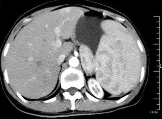
Splenik marjinal zon lenfoması genellikle 50 yaşından sonra görülen, indolent seyirli bir B hücreli lenfomadır ve çoğunlukla belirgin splenomegaliyle ortaya çıkar. BT, dalak boyutunu ve odaklarını haritalar; özellikle splenik hilus dışındaki nodal ya da ekstranodal yayılımı arayarak evreleme değerlendirmesine katkı verir. Dalakta belirgin büyüme olmasına karşın parankim homojen kalabileceğinden, normal görünümlü parankim hastalığı dışlamaz. Fokal lezyonları enfeksiyon veya infarkta bağlamak, yayılımı ve olası komplikasyonları gözden kaçırabilir.
Faz ve pencere
- Portal tanısal
- Normal dalak portal venöz fazda homojen kontrastlanır; SMZL çoğunlukla fokal kitle yerine diffüz splenomegaliyle ortaya çıkar. Daha seyrek olarak hipoattenüe odaklar seçilir; aynı faz hiler nodları ve karın içi yayılımı karşılaştırmaya elverişlidir.
Önerilen pencereler: Batın (G 400 / M 50), Yumuşak doku (G 400 / M 40).
BT bulguları
- Dalak uzun ekseni ve hacminde artış; infiltrasyon diffüz ise belirgin sınırlı kitle olmayabilir.
- Dalakta fokal lezyon görülmemesi splenik lenfomayı dışlamaz; lenfoma splenomegaliyle birlikte odaksız görünebilir.
- SMZL çoğunlukla diffüz splenomegaliyle ortaya çıkar; nadiren fokal splenik kitle şeklinde de görülebilir.
- Splenik hilus çevresindeki lenf nodlarını, splenik lenfomaya eşlik edebilen bir bulgu olarak incele.
- Hiler bölge dışındaki abdominal nodal istasyonlar ve karaciğer büyümesi eşlik edebilir.
- BT bulguları SMZL'yi tek başına doğrulamaz; klinik, kan ve doku incelemesiyle birlikte yorumlanır.
Ölçütler ve sınıflamalar
- WHO-HAEM5 histolojik sınıflaması
- Splenik marjinal zon lenfoması WHO-HAEM5'te splenik B hücreli lenfomalar ve lösemiler başlığı altında ayrı bir antite olarak yer alır; görüntüleme tanıyı tek başına kesinleştirmez.
Normalde
Normal dalak portal venöz BT'de homojen atenuasyon gösterir; dalak boyutu ve şekli değişkenlik gösterebilir, şekli komşu organlardan etkilenir. Dalak boyutu ve şekli değişkenlik gösterir; splenik lenfoma odaksız splenomegali şeklinde görülebilir. Dalak boyutunun normal olup olmadığını belirlemek güç olabilir; aksiyel ölçüler için önerilmiş referanslar bulunsa da tek bir çapı kesin eşik olarak yorumlama.
Normal BT ile kıyasla
Aynı bölgenin, kaynak veri setinde patoloji içermediği belirtilen bir BT incelemesini kesit kesit kaydırın; organ sınırlarını açarak anatomiyi hasta görüntüsüyle karşılaştırın. Küçük rastlantısal bulgular tümüyle dışlanmamıştır.
Kaynak: TotalSegmentator v2.01 veri seti (Wasserthal ve ark., Radiology: Artificial Intelligence 2023), CC BY 4.0, vaka s0541; organ sınırları veri setinin otomatik segmentasyonundan, birleştirilerek sadeleştirildi. Görüntüler 2,5 mm kesit aralığına yeniden örneklendi.
Ayırıcı tanı
- Dalak diffüz büyük B hücreli lenfoması
- daha sık kitle oluşturan veya belirgin fokal infiltratif görünüm ve hızlı klinik gidişle ayrılır.
- KLL'de splenomegali ve sitopeni görülebilir; lenfositoz ve lenfadenopati de klinik tabloya eşlik edebilir.
- Splenik sarkoidoz
- hiler nodlarla birliktelik olabilir; akciğer ve karaciğer bulguları destekleyebilir.
- Fungal dalak apseleri çoğunlukla çok odaklıdır, 2 cm'den küçüktür ve kontrastlı BT'de çevresel kontrastlanma göstermez.
- Splenik infarkt
- periferik, kama biçimli ve kapsüle taban veren perfüzyon kusuru beklenir.
Tuzaklar
- Splenik hilusta lenfadenopati splenik lenfomayı destekleyebilir ancak özgül değildir; splenik lenfomada lenfadenopati bulunmayabilir de.
- Dalak arteriyel fazda fizyolojik olarak trabeküler veya yılanımsı heterojen kontrastlanabilir; portal venöz fazda ise homojen görünüm beklenir.
- Homojen dalak görünümünü lenfoma yokluğu olarak raporlama; diffüz infiltrasyon kitle oluşturmadan yalnız splenomegali yapabilir.
Kendini dene
Splenomegalisi olan erişkinde portal fazda parankim neredeyse homojen; hiler nodlar mevcut. Hangi tanı olasılıklardan biridir?
Cevabı göster
Splenik marjinal zon lenfoması. SMZL dalakta kitle yapmadan büyümeye neden olabilir ve hiler nodlar eşlik edebilir. İnfarkt kama biçimli perfüzyon kaybı, apse sıvı koleksiyonu, hemanjiyom ise vasküler odak paterni bekletir; BT alt tipi kesinleştirmez.
SMZL'nin tipik dalak görüntüleme görünümü hangisidir?
Cevabı göster
Diffüz splenomegali ve genellikle fokal kitle olmaması. SMZL tipik olarak fokal splenik kitle yerine diffüz splenomegaliyle görülür. Kapsüle taban veren kama biçimli hipodens alan infarktı düşündürür; fungal dalak apseleri ise çoğunlukla çok odaklıdır, 2 cm'den küçüktür ve çevresel kontrastlanma göstermez.
İlgili konular
- Lenfomada dalak tutulumu
- Dalak boyutunun BT değerlendirmesi ve splenomegali
- Dalakta diffüz büyük B hücreli lenfoma
Kaynaklar
Bu sayfadaki 37 cümle ve 2 quiz sorusu aşağıdaki kaynaklardan alıntılarla otomatik olarak karşılaştırıldı; 17 cümle bu karşılaştırmada düzeltildi (2026-10-09).
- The 5th edition of the World Health Organization Classification of Haematolymphoid Tumours: Lymphoid Neoplasmspmc.ncbi.nlm.nih.gov
- Computed tomography of the spleen: how to interpret the hypodense lesionpmc.ncbi.nlm.nih.gov
- Antiphospholipid antibodies in malignancy: are these pathogenic or epiphenomena?pubmed.ncbi.nlm.nih.gov
- Chronic kidney disease linked to SARS-CoV-2 infection: a case report.pubmed.ncbi.nlm.nih.gov
- [Splenic marginal zone lymphoma].pubmed.ncbi.nlm.nih.gov
- Sarcoidosis-lymphoma syndrome: a diagnostic dilemma.pubmed.ncbi.nlm.nih.gov
- Impact of splenectomy on prognosis in lymphoma with splenic involvement.pubmed.ncbi.nlm.nih.gov
- Follicular lymphoma presenting with marked splenomegaly: report of three cases.pubmed.ncbi.nlm.nih.gov
- 18F-FDG PET/CT in splenic marginal zone lymphoma.pubmed.ncbi.nlm.nih.gov
- Successful Surgical Removal of the Largest Known Spleen.pubmed.ncbi.nlm.nih.gov
- Splenic Marginal Zone Lymphoma in the Setting of Noncirrhotic Portal Hypertension.pubmed.ncbi.nlm.nih.gov
- Splenic marginal zone lymphoma: a case report and literature review.pubmed.ncbi.nlm.nih.gov
- Massive splenomegaly requiring differential diagnosis of hematologic malignancies.pubmed.ncbi.nlm.nih.gov
- Synchronous rectal adenocarcinoma and splenic marginal zone lymphoma.pubmed.ncbi.nlm.nih.gov
- Pediatric presentation of splenic marginal zone lymphoma on FDG PET/CT scan.pubmed.ncbi.nlm.nih.gov
- Marginal zone lymphoma presenting with chylous ascites: A rare clinical manifestation and successful treatment with orelabrutinib-rituximab.pubmed.ncbi.nlm.nih.gov
- Coexistence of Microscopic Polyangiitis, Systemic Lupus Erythematosus, and Transthyretin Amyloidosis in an Elderly Japanese Patient With Splenic Marginal Zone Lymphoma: An Autopsy Case Report.pubmed.ncbi.nlm.nih.gov
- Classic Hairy Cell Leukemia and Related Disorders: An Updated Review of Molecular Features and Personalized Therapies.pubmed.ncbi.nlm.nih.gov
- Patterns of injury and violence in Yaoundé Cameroon: an analysis of hospital data.pubmed.ncbi.nlm.nih.gov
- Splenic Marginal Zone Lymphoma as a Vascular-Appearing Mass in a Patient With Sustained Virologic Response to Hepatitis C Therapy.pubmed.ncbi.nlm.nih.gov
- Multimodality Imaging Features of Various Splenic Lesions: Clinical and Histopathologic Correlation.pubmed.ncbi.nlm.nih.gov
- Machine Learning Radiomics Signature for Differentiating Lymphoma versus Benign Splenomegaly on CT.pubmed.ncbi.nlm.nih.gov
- Typical and Atypical Imaging Features of Malignant Lymphoma in the Abdomen and Mimicking Diseases.pubmed.ncbi.nlm.nih.gov
- A Recently Recognized and Underdiagnosed Entity of Splenic Diffuse Red Pulp Small B-Cell Lymphoma: A Report of Two Cases.pubmed.ncbi.nlm.nih.gov
- Case Report: Diagnosis of splenic marginal zone lymphoma by ultrasound and contrast- enhanced ultrasound.pubmed.ncbi.nlm.nih.gov
- Different facets of intracranial central nervous system lymphoma and its imaging mimics.pubmed.ncbi.nlm.nih.gov
- Special Issue: EACR 2026 Congress: Innovative Cancer Science, 8-11 June 2026.pubmed.ncbi.nlm.nih.gov
- Radiologic and pathologic correlation of anterior mediastinal lesions.pubmed.ncbi.nlm.nih.gov
- Long-term safety and effectiveness of the "OptEase" vena cava filter.pubmed.ncbi.nlm.nih.gov
- Management of marginal zone lymphomas.pubmed.ncbi.nlm.nih.gov
- Hairy cell leukemia in a 51-year-old Syrian male: a case report.pubmed.ncbi.nlm.nih.gov
- Unicentric Castleman disease of plasma cell type in the adrenal region: a case report and literature review.pubmed.ncbi.nlm.nih.gov
- <sup>68</sup>Ga-pentixafor PET pulmonary uptake in patients with suspected primary aldosteronism: incidence and etiology.pubmed.ncbi.nlm.nih.gov
- Splenoses and Other Ectopic and Heterotopic Splenic Tissue: The Use of Long-Lasting Enhancement in Contrast-Enhanced Ultrasound to Avoid Unnecessary Intervention.pubmed.ncbi.nlm.nih.gov
- Multimodal imaging features to diagnose abdominal complications of sickle cell disease.pubmed.ncbi.nlm.nih.gov
- Case Report: A rare case of hemorrhagic cystic IPAS masquerading as pancreatic neoplasm.pubmed.ncbi.nlm.nih.gov
- CT, MRI, and FDG PET/CT in the Assessment of Lymph Node Involvement in Pediatric Hodgkin Lymphoma: An Expert Consensus Definition by an International Collaboration on Staging Evaluation and Response Criteria Harmonization for Children, Adolescent, and Young Adult Hodgkin Lymphoma (SEARCH for CAYAHL).pubmed.ncbi.nlm.nih.gov
- Tips and Tricks in Thoracic Radiology for Beginners: A Findings-Based Approach.pubmed.ncbi.nlm.nih.gov
- CT Findings in Pulmonary and Abdominal Sarcoidosis. Implications for Diagnosis and Classification.pubmed.ncbi.nlm.nih.gov
- Comparison of canine adrenal gland size assessment using ultrasonography and computed tomography.pubmed.ncbi.nlm.nih.gov
- Establishing selection criteria for male New Zealand white rabbits in interventional radiology research using organ volumes and vessel diameters.pubmed.ncbi.nlm.nih.gov
- Time-Dependent Association of Thoracic Anthropometric Parameters with Survival After In-Hospital Cardiac Arrest: A Retrospective Single-Center Observational Study.pubmed.ncbi.nlm.nih.gov
- Spectrum of Neuroimaging Findings in CNS Lymphoproliferative Disorders.pubmed.ncbi.nlm.nih.gov
- Real-World Application of the WHO Reporting System for Lymph Node Cytopathology in the Context of the 5th Edition of the WHO Classification of Hematolymphoid Neoplasms.pubmed.ncbi.nlm.nih.gov
- Comparison of the World Health Organisation and International Consensus Classification of haematolymphoid tumours.pubmed.ncbi.nlm.nih.gov
- The rare phenomenon of glomerulonephritis presentation in extra-nodal marginal zone lymphoma: summary of two cases and review of the literature.pubmed.ncbi.nlm.nih.gov
- PET-CT in Clinical Adult Oncology: I. Hematologic Malignancies.pubmed.ncbi.nlm.nih.gov
- Concurrent hepatic cirrhosis and chronic lymphocytic leukemia: a challenging coexistence: a case report.pubmed.ncbi.nlm.nih.gov
- Bruton Tyrosine Kinase Inhibition in Combination With Bispecific Antibodies for Richter's Syndrome: A Report of Two Cases and Mini Review.pubmed.ncbi.nlm.nih.gov
- A Diagnostic Challenge in Regional Australia: Concurrent <i>Mycobacterium avium</i> Complex Lymphadenitis and Hairy Cell Leukemia.pubmed.ncbi.nlm.nih.gov
- Systemic chronic beryllium disease: beyond lung disease.pubmed.ncbi.nlm.nih.gov
- Multisystem FDG-avid sarcoidosis with osseous involvement mimicking disseminated metastatic malignancy.pubmed.ncbi.nlm.nih.gov
- Diagnostic Journey to Granulomatosis with Polyangiitis with Rare Hepatic and Suspected Splenic Involvement: A Case Report.pubmed.ncbi.nlm.nih.gov
- Hepatic cysts: a survival guide.pubmed.ncbi.nlm.nih.gov
- Cross-sectional imaging findings of splenic infections: is differential diagnosis possible?pubmed.ncbi.nlm.nih.gov
- Imaging of abdominal and pelvic infections in the cancer patient.pubmed.ncbi.nlm.nih.gov
- Adult Hepatic Infarction for Internists: A Practical Diagnostic and Management Pathway to Avoid Misdiagnosis, Unnecessary Drainage, and Delayed Vascular Recognition.pubmed.ncbi.nlm.nih.gov
- Infarcts and ischemia in the abdomen: an imaging perspective with an emphasis on cross-sectional imaging findings.pubmed.ncbi.nlm.nih.gov
- Contrast-enhanced ultrasound performed under urgent conditions. Indications, review of the technique, clinical examples and limitations.pubmed.ncbi.nlm.nih.gov
- The Spectrum of Solitary Benign Splenic Lesions-Imaging Clues for a Noninvasive Diagnosis.pubmed.ncbi.nlm.nih.gov
- Liver: glucose metabolism and 18F-fluorodeoxyglucose PET findings in normal parenchyma and diseases.pubmed.ncbi.nlm.nih.gov
- Dual-Energy CT Images: Pearls and Pitfalls.pubmed.ncbi.nlm.nih.gov
- Clinicopathological Pearls and Diagnostic Pitfalls in IgG4-Related Disease: Challenging Case Series and Literature Review.pubmed.ncbi.nlm.nih.gov
- Recommendations for the Management of Patients with Hairy-Cell Leukemia and Hairy-Cell Leukemia-like Disorders: A Work by French-Speaking Experts and French Innovative Leukemia Organization (FILO) Group.pubmed.ncbi.nlm.nih.gov
- Imaging and pathological characteristics of hepatosplenic EBV-positive inflammatory follicular dendritic cell sarcoma.pubmed.ncbi.nlm.nih.gov
- The Blood Supply of the Stomach: Anatomical and Surgical Considerations.pubmed.ncbi.nlm.nih.gov
- DescriptorMedSAM: language-image fusion with multi-aspect text guidance for medical image segmentation.pubmed.ncbi.nlm.nih.gov
- Efficacy of an Abdominal Surgery Simulator in Didactic Medical Training: A Randomized Controlled Trial.pubmed.ncbi.nlm.nih.gov
- International Harmonization of Nomenclature and Diagnostic Criteria (INHAND): Non-proliferative and Proliferative Lesions of the Non-human Primate ( M. fascicularis ).pubmed.ncbi.nlm.nih.gov
- Tumours of the spleen.pubmed.ncbi.nlm.nih.gov
- Postoperative recurrence patterns and anatomy-stratified clinical target volume delineation in pancreatic cancer.pubmed.ncbi.nlm.nih.gov
- An update on pancreatic cancer imaging, staging, and use of the PACT-UK radiology template pre- and post-neoadjuvant treatment.pubmed.ncbi.nlm.nih.gov
- The Landmark Series: Minimally Invasive Pancreatic Resection for Ductal Adenocarcinoma, Updates, Trends, and Future Considerations.pubmed.ncbi.nlm.nih.gov
- Complications of lymphoma in the abdomen and pelvis: clinical and imaging review.pubmed.ncbi.nlm.nih.gov
- Splenic marginal zone lymphoma: a literature review of diagnostic and therapeutic challenges.pubmed.ncbi.nlm.nih.gov
- Case Report: Rare spontaneous hepatic rupture hemorrhage complicated by autoimmune liver disease.pubmed.ncbi.nlm.nih.gov
- A comprehensive update of extracutaneous involvement of mycosis fungoides: A narrative review of literature.pubmed.ncbi.nlm.nih.gov
- Malignancy in mirror image anatomy reveals situs inversus totalis as an anatomic modifier of oncologic care.pubmed.ncbi.nlm.nih.gov
- Tumor-Induced Rewiring of Splenic Niches: from Immune Organ to Cancer Accomplice.pubmed.ncbi.nlm.nih.gov
- The Diagnostic Dilemma of Splenic Non-Hodgkin's Lymphoma and Splenic Abscess: A Narrative Review.pubmed.ncbi.nlm.nih.gov
- Commentary: an eye on PET quantification.pubmed.ncbi.nlm.nih.gov
- Acquired Hemophilia A Revealing Occult Splenic Marginal Zone Lymphoma.pubmed.ncbi.nlm.nih.gov
- Wandering Lymphoma: A Case of Wandering Spleen Secondary to Splenic Marginal Zone Lymphoma.pubmed.ncbi.nlm.nih.gov
- A hidden clue behind angioedema in an elderly patient.pubmed.ncbi.nlm.nih.gov
- Primary Spinal Marginal Zone Lymphoma Presenting With Spinal Cord Compression: A Case Report.pubmed.ncbi.nlm.nih.gov
- Clinical relevance of the compartments and lymphocyte subsets in the human spleen.pubmed.ncbi.nlm.nih.gov
- Gastric plasmacytoma with loss of Cd138 expression mimicking extranodal marginal zone lymphoma of mucosa-associated lymphoid tissue: a case report.pubmed.ncbi.nlm.nih.gov
- Clonal B-Cell Lymphocytosis of Marginal Zone Origin: Presenting Features, Clinical Evolution and Prognostic Factors.pubmed.ncbi.nlm.nih.gov
- Macrophage heterogeneity in human secondary lymphoid organs.pubmed.ncbi.nlm.nih.gov
- Persistent Pyelonephritis and Renal Abscess due to <i>Mycoplasma hominis</i> in an Immunocompromised Patient: A Case Report.pubmed.ncbi.nlm.nih.gov
- Multimodality imaging of renal lymphoma and its mimics.pubmed.ncbi.nlm.nih.gov
- Atraumatic Splenic Rupture as the Initial Manifestation of Splenic Marginal Zone Lymphoma: A Case Report.pubmed.ncbi.nlm.nih.gov
- <i>Atractylodes chinensis</i> polysaccharides downregulate RHEB to confer splenoprotective effects against aging-associated disorders.pubmed.ncbi.nlm.nih.gov
- Splenic FDG PET uptake and CT volume as prognostic biomarkers in diffuse large B cell lymphoma.pubmed.ncbi.nlm.nih.gov
- ESR Essentials: imaging of lymphoma-practice recommendations by the European Society of Oncologic Imaging.pubmed.ncbi.nlm.nih.gov
- Diagnostic Approach to Pulmonary B-Cell Lymphomas in Small Biopsies, with Practical Recommendations to Avoid Misinterpretation.pubmed.ncbi.nlm.nih.gov
- Lymph node metastasis in cancer progression: molecular mechanisms, clinical significance and therapeutic interventions.pubmed.ncbi.nlm.nih.gov
- Small B-cell lymphoma highly suspicious for splenic marginal zone lymphoma with <i>MYD88</i> L265P mutation and <i>MYC</i> copy-number gain presenting as severe autoimmune hemolytic anemia: a case report with literature review.pubmed.ncbi.nlm.nih.gov
- SPEN Loss Drives Extrafollicular Diffuse Large B-cell Lymphoma with Female-Specific Lethality and Therapeutic Vulnerabilities.pubmed.ncbi.nlm.nih.gov
- From CT to Microscopy: Radiological-Histopathological Correlation for Understanding Abdominal Lymphomas.pubmed.ncbi.nlm.nih.gov
- Real-world lymphoma cohort in an HIV-endemic setting: cohort design, epidemiological features and impact of implementing novel classification standards.pubmed.ncbi.nlm.nih.gov
- Splenic, Nonhairy, B-Cell Lymphoma Presenting as Fever of Unknown Origin.pubmed.ncbi.nlm.nih.gov
- Leukemic Non-Nodal Mantle Cell Lymphoma Presenting with Traumatic Splenic Rupture.pubmed.ncbi.nlm.nih.gov
- Notch2, a Key Player in Chronic Lymphocytic Leukemia: Mechanism, Microenvironment Interactions, and Therapeutic Implications.pubmed.ncbi.nlm.nih.gov
- EBUS-guided Cryobiopsy-Proven Anthracotic Mediastinal Lymph Node Mimicking Malignancy on FDG PET-CT: A Case Report.pubmed.ncbi.nlm.nih.gov
- Primary Splenic Follicular Lymphoma Presenting As Isolated Splenomegaly: A Case Report.pubmed.ncbi.nlm.nih.gov
- Coombs-Negative Hemolytic Anemia and Thrombocytopenia Associated With Sarcoidosis Confined to the Spleen and Bone Marrow: A Case Report.pubmed.ncbi.nlm.nih.gov
- Functional Heterogeneity of Immune Cell Subpopulations in Atherosclerosis: From Basic Mechanisms to Precision Therapy.pubmed.ncbi.nlm.nih.gov
- Atlas of gross, histologic, and immunohistologic anatomy of the lymphoid system in Egyptian rousettes (Rousettus aegyptiacus).pubmed.ncbi.nlm.nih.gov
Eğitim amaçlıdır; hasta başında tanı ve tedavi kararının yerine geçmez, tıbbi tavsiye değildir. Metinler yapay zekâ desteğiyle yazıldı ve otomatik bir adımda kaynak alıntılarıyla karşılaştırıldı; bu bir tıbbi doğrulama değildir ve içerik uzman radyolog incelemesinden geçmedi. Klinik kararlarda güncel kılavuzları, kurum protokollerini ve radyoloji raporunu esas alın.