
Özet
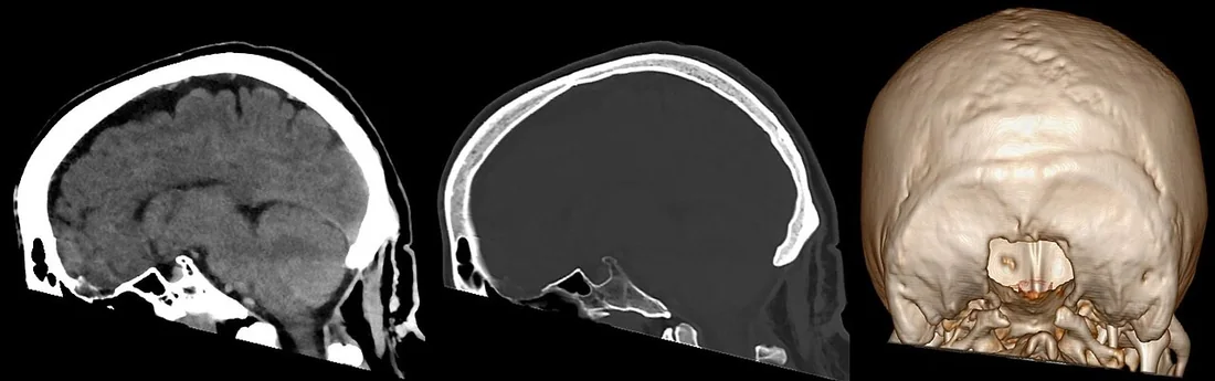
Chiari I, serebellar tonsillerin foramen magnumun altına uzandığı ve kraniyoservikal bileşkede kalabalıklaşmanın eşlik edebildiği bir malformasyondur. Kafa BT'si çoğu kez başka nedenle çekilir; sagittal rekonstrüksiyon tonsillerin aşağı konumunu gösterebilir, ancak yumuşak doku ve BOS akımını değerlendirmede MR üstündür. Tonsil konumu tek başına semptomatik Chiari tanısı veya klinik önem anlamına gelmez. Foramen magnum düzeyindeki bası, eşlik eden hidrosefali ya da syrinks şüphesi bildirilmezse ileri değerlendirme gereksinimi gözden kaçabilir.
Faz ve pencere
- Kontrastsız tanısal
- Sagittal kemik ve yumuşak doku rekonstrüksiyonlarında serebellar tonsillerin opisthion-basion (McRae) çizgisine göre aşağı yerleşimi ve foramen magnum çevresindeki kalabalıklaşma izlenebilir. ≥5 mm gibi milimetrik eşiğin doğrulanması için MRI morfometrisi gereklidir. Rutin kontrast fazı gerekli değildir.
Önerilen pencereler: Beyin (G 80 / M 40), Kemik (G 1800 / M 400), Yumuşak doku (G 400 / M 40).
BT bulguları
- Düşük yerleşimli tonsiller — sagittal kesitte serebellar tonsil alt ucu foramen magnum referans çizgisinin kaudalinde izlenebilir.
- Foramen magnum kalabalıklaşması — tonsiller ve alt vermis çevresindeki BOS aralığı daralmış görünebilir.
- Serebellomedüller bileşke sıkışıklığı — medulla ile tonsiller arasındaki normal sisternal boşluk silikleşebilir.
- Tonsil şekli ayırıcı tanıda yer alır; edinilmiş ektopide (spontan intrakraniyal hipotansiyon gibi) tonsiller yuvarlak konturlarını korurken, konjenital olgularda farklılaşabilir; BT ise yumuşak doku ayrımında sınırlıdır.
- Dördüncü ventrikül çıkışları — belirgin bası veya eşlik eden obstrüktif genişleme açısından kontrol edilir.
- Ventriküler boyut — lateral ve üçüncü ventrikül genişlemesi varsa BOS akım engeli araştırılır.
- Kafa tabanı ve C1 kemik ilişkisi — basion, opisthion ve atlas arkı kraniyoservikal bileşke anatomisini ortaya koyar.
Ölçütler ve sınıflamalar
- Sık kullanılan MRI morfometrik tonsil inişi eşiği
- ≥5 mm
Normalde
Normal kraniyoservikal bileşkede serebellar tonsiller foramen magnum düzeyinin üstünde ya da hemen komşuluğunda görünür ve tonsiller çevresinde sisternal BOS alanı korunur. Orta hat sagittal MRI'da opisthion-basion çizgisini, tonsil alt ucunu, medullayı ve dördüncü ventrikül çıkışını birlikte değerlendirin; milimetrik konum farkı ve BOS kanalları anatomik detayla net izlenir.
Normal BT ile kıyasla
Aynı bölgenin, kaynak veri setinde patoloji içermediği belirtilen bir BT incelemesini kesit kesit kaydırın; organ sınırlarını açarak anatomiyi hasta görüntüsüyle karşılaştırın. Küçük rastlantısal bulgular tümüyle dışlanmamıştır.
Kaynak: TotalSegmentator v2.01 veri seti (Wasserthal ve ark., Radiology: Artificial Intelligence 2023), CC BY 4.0, vaka s0103; organ sınırları veri setinin otomatik segmentasyonundan, birleştirilerek sadeleştirildi. Görüntüler 2,5 mm kesit aralığına yeniden örneklendi.
Ayırıcı tanı
- Düşük yerleşimli tonsiller varyantı
- belirgin foramen magnum kalabalıklaşması olmadan hafif kaudal konum görülebilir.
- Edinsel tonsiller herniasyon
- kafa içi hipotansiyon, kitle veya hidrosefali bağlamında gelişen aşağı yer değiştirmede altta yatan neden araştırılır.
- Chiari II malformasyonu
- küçük posterior fossa, aşağı yer değiştirmiş beyin sapı ve ek arka beyin deformiteleri eşlik eder.
- Posterior fossa kitle etkisi
- tonsillerin kaudal hareketine kitle, sisterna silinmesi veya hidrosefali neden olabilir.
- İntrakraniyal hipotansiyon
- beyin sarkmasıyla birlikte bazal sisternal daralma ve diğer kafa içi hipotansiyon bulguları beklenir.
Tuzaklar
- Tonsil alt ucunun tam konumunu değerlendirmek için orta hat sagittal ve çok düzlemlili rekonstrüksiyonlar kullanılmalıdır; tek bir düzlemde eksik izlenme yanıltıcı olabilir.
- Sık kullanılan ≥5 mm eşik değeri MRI morfometrisi ile doğrulanmış kriterdir; bu anatomik bulgu, hastanın klinik semptomları ve BOS dinamikleriyle ilişkilendirilmelidir.
- Tonsiller ektopiyi otomatik olarak konjenital Chiari I saymayın; edinsel beyin sarkması ve posterior fossa kitle etkisini dışlayın.
Kendini dene
Sagittal BT'de tonsiller foramen magnum altında; belirgin kitle yok. Ölçüm nasıl yorumlanmalı?
Cevabı göster
MR ve klinikle doğrulanır. BT tonsil konumunu düşündürebilir, ancak ölçüm düzlemi ve yumuşak doku çözünürlüğü sınırlıdır; klinik anlam ve eşlik eden BOS akımı için MR gerekir. Bu görünüm Chiari II'yi veya enfarktı tanımlamaz.
Tonsiller aşağıda, fakat beyin sapı ve posterior fossa da kitleyle basılı. Önce ne düşünülür?
Cevabı göster
Edinsel herniasyon. Posterior fossa kitle etkisi tonsilleri aşağı itebilir; bu nedenle görünümü doğrudan konjenital Chiari I olarak adlandırmak doğru değildir. Diğer seçenekler bu bası mekanizmasını açıklamaz.
İlgili konular
Kaynaklar
Bu sayfadaki 38 cümle ve 2 quiz sorusu aşağıdaki kaynaklardan alıntılarla tek tek karşılaştırıldı; 4 cümle bu karşılaştırmada düzeltildi (2026-10-08).
- Overview of Spontaneous Intracranial Hypotension and Differential Diagnosis with Chiari I Malformationpmc.ncbi.nlm.nih.gov
- Is Cervical Stabilization for All Cases of Chiari-I Malformation an Overkill? Evidence Speaks Louder Than Words!pmc.ncbi.nlm.nih.gov
- Diprosopus: A Rare Case of Craniofacial Duplication and a Systematic Review of the Literaturepmc.ncbi.nlm.nih.gov
- Advances in Magnetic Resonance Imaging of the Skull Basepmc.ncbi.nlm.nih.gov
- Kyphoscoliosis with Klippel-Trenaunay syndrome: a case report and literature reviewpmc.ncbi.nlm.nih.gov
- Headache in Chiari Malformation.pubmed.ncbi.nlm.nih.gov
- The Chiari I malformation.pubmed.ncbi.nlm.nih.gov
- Chiari I malformation.pubmed.ncbi.nlm.nih.gov
- Management of Chiari I malformations: a paradigm in evolution.pubmed.ncbi.nlm.nih.gov
- Imaging in Chiari I Malformation.pubmed.ncbi.nlm.nih.gov
- Chiari I malformation in patients with RASopathies.pubmed.ncbi.nlm.nih.gov
- "Growing cerebellum" requiring operative decompression following perinatal ventriculoperitoneal shunting.pubmed.ncbi.nlm.nih.gov
- Cerebrospinal Fluid Flow Studies and Recent Advancements.pubmed.ncbi.nlm.nih.gov
- Prevalence of Chiari I Malformation and Syringomyelia.pubmed.ncbi.nlm.nih.gov
- MRI Findings After Surgery for Chiari Malformation Type I.pubmed.ncbi.nlm.nih.gov
- Pathogenesis and Cerebrospinal Fluid Hydrodynamics of the Chiari I Malformation.pubmed.ncbi.nlm.nih.gov
- Functional and morphological changes in hypoplasic posterior fossa.pubmed.ncbi.nlm.nih.gov
- Definitions and Anatomic Considerations in Chiari I Malformation and Associated Syringomyelia.pubmed.ncbi.nlm.nih.gov
- Spontaneous resolution of a Chiari malformation with syringomyelia.pubmed.ncbi.nlm.nih.gov
- Headache, Chiari I malformation and foramen magnum decompression.pubmed.ncbi.nlm.nih.gov
- Consensus conference on Chiari: a malformation or an anomaly? Scoliosis and others orthopaedic deformities related to Chiari 1 malformation.pubmed.ncbi.nlm.nih.gov
- Embolization of CSF-venous fistula for syrinx resolution in acquired Chiari malformation: illustrative case.pubmed.ncbi.nlm.nih.gov
- Posterior C1-C2 screw-rod salvage for Chiari I malformation with atlantoaxial instability after failed foramen magnum decompression.pubmed.ncbi.nlm.nih.gov
- Case Report: Reversible acquired Chiari-like state and upper cervical syringomyelia caused by a giant posterior tentorial tumor confined to the supratentorial compartment.pubmed.ncbi.nlm.nih.gov
- Bilateral Papilledema without Evident Intracranial Hypertension in Pediatric Chiari Malformation Type I: A Case Report.pubmed.ncbi.nlm.nih.gov
- A rare case of near-complete resolution of extensive cervicothoracic syrinx following bilateral cerebellar tonsillar resection in Chiari malformation type I.pubmed.ncbi.nlm.nih.gov
- Imaging and physiology across the high-low cerebrospinal fluid pressure spectrum: Navigating diagnostic uncertainty in headache practice.pubmed.ncbi.nlm.nih.gov
- Chiari malformation type 1 is associated with a smaller fourth ventricle volume - a multi-cohort replication study.pubmed.ncbi.nlm.nih.gov
- Concurrent posterior vault expansion and extradural Chiari decompression in syndromic and non-syndromic craniosynostosis: a case series.pubmed.ncbi.nlm.nih.gov
- Characterizing symptom presentation in Chiari malformation type-1 in two subpopulations of pediatric patients who underwent decompression.pubmed.ncbi.nlm.nih.gov
- Bulbar "Red Flag" Symptoms and a 21-Year Diagnostic Delay in Adult Arnold-Chiari Type II Malformation With Tracheoesophageal Fistula and Syringomyelia: A Case Report.pubmed.ncbi.nlm.nih.gov
- Foramen Magnum Decompression for Retro-Odontoid Pseudotumor in an Elderly Patient With Chiari Malformation Type I: A Case Report.pubmed.ncbi.nlm.nih.gov
- Pediatric complex Chiari I Malformation-how complex is it?pubmed.ncbi.nlm.nih.gov
- Surgical Management of Chiari 1.5 in Children: A Truly Different Disease?pubmed.ncbi.nlm.nih.gov
- Comprehensive comparative study of Chiari-like malformation in veterinary and human medicine.pubmed.ncbi.nlm.nih.gov
- A Critical Update of the Classification of Chiari and Chiari-like Malformations.pubmed.ncbi.nlm.nih.gov
- Clinical applications of 4D flow magnetic resonance imaging in hepatic vascular disorders: practical biomarkers, protocol design, and translational implementation.pubmed.ncbi.nlm.nih.gov
- Society for Cardiovascular Magnetic Resonance 2024 Cases of SCMR case series.pubmed.ncbi.nlm.nih.gov
- Novel Insights into the Management of Painful Trigeminal Neuropathy: Targeting Peripheral and Central Pathomechanisms.pubmed.ncbi.nlm.nih.gov
- Chiari Malformation and Hindbrain Descent: Characterization and New Classification Based on Mechanism and Pathogenesis, and Surgical Management.pubmed.ncbi.nlm.nih.gov
- Variability in the circulation of cerebrospinal fluid: causes and clinical implications for intraventricular drug delivery.pubmed.ncbi.nlm.nih.gov
- Identifying central positional vertigo to prevent diagnostic pitfalls: A case series and review of literature.pubmed.ncbi.nlm.nih.gov
- Evaluation and Treatment of Patients with Small Posterior Cranial Fossa and Chiari Malformation, Types 0 and 1.pubmed.ncbi.nlm.nih.gov
- Magnetic Resonance Imaging-Related Anatomic and Functional Parameters for the Diagnosis and Prognosis of Chiari Malformation Type I: A Systematic Review and Meta-analysis.pubmed.ncbi.nlm.nih.gov
- EACR 2025 Congress: Innovative Cancer Science, 16-19 June 2025.pubmed.ncbi.nlm.nih.gov
- Synergistic use of high-resolution flow-sensitive and flow-compensated MRI sequences for the evaluation of cerebrospinal fluid flow disorders.pubmed.ncbi.nlm.nih.gov
- Idiopathic Syringomyelia: A Systematic Scoping Review.pubmed.ncbi.nlm.nih.gov
- Pulsatile intraspinal pressure drives post-traumatic syringomyelia: evidence supporting the CSF pulsatile pressure dynamic equilibrium hypothesis.pubmed.ncbi.nlm.nih.gov
- Comprehensive review of the cerebral aqueduct: anatomy, embryology, imaging, genetics, molecular, and pathology.pubmed.ncbi.nlm.nih.gov
- Hydrocephalus-associated trigeminal neuralgia.pubmed.ncbi.nlm.nih.gov
- Novel endoscopic posterior fossa cyst fenestration via an occipital meningocele defect in neonatal Dandy-Walker malformation: illustrative case.pubmed.ncbi.nlm.nih.gov
- A review and commentary on congenital anomalies of the craniocervical junction.pubmed.ncbi.nlm.nih.gov
- An examination of atlanto-occipital curvature in adult Chiari malformation type 1 and control groups.pubmed.ncbi.nlm.nih.gov
- Atlanto-occipital assimilation: the role of ex-, basi-, and suboccipital bones in the physiopathology of type I basilar invagination. Patient series.pubmed.ncbi.nlm.nih.gov
- Evaluating CSF Flow (Cine MR) and Clinical Outcomes in Patients with Chiari Type 1 Who Underwent Dural Relaxation or Durotomy.pubmed.ncbi.nlm.nih.gov
- Deep Feature-Based Detection of Chiari Malformation Type I from Sagittal T2-Weighted MRI Using a Hybrid CNN-Machine Learning Framework.pubmed.ncbi.nlm.nih.gov
- MRI-morphometric characterization of Chiari malformation types 0 and 1 with syringomyelia: implications for diagnosis and pathogenesis.pubmed.ncbi.nlm.nih.gov
- The myodural bridge complex: a comprehensive review of morphology, physiology, developmental biology and pathology.pubmed.ncbi.nlm.nih.gov
- Association of Chiari malformation type I and tethered cord syndrome: preliminary results of sectioning filum terminale.pubmed.ncbi.nlm.nih.gov
- The Small Posterior Cranial Fossa Syndrome and Chiari Malformation Type 0.pubmed.ncbi.nlm.nih.gov
- Resolution of tonsillar herniation and syringomyelia after resection of supratentorial large meningioma.pubmed.ncbi.nlm.nih.gov
- Is foramen magnum decompression for acquired Chiari I malformation like putting a finger in the dyke? - A simplistic overview of artificial intelligence in assessing critical upstream and downstream etiologies.pubmed.ncbi.nlm.nih.gov
- Chiari III malformation in a neonate.pubmed.ncbi.nlm.nih.gov
- The Pathogenesis of Chiari Malformation and Syringomyelia: A Case Report and Systematic Review of Current Theories.pubmed.ncbi.nlm.nih.gov
- Acute fulminant cerebellitis in children: a case series and literature review on diagnostic challenges and management strategies.pubmed.ncbi.nlm.nih.gov
- Postpartum Vertebral Artery Dissection After Neck Massage Resulting in a Right Cerebellar Infarct: A Case Report and Literature Review.pubmed.ncbi.nlm.nih.gov
- Movement Disorders in Brain Sagging Syndrome Due To Spontaneous Intracranial Hypotension: A Review.pubmed.ncbi.nlm.nih.gov
- Spontaneous Intracranial Hypotension: A Review of Neuroimaging and Current Concepts.pubmed.ncbi.nlm.nih.gov
- Immersive Surgical Anatomy of the Craniometric Points.pubmed.ncbi.nlm.nih.gov
- Brain Shift Patterns: Upward, Lateral and Downward Herniation, Its Correlation with Clinical Patterns in Acute Intracranial Pathologies and Neurosurgical Management.pubmed.ncbi.nlm.nih.gov
- Anatomically Precise Microsurgical Resection of a Posterior Fossa Cerebellar Metastasis in an Elderly Patient with Preservation of Venous Outflow, Dentate Nucleus, and Cerebrospinal Fluid Pathways.pubmed.ncbi.nlm.nih.gov
- Individualized Functional Decompression Options for Adult Chiari Malformation With Syringomyelia and A Novel Scale for Syringomyelia Resolution: A Single-Center Experience.pubmed.ncbi.nlm.nih.gov
- Treatment strategies and prognostic outcomes in craniosynostosis with concurrent hydrocephalus: a case series.pubmed.ncbi.nlm.nih.gov
- An Uncommon Occipital Horn Arachnoid Cyst in a Newborn.pubmed.ncbi.nlm.nih.gov
- Thalamic Tuberculoma Presenting as Isolated Gait Disturbance Without Systemic or Meningeal Symptoms in a 2-Year-Old Child: A Case Report.pubmed.ncbi.nlm.nih.gov
- Duraplasty Versus Bony Decompression in Adult Chiari I: Comparative Clinical and Morphometric Analysis.pubmed.ncbi.nlm.nih.gov
- Coexistence of acid sphingomyelinase deficiency type A/B and Arnold-Chiari malformation: a novel case report.pubmed.ncbi.nlm.nih.gov
- Association of Chiropractic Colleges Educational Conference and Research Agenda Conference 2026: Connected Care.pubmed.ncbi.nlm.nih.gov
- Conference Proceedings: Select Abstracts Presented at the 2025 Advocate Aurora Scientific Day.pubmed.ncbi.nlm.nih.gov
- A review of the oral health of individuals with disabilities in Puerto Rico and among U.S. Hispanics.pubmed.ncbi.nlm.nih.gov
- Venous-significant intracranial hemorrhage in neonatal HIE treated with therapeutic hypothermia: imaging phenotypes, diagnostic pitfalls, and prognostic implications.pubmed.ncbi.nlm.nih.gov
- Spinal lesions: a comprehensive radiologic overview.pubmed.ncbi.nlm.nih.gov
- Imaging in Vascular Liver Diseases.pubmed.ncbi.nlm.nih.gov
- Morphometric assessment of the posterior cranial fossa and its contents in patients with chiari malformation type I and type 0.pubmed.ncbi.nlm.nih.gov
- From Sound to Silence: Cerebellar Abscesses and Herniation Due to Cochlear Implant Infection With <i>Escherichia coli</i> and <i>Bacteroides fragilis</i>.pubmed.ncbi.nlm.nih.gov
- The value of cerebrospinal fluid cytology in the diagnosis of atypical medulloblastoma: a case report and review of the literature.pubmed.ncbi.nlm.nih.gov
- Volumetric segmentation in the context of posterior fossa-related pathologies: a systematic review.pubmed.ncbi.nlm.nih.gov
- Aortoduodenal syndrome caused by infrarenal abdominal aortic aneurysm: Evolution of CT findings and early imaging clues.pubmed.ncbi.nlm.nih.gov
- Case Report: Refractory life-threatening post-biopsy hemorrhage in a neonate successfully controlled with Ankaferd Blood Stopper.pubmed.ncbi.nlm.nih.gov
- Amylin: A Multi-Functional Pancreatic Hormone-A Review.pubmed.ncbi.nlm.nih.gov
- Lumbar Puncture and Brain Herniation in Acute Bacterial Meningitis: An Updated Narrative Review.pubmed.ncbi.nlm.nih.gov
- Clinical characteristics of postoperative anterior arch fracture of the atlas following C1 laminectomy: 14-year experience of a single neurosurgical center.pubmed.ncbi.nlm.nih.gov
- Neonatal Arnold-Chiari II Malformation: An Imaging-Focused Case Report.pubmed.ncbi.nlm.nih.gov
- Duropathies as Unifying Concept-Part Two: A Narrative Overview of Clinical and Neuroradiological Features.pubmed.ncbi.nlm.nih.gov
- High-Grade Astrocytoma With Piloid Features: An Aggressive Clinicogenomic Entity Distinct From Pilocytic Astrocytoma.pubmed.ncbi.nlm.nih.gov
- Decoding Chiari Malformation and Syringomyelia: From Epidemiology and Genetics to Advanced Diagnosis and Management Strategies.pubmed.ncbi.nlm.nih.gov
- Foramen Magnum Decompression with Outer Dura Matter Layer Resection for Chiari Type I Malformation: Factors Correlated with Syrinx Reduction.pubmed.ncbi.nlm.nih.gov
- Spontaneous Intracranial Hypotension: Atypical Radiologic Appearances, Imaging Mimickers, and Clinical Look-Alikes.pubmed.ncbi.nlm.nih.gov
Eğitim amaçlıdır; hasta başında tanı ve tedavi kararının yerine geçmez. İçerik yapay zekâ destekli bir süreçle güncel literatüre göre cümle cümle kaynaklarla karşılaştırıldı, ancak uzman radyolog incelemesinden geçmedi. Klinik kararlarda güncel kılavuzları, kurum protokollerini ve radyoloji raporunu esas alın.