Özet
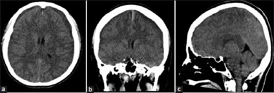
Spontan intrakraniyal hipotansiyon çoğunlukla spinal BOS kaçağıyla ilişkili düşük BOS hacmi sendromudur ve BOS açılış basıncı normal olabilir; spinal manipülasyon veya şant aşırı drenajı sonrasında da gelişebilir. Kontrastsız beyin BT tanısal duyarlılığı sınırlı olan bir incelemedir; subdural koleksiyon, bazal sistern silinmesi, posterior fossa kalabalıklaşması ve yalancı subaraknoid kanama görünümü gösterebilir. BT normal olsa bile tipik ortostatik baş ağrısı ve klinik şüphe varsa hastalık dışlanmış olmaz; beyin ve omurga MR değerlendirmesi gerekebilir. Tanının atlanması devam eden BOS kaybının ve subdural kanama gibi komplikasyonların geç fark edilmesine yol açabilir.
Faz ve pencere
- Kontrastsız tanısal
- Bilateral ince subdural sıvı/kan koleksiyonları, ventrikül ve sisternlerde görece küçülme, beyin sapı çevresinde kalabalıklaşma görülebilir. Bazal sisternlerde artmış atenüasyon gerçek kan olmadan yalancı subaraknoid kanamayı taklit edebilir; normal BT intrakraniyal hipotansiyonu dışlamaz.
Önerilen pencereler G genişlik, M merkez; değerler Hounsfield birimi (HU)
- BeyinG 80 / M +40
- SubduralG 200 / M +75
BT bulguları
- Bilateral, ince ve hilal biçimli subdural koleksiyonlar; çoğu BOS benzeri düşük atenüasyonlu, kanama eşlik ederse heterojen olabilir.
- Suprasellar, interpedinküler ve prepontin sisternlerde daralma veya silinme; beyin sarkmasıyla beraber yorumlanır.
- Posterior fossa kalabalıklaşması, pons ön yüzünde basıklık ve serebellar tonsillerin aşağı konumu, tanısal süreçte tipik olarak manyetik rezonans görüntülemede izlenir.
- BOS hacmi azaldığı için ventriküller ve sulkuslar küçülebilir; ancak bu bulgular tek başına özgül değildir ve MR'da genellikle direkt kaçağı göstermeyen dolaylı bulgulardır.
- Bazal sisternlerde ve tentoryum çevresinde yüksek atenüasyon yalancı subaraknoid kanama görünümü oluşturabilir.
- Kontrastlı beyin MR’ında yaygın pakimeningeal kontrastlanma görülebilir.
- Kontrastlı beyin MR’ında yaygın pakimeningeal kontrastlanma, venöz sinüslerde genişleme ve hipofiz bezinde büyüme görülebilir; bu bulgular beyin sarkması ve subdural koleksiyonlarla birlikte değerlendirilir.
Normalde
Normal hacim durumunda suprasellar ve prepontin sisternler açık BOS aralıkları olarak seçilir; pons ve serebellar tonsiller beklenen konumda, posterior fossa kalabalıklaşmamıştır. Aynı düzeylerde sisternal açıklık, pons ön konturu, tonsillerin foramen magnuma göre konumu ve bilateral subdural aralık karşılaştırılır.
Normal BT ile kıyasla
Aynı bölgenin, kaynak veri setinde patoloji içermediği belirtilen bir BT incelemesini kesit kesit kaydırın; organ sınırlarını açarak anatomiyi hasta görüntüsüyle karşılaştırın. Küçük rastlantısal bulgular tümüyle dışlanmamıştır.
Kaynak: TotalSegmentator v2.01 veri seti (Wasserthal ve ark., Radiology: Artificial Intelligence 2023), CC BY 4.0, vaka s0103; organ sınırları veri setinin otomatik segmentasyonundan, birleştirilerek sadeleştirildi. Görüntüler 2,5 mm kesit aralığına yeniden örneklendi.
Ayırıcı tanı
- Anevrizmal subaraknoid kanama
- sistern ve sulkuslarda kan yoğunluğu, klinik başlangıç ve damar görüntülemesiyle değerlendirilir; hipotansiyonda kalabalıklaşmış sisternler kanı taklit edebilir.
- Chiari I malformasyonu
- tonsiller ektopi temel bulgu olabilir; beyin sapı sarkması, sisternal silinme ve subdural koleksiyonların birlikteliği düşük BOS hacmini destekler.
- Menenjit
- klinik enfeksiyon ve MR’daki leptomeningeal bulgular yardımcıdır; BT’de sisternal yüksek atenüasyon tek başına özgül değildir.
- Travmatik subdural hematom
- travma ve fokal/heterojen kan yoğunluğu ayırıcıdır; spontan veya iatrojenik hipotansiyonla aynı anda bulunabileceği unutulmamalıdır.
Tuzaklar
- BT’nin normal olması hastalığı dışlamaz; BT, beyin sarkması ve dural bulgular için MR’dan daha az duyarlıdır.
- Bazal sistern hiperdensitesi yalancı subaraknoid kanama görünümü olabilir; yoğunluk ve anatomik dağılım gerçek kanama ile klinik bağlamda karşılaştırılır.
- Tonsillerin aşağı yerleşimini tek başına Chiari I olarak yorumlamayın; beyin sapı sarkması, pons biçimi ve sisternal açıklık da önemlidir.
- Subdural kanama ya da higroma yalnızca yaşlılık/trauma ile açıklanmayabilir; özellikle ortostatik baş ağrısında BOS kaybı nedenini düşünün.
- Spontan intrakraniyal hipotansiyon şüphesinde normal BOS açılış basıncı tanıyı dışlamaz.
Kendini dene
Ortostatik baş ağrısı olan hastada kontrastsız BT normal raporlanmış. Klinik şüphe sürüyor. Bu BT için en doğru yorum nedir?
Cevabı göster
Hastalığı dışlamaz. Kontrastsız BT intrakraniyal hipotansiyonu dışlamak için duyarlı değildir; normal görünüm klinik şüpheyi ortadan kaldırmaz. Kaçak yeri ve BOS basıncı BT baş incelemesinden doğrudan belirlenemez.
İntrakraniyal hipotansiyonda serebellar tonsillerin aşağı yer değiştirmesi hangi yapısal anomaliyle karışabilir?
Cevabı göster
Chiari I malformasyonu. İntrakraniyal hipotansiyon ve Chiari I malformasyonu bazı MR bulgularını, özellikle serebellar tonsillerin aşağı yerleşimini paylaşır.
İlgili konular
- Şant aşırı drenajı ve intrakraniyal hipotansiyon bulguları
- Şant ilişkili subdural higroma veya hematom
- Slit ventrikül örüntüsü ve şant komplikasyonu
Kaynaklar
Bu sayfadaki 36 cümle ve 2 quiz sorusu aşağıdaki kaynaklardan alıntılarla otomatik olarak karşılaştırıldı; 7 cümle bu karşılaştırmada düzeltildi (2026-10-09).
- Intracranial venous thrombosis imaging by anatomical location: an educational review with mimics and diagnostic pitfallspmc.ncbi.nlm.nih.gov
- Transethmoidal encephalocele associated with granulomatosis with polyangiitis: A case reportpmc.ncbi.nlm.nih.gov
- Lumbar microspur-related ventral cerebrospinal fluid leak localized by digital subtraction myelography: A case reportpmc.ncbi.nlm.nih.gov
- Pneumocephalus with suspected cerebrospinal fluid leakage after epiduroscopic epidural neuroplasty: a case reportpmc.ncbi.nlm.nih.gov
- Delayed Traumatic Subarachnoid Hemorrhage Following Initially Negative Computed Tomography (CT) Imaging in a Patient With a Mechanical Mitral Valve and Markedly Elevated International Normalized Ratio (INR)pmc.ncbi.nlm.nih.gov
- CSF-Venous Fistula.pubmed.ncbi.nlm.nih.gov
- [Intracranial hypotension].pubmed.ncbi.nlm.nih.gov
- Spontaneous Intracranial Hypotension.pubmed.ncbi.nlm.nih.gov
- Spontaneous Intracranial Hypotension.pubmed.ncbi.nlm.nih.gov
- Thunderclap Headache.pubmed.ncbi.nlm.nih.gov
- Spontaneous Intracranial Hypotension.pubmed.ncbi.nlm.nih.gov
- Spontaneous intracranial hypotension: diagnostic and therapeutic workup.pubmed.ncbi.nlm.nih.gov
- Chronic subdural hematoma.pubmed.ncbi.nlm.nih.gov
- Thunderclap headache.pubmed.ncbi.nlm.nih.gov
- Thunderclap headache: an update.pubmed.ncbi.nlm.nih.gov
- Traumatic brain injury in adults.pubmed.ncbi.nlm.nih.gov
- CT-guided Fibrin Glue Occlusion of Cerebrospinal Fluid-Venous Fistulas.pubmed.ncbi.nlm.nih.gov
- Spontaneous intracranial hypotension with negative brain MRI findings: a systematic review of diagnostic strategies and clinical outcomes.pubmed.ncbi.nlm.nih.gov
- Predictive factors of epidural blood patch success in intracranial hypotension syndrome.pubmed.ncbi.nlm.nih.gov
- Cervical Osteophyte as a Rare Etiology of Spontaneous Intracranial Hypotension With Bilateral Subdural Hematomas: A Case Report and Literature Review.pubmed.ncbi.nlm.nih.gov
- Risk factors for subdural hematoma in spontaneous intracranial hypotension are male sex, longer disease duration, and repeated epidural blood patches.pubmed.ncbi.nlm.nih.gov
- Intracranial Hypotension Following Spinal Manipulation: A Case Report and Scoping Review of the Literature.pubmed.ncbi.nlm.nih.gov
- Bilateral subdural hematomas in spontaneous intracranial hypotension: A systematic review with case illustration.pubmed.ncbi.nlm.nih.gov
- Intracranial venous thrombosis imaging by anatomical location: an educational review with mimics and diagnostic pitfalls.pubmed.ncbi.nlm.nih.gov
- Sacral CSF-venous fistula: a rare entity. Illustrative case.pubmed.ncbi.nlm.nih.gov
- Embolization of CSF-venous fistula for syrinx resolution in acquired Chiari malformation: illustrative case.pubmed.ncbi.nlm.nih.gov
- Traumatic intracranial pseudoaneurysms of cortical middle cerebral artery and posterior cerebral artery branches presenting as subdural hematomas in elderly patients: A two-case report.pubmed.ncbi.nlm.nih.gov
- Spontaneous Intracranial Hypotension: Case Report and Update on Diagnosis and Treatment.pubmed.ncbi.nlm.nih.gov
- Spontaneous Intracranial Hypotension: A Review of Neuroimaging and Current Concepts.pubmed.ncbi.nlm.nih.gov
- Superior Sagittal Sinus Thrombosis in a Patient With Iatrogenic Intracranial Hypotension: A Case Report.pubmed.ncbi.nlm.nih.gov
- Case Report: Endoscopic-assisted cistern ostomy with Ommaya reservoir insertion for trapped temporal horn: surgical technique and case illustration.pubmed.ncbi.nlm.nih.gov
- Parasagittal subdural space: a novel quantitative marker of spontaneous intracranial hypotension syndrome-induced chronic subdural hematoma.pubmed.ncbi.nlm.nih.gov
- Subarachnoid Hemorrhage Confirmed by Magnetic Resonance Imaging in a Patient with Brain Death owing to Hypoxic Encephalopathy Following Suicide by Hanging.pubmed.ncbi.nlm.nih.gov
- Acute subdural hematoma in spontaneous intracranial hypotension: illustrative case and review of the literature.pubmed.ncbi.nlm.nih.gov
- Surgical management of warfarin-associated spontaneous bilateral subdural hematoma in a mechanical heart valve patient: a rare case report.pubmed.ncbi.nlm.nih.gov
- Continuous Epidural Infusion of Hydroxyethyl Starch for Spontaneous Intracranial Hypotension: A Retrospective Case Series.pubmed.ncbi.nlm.nih.gov
- Brain Shift Patterns: Upward, Lateral and Downward Herniation, Its Correlation with Clinical Patterns in Acute Intracranial Pathologies and Neurosurgical Management.pubmed.ncbi.nlm.nih.gov
- Acute Spontaneous Paradoxical Herniation After Large Cerebral Meningioma Resection and Decompressive Craniectomy: A Case Report and Systematic Review of the Literature.pubmed.ncbi.nlm.nih.gov
- Successful Treatment of Spontaneous Intracranial Hypotension With Subdural Hematoma With Cervical Epidural Blood Patch: A Case Report.pubmed.ncbi.nlm.nih.gov
- Diagnostic Challenges and Surgical Outcomes of Miyazaki Syndrome: A Report of Two Cases and a Systematic Review.pubmed.ncbi.nlm.nih.gov
- Spontaneous Intracranial Hypotension with a Reversible Splenial Lesion after Swimming.pubmed.ncbi.nlm.nih.gov
- Imaging Evaluation of Acute Traumatic Brain Injury.pubmed.ncbi.nlm.nih.gov
- Lumbar microspur-related ventral cerebrospinal fluid leak localized by digital subtraction myelography: A case report.pubmed.ncbi.nlm.nih.gov
- Resolution of spontaneous intracranial hypotension due to a spinal CSF-venous fistula after a dynamic myelogram: illustrative case.pubmed.ncbi.nlm.nih.gov
- Imaging and physiology across the high-low cerebrospinal fluid pressure spectrum: Navigating diagnostic uncertainty in headache practice.pubmed.ncbi.nlm.nih.gov
- Endoscopic endonasal intradural pituitary transposition for resecting retroinfundibular lesions: technique notes and a single institute experience.pubmed.ncbi.nlm.nih.gov
- Diagnostic Value of Flow-Void Sign for Localizing Ventral Dural Defects in Spine MRI.pubmed.ncbi.nlm.nih.gov
- Spontaneous intracranial hypotension complicated by diffuse cerebral edema and episodes of severely elevated intracranial pressure: illustrative case.pubmed.ncbi.nlm.nih.gov
- Atypical Presentations of Intracranial Hypotension: Comparison with Classic Spontaneous Intracranial Hypotension.pubmed.ncbi.nlm.nih.gov
- Evaluation of hydrocephalus and other cerebrospinal fluid disorders with MRI: An update.pubmed.ncbi.nlm.nih.gov
- When headache meets visual disturbance: An evidence-based review of common and critical entities.pubmed.ncbi.nlm.nih.gov
- Reversible cerebral vasoconstriction syndrome: A narrative review.pubmed.ncbi.nlm.nih.gov
- Vasospasm following resection of olfactory groove meningiomas via orbitozygomatic craniotomy: illustrative cases.pubmed.ncbi.nlm.nih.gov
- Beyond the Headaches: Severe Dysphagia As the Dominant Symptom of Spontaneous Intracranial Hypotension.pubmed.ncbi.nlm.nih.gov
- Acquired tonsillar herniation related to spontaneous intracranial hypotension: case reports.pubmed.ncbi.nlm.nih.gov
- Hallmarks of headache disorders: part 5 - secondary headaches.pubmed.ncbi.nlm.nih.gov
- Infectious meningitis and encephalitis: A primer for acute care practitioners.pubmed.ncbi.nlm.nih.gov
- Multidisciplinary Management of Pediatric Craniopharyngioma: From Diagnosis to Long-Term Outcomes.pubmed.ncbi.nlm.nih.gov
- Spontaneous Intracranial Hypotension in Pregnancy with Aggravated Comorbidity: A Case Report and Review of Diagnostic and Management Challenges.pubmed.ncbi.nlm.nih.gov
- Retroclival Epidural Hematoma in Pediatric Head Trauma: A Case Report and Comprehensive Literature Review.pubmed.ncbi.nlm.nih.gov
- High risk and low incidence diseases: Aneurysmal subarachnoid hemorrhage.pubmed.ncbi.nlm.nih.gov
- Post-Traumatic Cerebral Infarction: A Narrative Review of Pathophysiology, Diagnosis, and Treatment.pubmed.ncbi.nlm.nih.gov
- Multimodal Monitoring in Eclamptic Intracerebral Hemorrhage Complicated by Reversible Cerebral Vasoconstriction Syndrome (RCVS): A Case Report.pubmed.ncbi.nlm.nih.gov
- Cerebral venous thrombosis revealed by psychiatric disorders: a diagnosis not to be missed.pubmed.ncbi.nlm.nih.gov
- Catastrophic Hypoxic-Ischemic Brain Injury Following Influenza A Illness in a Young Woman: A Case Report.pubmed.ncbi.nlm.nih.gov
- Update on hypoxic-ischemic brain injury: Prognosis and management.pubmed.ncbi.nlm.nih.gov
- Neonatal Spontaneous Abdominal Aortic Thrombosis Associated With Hypernatremic Dehydration and Acute Renal Failure: A Case Series.pubmed.ncbi.nlm.nih.gov
- Overview of Spontaneous Intracranial Hypotension and Differential Diagnosis with Chiari I Malformation.pubmed.ncbi.nlm.nih.gov
- Diagnosis and Treatment of Spontaneous Intracranial Hypotension: Role of Epidural Blood Patching.pubmed.ncbi.nlm.nih.gov
- Surgical Outcomes in Chiari 1 and Chiari 1.5 Malformation Treated by Posterior Fossa Reconstruction: A Comprehensive Analysis of 110 Pediatric Cases and Literature Review.pubmed.ncbi.nlm.nih.gov
- Seizure following epidural blood patch in spontaneous intracranial hypotension complicated by subdural hematoma: Illustrated case and literature review.pubmed.ncbi.nlm.nih.gov
- Resolution of pachymeningeal enhancement is a radiographic marker for effective treatment of spontaneous intracranial hypotension: patient series.pubmed.ncbi.nlm.nih.gov
- Frequency and Diagnostic Utility of Clinical and Radiological Features of Spontaneous Intracranial Hypotension in Postpartum Patients with Neurological Symptoms: A Cross-Sectional Study.pubmed.ncbi.nlm.nih.gov
- Duropathies as Unifying Concept-Part Two: A Narrative Overview of Clinical and Neuroradiological Features.pubmed.ncbi.nlm.nih.gov
- Case Report: Reversible acquired Chiari-like state and upper cervical syringomyelia caused by a giant posterior tentorial tumor confined to the supratentorial compartment.pubmed.ncbi.nlm.nih.gov
- Chiari Malformation and Hindbrain Descent: Characterization and New Classification Based on Mechanism and Pathogenesis, and Surgical Management.pubmed.ncbi.nlm.nih.gov
- Clinical recovery of severe infantile <i>Angiostrongylus cantonensis</i> meningoencephalomyelitis treated with pulse methylprednisolone: A case report and review of literature.pubmed.ncbi.nlm.nih.gov
- Progressive loss of hearing and balance in superficial siderosis due to occult spinal dural defects.pubmed.ncbi.nlm.nih.gov
- A Practical Diagnostic Approach to Pediatric Episodic Vestibular Syndrome.pubmed.ncbi.nlm.nih.gov
- Adrenal causes of endocrine hypertension in childhood or adolescence.pubmed.ncbi.nlm.nih.gov
- High-Throughput Computing to Automate Population-Based Studies to Detect the 30-Day Risk of Adverse Outcomes After New Outpatient Medication Use in Older Adults with Chronic Kidney Disease: A Clinical Research Protocol.pubmed.ncbi.nlm.nih.gov
- Optical imaging and spectroscopy for the study of the human brain: status report.pubmed.ncbi.nlm.nih.gov
- The onset of posterior reversible encephalopathy syndrome (PRES) and reversible cerebral vasoconstriction syndrome (RCVS) with the use of decongestant agents: a European pharmacovigilance analysis.pubmed.ncbi.nlm.nih.gov
- Repeated transitions between intracranial hypotension and intracranial hypertension in Gorham-Stout disease: illustrative case.pubmed.ncbi.nlm.nih.gov
- The Duke CSF-Venous Fistula Confidence Score: Real-World Application on CT Myelography.pubmed.ncbi.nlm.nih.gov
- Occipital shuntalgia: Rethinking post-shunt occipital headache etiology and care.pubmed.ncbi.nlm.nih.gov
- Non-invasive intracranial pressure estimation in the intensive care unit: narrative review of methods and clinical applications.pubmed.ncbi.nlm.nih.gov
- Foetal disruptive brain injuries: Diagnosing the underlying pathogenetic mechanisms with cranial ultrasonography.pubmed.ncbi.nlm.nih.gov
- The interdural hematoma: A subtype of convexity subdural/dural hematoma with specific radioanatomical characteristics.pubmed.ncbi.nlm.nih.gov
- Development and Proof-of-Concept Evaluation of a Structured Reporting Template for Emergency Radiology Using Synthetic Cases.pubmed.ncbi.nlm.nih.gov
- How the heterogeneity of the severely injured brain affects hybrid diffuse optical signals: case examples and guidelines.pubmed.ncbi.nlm.nih.gov
- Surgical Treatment of Spontaneous Intracranial Hypotension: Clinical Characteristics and Outcomes in a Surgically Treated Cohort of Type 1 and Type 3 Leaks.pubmed.ncbi.nlm.nih.gov
- Frequency and characteristics of multi-domain autonomic dysfunction in patients with spontaneous intracranial hypotension.pubmed.ncbi.nlm.nih.gov
- Targeted repair of a refractory ventral cerebrospinal fluid leak identified by dynamic computed tomography myelography: illustrative case.pubmed.ncbi.nlm.nih.gov
- Contrast-Enhanced Brain MRI for Ambulatory Evaluation of Headache: Comprehensive or Excessive?pubmed.ncbi.nlm.nih.gov
- Idiopathic Intracranial Hypertension in a Child with Marfan Syndrome: Clinical, Neuroimaging, and Biomarker Findings from a Case Report.pubmed.ncbi.nlm.nih.gov
Eğitim amaçlıdır; hasta başında tanı ve tedavi kararının yerine geçmez, tıbbi tavsiye değildir. Metinler yapay zekâ desteğiyle yazıldı ve otomatik bir adımda kaynak alıntılarıyla karşılaştırıldı; bu bir tıbbi doğrulama değildir ve içerik uzman radyolog incelemesinden geçmedi. Klinik kararlarda güncel kılavuzları, kurum protokollerini ve radyoloji raporunu esas alın.