Özet
BOS şantı bulunan hastada aşırı drenaj, özellikle ortostatik baş ağrısı veya yeni nörolojik yakınmalarla başvurduğunda akla gelmelidir. Kontrastsız beyin BT, ventrikül küçülmesini ve buna bağlı subdural sıvı veya kan birikimlerini gösterir; beyin sarkması bulgularının güvenilir değerlendirmesi için MR gereklidir. Ventriküllerin küçük olması tek başına şantın aşırı çalıştığını kanıtlamaz; hastanın önceki BT’si ve klinik seyir belirleyicidir. Koleksiyonun büyümesi veya kitle etkisinin gözden kaçması nörolojik kötüleşmeyi ve herniasyonu kaçırabilir.
Faz ve pencere
- Kontrastsız tanısal
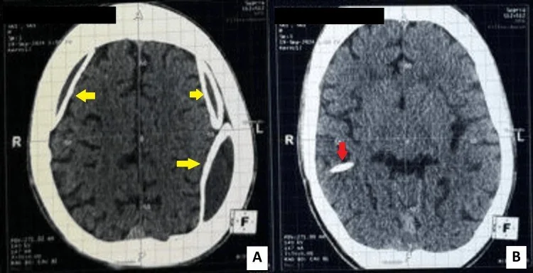
- Ventriküller küçülmüş veya çökmüş olabilir; altta yatan CSF hipovolemisine bağlı subdural sıvı birikimleri görülebilir ve BT ile hematomun yaşı değerlendirilebilir. Bazal sistern daralması ve beyin sapı/tonsil aşağı yer değiştirmesi ileri hacim kaybını düşündürür; rutin IV kontrast tanı için gerekli değildir.
Önerilen pencereler: Beyin (G 80 / M 40), Subdural (G 200 / M 75).
BT bulguları
- Önceki BT’ye göre lateral ve üçüncü ventrikül kalibresinde azalma; tek ölçüm yerine seri değişim önemlidir.
- İnce, çoğu kez bilateral frontoparietal subdural sıvı hilalleri; belirgin kitle etkisi olmayabilir.
- Koleksiyonda hematom gelişimi ve BT ile hematomun yaşı değerlendirilebilir.
- Suprasellar, prepontin ve perimezensefalik sisternlerde daralma; beyin sapı çevresindeki BOS aralığının kaybı.
- Beyin sarkması: mezensefalon ve diensefalonun aşağı konumu, pons ön konturunda basıklık veya tonsillerin aşağı yer değiştirmesi; BT’de duyarlılık sınırlıdır.
- Ventrikül içi kateterin seyri, uç konumu, rezervuar ve görünür bağlantı hattı; kraniyal BT hattın tüm devamlılığını tek başına değerlendirmez.
Normalde
Aynı aksiyel düzeyde normal kalibrasyondaki lateral ventrikül gövdeleri seçilir; kortikal sulkuslar ve bazal sisternler açıktır. Önceki aynı düzeyli BT ile ventrikül genişliği, subdural aralığın simetrisi ve prepontin/suprasellar BOS boşluklarının korunup korunmadığı karşılaştırılır.
Normal BT ile kıyasla
Aynı bölgenin, kaynak veri setinde patoloji içermediği belirtilen bir BT incelemesini kesit kesit kaydırın; organ sınırlarını açarak anatomiyi hasta görüntüsüyle karşılaştırın. Küçük rastlantısal bulgular tümüyle dışlanmamıştır.
Kaynak: TotalSegmentator v2.01 veri seti (Wasserthal ve ark., Radiology: Artificial Intelligence 2023), CC BY 4.0, vaka s0103; organ sınırları veri setinin otomatik segmentasyonundan, birleştirilerek sadeleştirildi. Görüntüler 2,5 mm kesit aralığına yeniden örneklendi.
Ayırıcı tanı
- Şant tıkanmasına bağlı ventrikül genişlemesi
- ventriküller büyür ve transependimal ödem eşlik edebilir; aşırı drenajdaki küçülmenin tersidir.
- Travmatik subdural hematom
- travma öyküsü ve hiperdens akut kan bileşeni destekler; şant aşırı drenajı ile birlikte de olabilir.
- Beyin atrofisine bağlı geniş BOS aralıkları
- sulkuslar ve ventriküller birlikte geniştir, kitle etkisi yoktur.
- Chiari I malformasyonu
- tonsiller ektopi tek başına düşük BOS hacmini kanıtlamaz; beyin sapı sarkması ve subdural koleksiyon birlikteliği aranır.
Tuzaklar
- Kronik şantlı hastada küçük ventriküller başlangıçtan beri mevcut olabilir; eski görüntüler olmadan bunu yeni aşırı drenaj diye adlandırma.
- Slit ventrikül görünümü şant işlevinin normal olduğunu göstermez; aralıklı kateter tıkanması benzer küçük ventriküllerle görülebilir.
- İntrakraniyal hipotansiyonun beyin sarkması ve dural kontrastlanma bulguları MR’da daha belirgindir; BT’nin olağan görünmesi şüpheyi dışlamaz.
Kendini dene
VP şantlı hastada ortostatik baş ağrısı var. BT'de ventriküller küçülmüş veya çökmüş, subdural sıvı birikimleri ve beyin sapı sarkması görülen hastada en olası tanı nedir? En olası örüntü nedir?
Cevabı göster
Şant aşırı drenajı. Küçülmüş ventriküller, subdural sıvı ve sisternal daralma düşük BOS hacmiyle uyumludur. Aşırı drenajda ventrikül çökmesi ve subdural sıvı birikimi tipiktir; tıkanma veya diğer patolojiler bu bulguyu açıklamaz.
Şantlı hastada ventriküller küçük; ancak karşılaştırmalı eski BT’de de aynı kalibrasyonda ve yeni subdural koleksiyon yok. Bu bulgu tek başına neyi kanıtlamaz?
Cevabı göster
Şant aşırı çalışmasını. Kronik küçük ventrikül tek başına aşırı drenaj tanısı koydurmaz; seri değişim ve eşlik eden bulgular aranmalıdır. Diğer seçenekler görüntüde doğrudan gözlenebilen özellikleri tarif eder.
Kaynaklar
Bu sayfadaki 35 cümle ve 2 quiz sorusu aşağıdaki kaynaklardan alıntılarla tek tek karşılaştırıldı; 7 cümle bu karşılaştırmada düzeltildi (2026-10-08).
- Slit ventricle syndrome: Historical considerations, diagnosis, pathophysiology, and treatment reviewpmc.ncbi.nlm.nih.gov
- Overdrainage of cerebrospinal fluid caused by detachment of the pressure control cam in a programmable valve after 3-tesla magnetic resonance imaging.pubmed.ncbi.nlm.nih.gov
- Overshunting-Associated Myelopathy (OSAM) in a Patient with Bilateral Jugular Vein Occlusion.pubmed.ncbi.nlm.nih.gov
- A new mechanism of cerebrospinal fluid leakage after lumboperitoneal shunt: a theory of shunt side hole--case report.pubmed.ncbi.nlm.nih.gov
- Diagnostic Pitfalls of Macrocephaly and Intracranial Dural Arteriovenous Fistulas: Connecting the Dots With the Red Flags.pubmed.ncbi.nlm.nih.gov
- Middle meningeal artery embolization as an alternative strategy for symptomatic CSF hypovolemia-related chronic subdural hematoma: A case series.pubmed.ncbi.nlm.nih.gov
- Rare case of intraperitoneal cerebrospinal fluid pseudocyst secondary to ventriculoperitoneal shunt: A case report and literature review.pubmed.ncbi.nlm.nih.gov
- Clinical profile and final diagnoses of hospitalized patients with a chief complaint of headache, an experience from a headache tertiary center in Iran.pubmed.ncbi.nlm.nih.gov
- Superb microvascular ultrasound is a promising non-invasive diagnostic tool to assess a ventriculoperitoneal shunt system function: a feasibility study.pubmed.ncbi.nlm.nih.gov
- Dural sac shrinkage signs on spinal magnetic resonance imaging indicate overdrainage after lumboperitoneal shunt for idiopathic normal pressure hydrocephalus.pubmed.ncbi.nlm.nih.gov
- Incidence and management of overdrainage in pediatric hydrocephalus patients exclusively treated with modern gravitational VP shunt valves.pubmed.ncbi.nlm.nih.gov
- Current Diagnostic, Counseling, and Treatment Options in Non-Severe and Severe Apparently Isolated Fetal Ventriculomegaly.pubmed.ncbi.nlm.nih.gov
- Reframing the clinical phenotype and management of cryptococcal meningitis.pubmed.ncbi.nlm.nih.gov
- Liver Transplantation Despite Active Intracranial Hemorrhage in Acetaminophen-Induced Acute Liver Failure: A Case Report.pubmed.ncbi.nlm.nih.gov
- Twist-Drill Craniostomy With Pressure-Controlled Fibrinolytic Irrigation Therapy for the Evacuation of Acute Traumatic Subdural Hematoma in Patients Not Requiring Immediate Craniotomy.pubmed.ncbi.nlm.nih.gov
- Malignant Cerebral Edema After Cranioplasty: A Case Report and Literature Insights.pubmed.ncbi.nlm.nih.gov
- Chronic Subdural Hematoma (cSDH): A review of the current state of the art.pubmed.ncbi.nlm.nih.gov
- Long-term safety and effectiveness of the "OptEase" vena cava filter.pubmed.ncbi.nlm.nih.gov
- New developments in the antiviral treatment of hepatitis C.pubmed.ncbi.nlm.nih.gov
- Spontaneous Intracranial Hypotension: A Review of Neuroimaging and Current Concepts.pubmed.ncbi.nlm.nih.gov
- Expert Strategies: Skull Base Reconstruction-Global Perspectives, Insights, and Algorithms through a Mixed Methods Approach.pubmed.ncbi.nlm.nih.gov
- Combination HSCT and intravenous AAV-mediated gene therapy in a canine model proves pivotal for translation of Krabbe disease therapy.pubmed.ncbi.nlm.nih.gov
- Role of Interventional Radiology in the Management of Non-aortic Thoracic Trauma.pubmed.ncbi.nlm.nih.gov
- Incidence and clinical analysis of asymptomatic intracranial hemorrhage in neonates with cerebral hypoxic-ischaemic risk based on multisequence MR images.pubmed.ncbi.nlm.nih.gov
- A systematic approach in the diagnosis of paediatric skull lesions: what radiologists need to know.pubmed.ncbi.nlm.nih.gov
- MDCT imaging of traumatic brain injury.pubmed.ncbi.nlm.nih.gov
- Classification of recurrence status after surgical treatment of chronic subdural hemorrhage - A machine learning approach.pubmed.ncbi.nlm.nih.gov
- Risk factors of recurrence in chronic subdural hematoma and a proposed extended classification of internal architecture as a predictor of recurrence.pubmed.ncbi.nlm.nih.gov
- Overview of Spontaneous Intracranial Hypotension and Differential Diagnosis with Chiari I Malformation.pubmed.ncbi.nlm.nih.gov
- Cerebrospinal Fluid Shunts: An Updated Radiologic Review of Devices, Malfunctions, and Complications.pubmed.ncbi.nlm.nih.gov
- Surgical Management of Breast Cancer Adjacent to a Calcified Ventriculoperitoneal Shunt: A Case Report.pubmed.ncbi.nlm.nih.gov
- International Society of Paediatric Surgical Oncology (IPSO) Surgical Practice Guidelines.pubmed.ncbi.nlm.nih.gov
- Basilar artery dolichoectasia presenting as obstructive hydrocephalus mimicking normal pressure hydrocephalus.pubmed.ncbi.nlm.nih.gov
- Point-of-Care Ultrasound Assessment of Ventriculoperitoneal Shunt Patency: A Case Report.pubmed.ncbi.nlm.nih.gov
- Prenatal diagnosis of infratentorial anomalies: ultrasound, fetal MRI, and implications for counseling.pubmed.ncbi.nlm.nih.gov
- Radiological assessment of dementia: the Italian inter-society consensus for a practical and clinically oriented guide to image acquisition, evaluation, and reporting.pubmed.ncbi.nlm.nih.gov
- Fetal Brain Development: Regulating Processes and Related Malformations.pubmed.ncbi.nlm.nih.gov
- A systematic review of surgical and interventional radiology procedures for pediatric idiopathic intracranial hypertension.pubmed.ncbi.nlm.nih.gov
- Acute subdural hematomas in shunted normal-pressure hydrocephalus patients - Management options and literature review: A case-based series.pubmed.ncbi.nlm.nih.gov
- Shunt-related cerebrospinal fluid overdrainage - a multicentre consensus definition.pubmed.ncbi.nlm.nih.gov
- Occipital shuntalgia: Rethinking post-shunt occipital headache etiology and care.pubmed.ncbi.nlm.nih.gov
- Advances in temporizing treatment strategies and associated complications for posthemorrhagic hydrocephalus in preterm infants.pubmed.ncbi.nlm.nih.gov
- Critical Reappraisal and Validation of the Bern Score System for Diagnosing Spontaneous Intracranial Hypotension.pubmed.ncbi.nlm.nih.gov
- Neurosarcoidosis: a review from diagnosis and future perspectives.pubmed.ncbi.nlm.nih.gov
- Spontaneous Intracranial Hypotension: Case Report and Update on Diagnosis and Treatment.pubmed.ncbi.nlm.nih.gov
- Advances in differential diagnosis of cerebrovascular diseases in magnetic resonance imaging: a narrative review.pubmed.ncbi.nlm.nih.gov
- Leptomeninges: Anatomy, Mechanisms of Disease and Neuroimaging.pubmed.ncbi.nlm.nih.gov
- Comprehensive review on rhino-neurosurgery.pubmed.ncbi.nlm.nih.gov
- Cervical epidural venous engorgement after cerebrospinal fluid shunting: clinical-radiological correlation and systematic review.pubmed.ncbi.nlm.nih.gov
- Headache and Neurological Symptoms Following Lumbar Puncture in a Patient With a Ventriculoperitoneal Shunt.pubmed.ncbi.nlm.nih.gov
- Diagnostic Challenges and Surgical Outcomes of Miyazaki Syndrome: A Report of Two Cases and a Systematic Review.pubmed.ncbi.nlm.nih.gov
- A case of idiopathic normal pressure hydrocephalus without enlarged ventricle and with negative tap test.pubmed.ncbi.nlm.nih.gov
- Regulation of brain fluid volumes and pressures: basic principles, intracranial hypertension, ventriculomegaly and hydrocephalus.pubmed.ncbi.nlm.nih.gov
- Guidelines for Management of Idiopathic Normal Pressure Hydrocephalus (Third Edition): Endorsed by the Japanese Society of Normal Pressure Hydrocephalus.pubmed.ncbi.nlm.nih.gov
- Surgery for post-traumatic hydrocephalus: lessons, challenges and future directions.pubmed.ncbi.nlm.nih.gov
- External Ventricular Drainage for Hydrocephalus Following Cerebellar Infarction: A Scoping Review.pubmed.ncbi.nlm.nih.gov
- Brain Midline Shift Measurement and Its Automation: A Review of Techniques and Algorithms.pubmed.ncbi.nlm.nih.gov
- Role of the "In-Window" Craniectomy in the Management of Serious Head Trauma: 12 Case Series, Technical Note, Literature Review, and Comparison With Other Surgical Managements.pubmed.ncbi.nlm.nih.gov
- Machine learning in developing a predictive model for chronic hydrocephalus following aneurysmal subarachnoid hemorrhage.pubmed.ncbi.nlm.nih.gov
- Post-traumatic hydrocephalus after decompressive craniectomy: a multidimensional analysis of clinical, radiological, and surgical risk factors.pubmed.ncbi.nlm.nih.gov
- AI Models for Surgical Decision Support in Spontaneous Intracerebral Hemorrhage: A Systematic Review in Relation to Trials and Guidelines.pubmed.ncbi.nlm.nih.gov
- Ventriculoperitoneal Shunt Treatment Increases 7 Alpha Hy-Droxy-3-Oxo-4-Cholestenoic Acid and 24-Hydroxycholesterol Concentrations in Idiopathic Normal Pressure Hydrocephalus.pubmed.ncbi.nlm.nih.gov
- ETV in infancy and childhood below 2 years of age for treatment of hydrocephalus.pubmed.ncbi.nlm.nih.gov
- Slit ventricle syndrome: Historical considerations, diagnosis, pathophysiology, and treatment review.pubmed.ncbi.nlm.nih.gov
- Sexual Dysregulation After Traumatic Brain Injury and Stroke: A Critical Narrative Review of Neurobiology, Clinical Phenotypes, and Management.pubmed.ncbi.nlm.nih.gov
- Optical digital twins for disease prevention, diagnosis, therapy, and intervention.pubmed.ncbi.nlm.nih.gov
- Network hierarchy in Alzheimer's and Lewy body spectrum disorders: functional gradients, cognitive phenotypes, and translational gaps.pubmed.ncbi.nlm.nih.gov
- Weight loss and a shunt on "off": an odd case of overshunting in an idiopathic intracranial hypertension patient. Illustrative case.pubmed.ncbi.nlm.nih.gov
- Continuum of Irreversible and Potentially Reversible Traumatic Brain Injury Sequelae: Axonal Injury and Hydrocephalus.pubmed.ncbi.nlm.nih.gov
- Progress in the diagnosis and treatment of post-traumatic hydrocephalus.pubmed.ncbi.nlm.nih.gov
- Recurrent Pneumocephalus due to over Draining Thecoperitoneal Shunt in a Patient with Multiple Skull base Defects.pubmed.ncbi.nlm.nih.gov
Eğitim amaçlıdır; hasta başında tanı ve tedavi kararının yerine geçmez. İçerik yapay zekâ destekli bir süreçle güncel literatüre göre cümle cümle kaynaklarla karşılaştırıldı, ancak uzman radyolog incelemesinden geçmedi. Klinik kararlarda güncel kılavuzları, kurum protokollerini ve radyoloji raporunu esas alın.