Özet
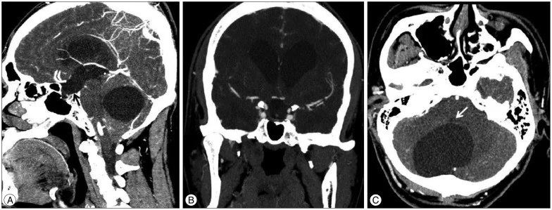
Posterior fossa araknoid kisti, çocukta gelişimsel değerlendirme sırasında veya erişkinde başka nedenle çekilen beyin BT'sinde tesadüfen saptanabilen, araknoid yapraklar arasında yer alan BOS içerikli ekstraaksiyel bir koleksiyondur. Çoğu belirti vermez; genişleyen kist serebellumu, beyin sapını ya da dördüncü ventrikülü sıkıştırarak denge bulgularına ve BOS akımında tıkanmaya yol açabilir. Kontrastsız BT koleksiyonun yerini, BOS benzeri atenüasyonunu, komşu yapılardaki basıyı ve hidrosefaliyi gösterir; epidermoid gibi bazı kistik taklitçileri kesin dışlayamaz. Basıyı veya ventrikül genişlemesini kaçırmak arka çukurda klinik açıdan önemli kitle etkisinin gözden kaçmasına, basit sisternal genişlemeyi kist diye adlandırmak ise yanlış tanısal çerçeveye neden olabilir.
Faz ve pencere
- Kontrastsız tanısal
- Araknoid kistler ekstraaksiyel ve BOS dansitesinde lezyonlardır; posterior fossada serebellum arkasında veya serebellopontin açı komşuluğunda yerleşebilir. Aynı kesitlerde serebellar foliaların silinmesi veya yer değiştirmesi, dördüncü ventrikülün itilmesi ve yukarı akım ventrikül genişlemesi aranır; kist için kontrast fazı rutin tanısal gereklilik değildir.
Önerilen pencereler: Beyin (G 80 / M 40), Kemik (G 1800 / M 400).
BT bulguları
- Posterior fossa araknoid kistleri retro-serebellar veya serebellopontin açı yerleşimli olabilir ve komşu serebellumu basıya uğratabilir.
- Serebellar bası — komşu serebellar hemisfer ve vermis dıştan basıya uğrayabilir veya yer değiştirebilir.
- Dördüncü ventrikül etkilenmesi — kist ventrikülü öne ya da yana itebilir veya lümeni daraltabilir; bası belirginse lateral ve üçüncü ventriküllerde hidrosefali eşlik edebilir.
- Araknoid kist içeriği BOS ile eşdeğer olabilir; posterior fossa epidermoidleri BT ve konvansiyonel MR'da benzer görünebildiğinden ayırıcı tanıda DWI ve FLAIR yararlıdır.
- Kronik kemik etkisi — büyük ve uzun süreli koleksiyon komşu oksipital kemiğin iç tablosunda düzgün çanaklaşma/remodeling oluşturabilir; bu bulgu her kistte bulunmaz.
- Komplikasyon — kist çevresinde yeni kanama veya komşu ekstraaksiyel kan/sıvı koleksiyonu varsa basit, komplikasyonsuz kist görünümünden sapma olarak tanımlanmalıdır.
Normalde
Normal aksiyel BT'de serebellar hemisferlerin foliaları ve arka konturları simetriktir; aralarında ya da arkalarında kitle etkisi yapan sınırlı bir BOS boşluğu bulunmaz. Aynı seviyede dördüncü ventrikülün orta hatta ve açık konturlu olmasını, serebellopontin açıların doluluğunu, vermisin konumunu ve oksipital iç tabulanın düzgün konturunu karşılaştırın.
Normal BT ile kıyasla
Aynı bölgenin, kaynak veri setinde patoloji içermediği belirtilen bir BT incelemesini kesit kesit kaydırın; organ sınırlarını açarak anatomiyi hasta görüntüsüyle karşılaştırın. Küçük rastlantısal bulgular tümüyle dışlanmamıştır.
Kaynak: TotalSegmentator v2.01 veri seti (Wasserthal ve ark., Radiology: Artificial Intelligence 2023), CC BY 4.0, vaka s0103; organ sınırları veri setinin otomatik segmentasyonundan, birleştirilerek sadeleştirildi. Görüntüler 2,5 mm kesit aralığına yeniden örneklendi.
Ayırıcı tanı
- Mega sisterna magna
- geniş retro-serebellar BOS aralığına rağmen vermis ve dördüncü ventrikül korunur, serebellumda itilme veya hidrosefali görülmez.
- Dandy–Walker malformasyonu, posterior fossada dördüncü ventrikülle devamlı kist, vermisin kısmi veya tam agenezisi ve serebellar tentoryumun yükselmesiyle tanımlanan farklı bir gelişimsel örüntüdür.
- Blake poş kisti posterior fossa kistik lezyonlarının ayırıcı tanısında yer alır ve dördüncü ventrikülle ileti gösterebilir.
- Posterior fossa epidermoid
- BT'de BOS yoğunluğuna yakın görünebilir; MR'da difüzyon kısıtlaması epidermoid lehine ayırıcı özelliktir.
- Serebellar kistik tümör
- mural nodül, solid bileşen, çevresel ödem veya kontrastlanma basit araknoid kist yerine neoplaziyi düşündürür.
- Dermoid kistlerin yüksek lipid içeriği, BT ve MR'da tanıya yardımcı olabilecek karakteristik görüntüleme özellikleri oluşturabilir.
Tuzaklar
- Sisterna magna genişliğini otomatik olarak araknoid kist saymayın; kistte dıştan serebellar bası veya yapıların yer değiştirmesi beklenebilir, basısız geniş sisternal boşluk mega sisterna magna lehinedir.
- BOS benzeri atenüasyon epidermoid için de görülebilir; BT'de lezyon atipikse veya tanı klinik açıdan sonuç doğuracaksa MR difüzyon ile ayrım gerekir.
- Dördüncü ventrikülün yer değiştirmesi ile kistin ventrikülle iletişimini aynı şey kabul etmeyin; gelişimsel kistik anomalilerden ayrım için sagittal MR anatomisi daha güvenilirdir.
- Posterior fossa kistik anomalilerinin ayrımında vermis morfolojisi ve kistik alanın dördüncü ventrikülle ilişkisi MR'da değerlendirilmelidir.
Kendini dene
Posterior fossa kistik lezyonlarının ayırıcı tanısında, difüzyon ağırlıklı görüntülemede tipik olarak difüzyon kısıtlanması gösteren lezyon hangisidir?
Cevabı göster
Epidermoid kist. Posterior fossa epidermoid kistleri difüzyon ağırlıklı görüntülemede tipik olarak difüzyon kısıtlanması gösterir.
Retro-serebellar BOS yoğunluklu lezyon MR difüzyonda belirgin kısıtlanıyor. En olası tanı nedir?
Cevabı göster
Epidermoid kist. Difüzyon kısıtlanması epidermoid kisti destekler. Araknoid kist ve mega sisterna magna BOS ile benzer difüzyon davranışı gösterir; Dandy–Walker malformasyonu ise vermis ve dördüncü ventrikül ilişkisiyle tanımlanan gelişimsel bir örüntüdür.
İlgili konular
Kaynaklar
Bu sayfadaki 36 cümle ve 2 quiz sorusu aşağıdaki kaynaklardan alıntılarla otomatik olarak karşılaştırıldı; 9 cümle bu karşılaştırmada düzeltildi (2026-10-09).
- Symptomatic posterior fossa arachnoid cyst treated with endoscopic cystoventriculostomy in an adultpmc.ncbi.nlm.nih.gov
- Reversible Cortical Visual Impairment in an Adolescent Due to a Posterior Fossa Arachnoid Cyst: A Case Reportpmc.ncbi.nlm.nih.gov
- Bobble-head doll syndrome secondary to a giant posterior fossa arachnoid cyst: A rare case reportpmc.ncbi.nlm.nih.gov
- Case Report: Sustained Cognitive Improvement and Reversible Brain Hypoperfusion in the Context of Posterior Fossa Arachnoid Cyst Shunting Procedure in KBG Syndromepmc.ncbi.nlm.nih.gov
- Arachnoid Cyst in the Posterior Fossa and Its Contested Association with Mania and Psychotic Symptoms in a Prepubertal Child: A Case Reportpmc.ncbi.nlm.nih.gov
- Posterior fossa malformations.pubmed.ncbi.nlm.nih.gov
- Fetal cerebellar disorders.pubmed.ncbi.nlm.nih.gov
- Main congenital cerebral anomalies: how prenatal imaging aids counseling.pubmed.ncbi.nlm.nih.gov
- Posterior fossa arachnoid cysts and cerebellar tonsillar descent: short review.pubmed.ncbi.nlm.nih.gov
- Neurodevelopmental and Psychiatric Symptoms in Patients with a Cyst Compressing the Cerebellum: an Ongoing Enigma.pubmed.ncbi.nlm.nih.gov
- COGNITIVE AND PSYCHOTIC SYMPTOMS IN A PATIENT WITH INFRATENTORIAL ARACHNOID CYST: CASE REPORT.pubmed.ncbi.nlm.nih.gov
- Twelfth nerve paresis induced by an unusual posterior fossa arachnoid cyst: case report and literature review.pubmed.ncbi.nlm.nih.gov
- Third ventricular diverticulum of the suprapineal recess can be a rare and confounding complication of chronic hydrocephalus: A case report with 2-year follow-up.pubmed.ncbi.nlm.nih.gov
- Advanced cone-beam CT venous angiographic imaging.pubmed.ncbi.nlm.nih.gov
- Corpus callosum disorders and associated malformations in paediatric epilepsy: MRI analytic study.pubmed.ncbi.nlm.nih.gov
- Neuroimaging findings in pediatric sports-related concussion.pubmed.ncbi.nlm.nih.gov
- Posterior Fossa Arachnoid Cyst in a Pediatric Population is Associated with Social Perception and Rest Cerebral Blood Flow Abnormalities.pubmed.ncbi.nlm.nih.gov
- Posterior fossa microcystic meningioma mimicking an arachnoid cyst.pubmed.ncbi.nlm.nih.gov
- Posterior fossa arachnoid cysts in adults: Surgical strategy: Case series.pubmed.ncbi.nlm.nih.gov
- Mega Cisterna Magna: Current Perspectives and Future Directions-A Literature Review.pubmed.ncbi.nlm.nih.gov
- Pediatric Arachnoid Cysts: A Comprehensive Systematic Review of Clinical Features and Therapeutic Approaches.pubmed.ncbi.nlm.nih.gov
- Sternberg's canal defect: meningitis as the initial presentation in a patient with choroid plexus cyst incidentaloma. Illustrative case.pubmed.ncbi.nlm.nih.gov
- Bobble-head doll syndrome secondary to a giant posterior fossa arachnoid cyst: A rare case report.pubmed.ncbi.nlm.nih.gov
- Primary Intracranial Cholesteatoma Presenting as a Cerebellar Mass Without Otologic Symptoms.pubmed.ncbi.nlm.nih.gov
- Diagnostic Challenges and Surgical Outcomes of Miyazaki Syndrome: A Report of Two Cases and a Systematic Review.pubmed.ncbi.nlm.nih.gov
- Recurrent multiloculated cystic variant of Lhermitte-Duclos disease managed with cyst fenestration and Ommaya reservoir placement: illustrative case.pubmed.ncbi.nlm.nih.gov
- Novel endoscopic posterior fossa cyst fenestration via an occipital meningocele defect in neonatal Dandy-Walker malformation: illustrative case.pubmed.ncbi.nlm.nih.gov
- Late finding of a dermoid cyst in a posterior fossa intra-axial lesion: A rare case report.pubmed.ncbi.nlm.nih.gov
- Arachnoid Cyst in the Posterior Fossa and Its Contested Association with Mania and Psychotic Symptoms in a Prepubertal Child: A Case Report.pubmed.ncbi.nlm.nih.gov
- Duropathies as Unifying Concept-Part Two: A Narrative Overview of Clinical and Neuroradiological Features.pubmed.ncbi.nlm.nih.gov
- Diagnosis and Management Approaches for Cerebellar Hydatid Cysts: A Systematic Review of Cases.pubmed.ncbi.nlm.nih.gov
- Surgical Outcomes in Chiari 1 and Chiari 1.5 Malformation Treated by Posterior Fossa Reconstruction: A Comprehensive Analysis of 110 Pediatric Cases and Literature Review.pubmed.ncbi.nlm.nih.gov
- Fourth Ventricle Epidermoid Cyst: Case Report of Precision Telovelar Microsurgery, Functional Preservation, and Lifelong Surveillance.pubmed.ncbi.nlm.nih.gov
- Telovelar Approach for Fourth-Ventricular Epidermoid Cyst: Anatomical Respect, Functional Recovery, and Long-Term Stability.pubmed.ncbi.nlm.nih.gov
- Bilateral lateral ventricular epidermoid cyst: A case report.pubmed.ncbi.nlm.nih.gov
- Multimodal imaging in neuro-ophthalmology.pubmed.ncbi.nlm.nih.gov
- Review of imaging modalities and radiological findings of calvarial lesions.pubmed.ncbi.nlm.nih.gov
- Unilateral Parietal Bone Thickening in a Young Female Donor With a History of a Malignant Brain Neoplasm.pubmed.ncbi.nlm.nih.gov
- Arachnoid Cyst in Middle Cranial Fossa With Intraorbital Cyst (Orbital Meningocele).pubmed.ncbi.nlm.nih.gov
- Revisiting the disabling potential of calcified ventriculoperitoneal shunt tubing causing intractable neck pain with a novel technique for operative excision: illustrative case.pubmed.ncbi.nlm.nih.gov
- Beyond mass effect: An age-stratified analysis of CSF circulation disturbance in symptomatic intracranial arachnoid cysts.pubmed.ncbi.nlm.nih.gov
- Extensive peritruncal subarachnoid hemorrhage: a risk factor for the development of spinal arachnoid cysts. Illustrative cases.pubmed.ncbi.nlm.nih.gov
- Acute Truncal Ataxia After a Minor Head Trauma Revealing a Pediatric Cerebellar Pilocytic Astrocytoma.pubmed.ncbi.nlm.nih.gov
- Age-Related Volumetric Changes of the Posterior Cranial Fossa and its Structures in Stroke Patients.pubmed.ncbi.nlm.nih.gov
- Suboccipital transhorizontal fissure approach for a deep-seated pediatric cerebellar tumor: illustrative case.pubmed.ncbi.nlm.nih.gov
- Identifying central positional vertigo to prevent diagnostic pitfalls: A case series and review of literature.pubmed.ncbi.nlm.nih.gov
- A Case Report of Mega Cisterna Magna with Extra-cranial Features of Dandy-Walker Malformation in an Adult: Implications for the Dandy-Walker Complex Continuum.pubmed.ncbi.nlm.nih.gov
- A radiological finding suggesting Blake's pouch cyst: A rare pediatric anomaly associated with hydrocephalus - A case report.pubmed.ncbi.nlm.nih.gov
- Dandy-Walker malformation in a bovine Angus calf: case report and literature review.pubmed.ncbi.nlm.nih.gov
- Prenatal Diagnosis of Keratitis-Ichthyosis-Deafness Syndrome With Dandy Walker Malformation: A Case Report.pubmed.ncbi.nlm.nih.gov
- Fetal Posterior Fossa Anomalies: Diagnosis-Specific Ultrasound-MRI Concordance and Divergent Perinatal Outcomes.pubmed.ncbi.nlm.nih.gov
- Prenatal Diagnosis and Neurodevelopmental Outcome of Children With Marked Opening of the Fourth Ventricle: Challenges and Pitfalls in MRI Diagnostic Criteria.pubmed.ncbi.nlm.nih.gov
- Radiological features and surgical outcome of a rare neurenteric cyst in Meckel's cave: illustrative case.pubmed.ncbi.nlm.nih.gov
- Imaging features of a frontal neuroglial cyst in an adult patient: A rare case report.pubmed.ncbi.nlm.nih.gov
- The foramen of Magendie: a comprehensive review of its history, anatomy, pathology, and surgical approaches.pubmed.ncbi.nlm.nih.gov
- Fourth ventricular pilocytic astrocytoma mimicking an arachnoid cyst: A case report and review of literature.pubmed.ncbi.nlm.nih.gov
- Spontaneous Rupture of Mature Teratoma at the Cerebellar Vermis Comorbid With Dermal Sinus Tract and Subcutaneous Lipoma.pubmed.ncbi.nlm.nih.gov
- Spinal teratoma in an adolescent: a rare case report with intradural presentation and review of management strategies.pubmed.ncbi.nlm.nih.gov
- Prenatal Diagnosis and Perinatal Outcomes of Posterior Fossa Anomalies in a Tertiary Referral Center: A Five-Year Experience.pubmed.ncbi.nlm.nih.gov
- Symptomatic posterior fossa arachnoid cyst treated with endoscopic cystoventriculostomy in an adult.pubmed.ncbi.nlm.nih.gov
- Role of the Ear in Meningitis: A Narrative Review of Neuroimaging.pubmed.ncbi.nlm.nih.gov
- Surgical treatment for arachnoid cysts in adults: Clinical, radiological, neurocognitive and patient-reported outcomes.pubmed.ncbi.nlm.nih.gov
- Case Report: Sustained Cognitive Improvement and Reversible Brain Hypoperfusion in the Context of Posterior Fossa Arachnoid Cyst Shunting Procedure in KBG Syndrome.pubmed.ncbi.nlm.nih.gov
- Bilateral frontal arachnoid cyst rupture associated with chronic subdural hematoma following traumatic brain injury: a case report and literature review.pubmed.ncbi.nlm.nih.gov
- Delayed mass effect following endovascular coiling of posterior inferior cerebellar artery aneurysms: Mechanism and treatment strategy: A case report.pubmed.ncbi.nlm.nih.gov
- Leptomeninges: Anatomy, Mechanisms of Disease and Neuroimaging.pubmed.ncbi.nlm.nih.gov
- Histogenesis of Atypical Teratoid Rhabdoid Tumors: Anatomical and Embryological Perspectives.pubmed.ncbi.nlm.nih.gov
- Clinicoradiological Dissociation in a Giant Lytic Posterior Fossa Epidermoid Cyst: A Case Report.pubmed.ncbi.nlm.nih.gov
- Cancer predisposition syndromes: an imaging review.pubmed.ncbi.nlm.nih.gov
- Posterior fossa arachnoid cysts in multiple system atrophy.pubmed.ncbi.nlm.nih.gov
- Progressive Quadriparesis of a Toddler with a Posterior Cranial Fossa Arachnoid Cyst (AC): Illustrative Case Report and Narrative Literature Review.pubmed.ncbi.nlm.nih.gov
- Arachnoid cyst in the pediatric patient: What the radiologist needs to know.pubmed.ncbi.nlm.nih.gov
- Giant Intradiploic Epidermoid Cyst of the Occipital Bone with Posterior Fossa Compression: A Case Report.pubmed.ncbi.nlm.nih.gov
- Enterogenous cyst at the cerebellopontine angle in an adult: the diagnostic challenge in a rare case report.pubmed.ncbi.nlm.nih.gov
- Cerebello-Pontine Angle Tumors in Children: An Update on Challenging Neoplasms.pubmed.ncbi.nlm.nih.gov
- Idiopathic Syringomyelia: A Systematic Scoping Review.pubmed.ncbi.nlm.nih.gov
- Comparison of image quality between Deep learning image reconstruction and Iterative reconstruction technique for CT Brain- a pilot study.pubmed.ncbi.nlm.nih.gov
- Imaging of posterior fossa epidermoid tumors.pubmed.ncbi.nlm.nih.gov
- Reversible Cortical Visual Impairment in an Adolescent Due to a Posterior Fossa Arachnoid Cyst: A Case Report.pubmed.ncbi.nlm.nih.gov
- Hydatid Disease: A Radiological Pictorial Review of a Great Neoplasms Mimicker.pubmed.ncbi.nlm.nih.gov
- A practical overview of CT and MRI features of developmental, inflammatory, and neoplastic lesions of the sphenoid body and clivus.pubmed.ncbi.nlm.nih.gov
- Identification of tubulin gene variants in patients with dandy-walker malformation: expanding the spectrum of tubulinopathies.pubmed.ncbi.nlm.nih.gov
- Microcephaly, Agenesis of the Corpus Callosum, and Suspected Blake Pouch Cyst Presenting With Failure to Thrive in an Infant.pubmed.ncbi.nlm.nih.gov
- Spontaneous rupture of a clinically recurrent intracranial dermoid cyst: a case report and narrative review.pubmed.ncbi.nlm.nih.gov
- Incidental nondysraphic cervical intradural extramedullary lipoma at C1-C2: CT and MRI findings.pubmed.ncbi.nlm.nih.gov
- A case report and literature review on imaging manifestations of immature teratoma in fetal oral cavity with concurrent intracranial abnormalities.pubmed.ncbi.nlm.nih.gov
- ESUR female pelvis group approach to cystic female pelvic lesions.pubmed.ncbi.nlm.nih.gov
- Rare anatomical localizations of hydatid cysts (2020-2025): Narrative review of presentation, imaging, and management.pubmed.ncbi.nlm.nih.gov
- Masses of developmental and genetic origin affecting the paediatric craniofacial skeleton.pubmed.ncbi.nlm.nih.gov
- Ruptured intracranial dermoid cyst as an incidental finding: Case report.pubmed.ncbi.nlm.nih.gov
- Bilateral intradiploic dermoid cysts with unilateral rupture causing hearing loss: illustrative case.pubmed.ncbi.nlm.nih.gov
- Incidentally discovered asymptomatic extrasinusal mucocele of the sphenoid greater wing: A case report and review of differential diagnoses.pubmed.ncbi.nlm.nih.gov
- Correlation of radiological features of white epidermoid cysts with histopathological findings.pubmed.ncbi.nlm.nih.gov
- evaluation of communication between intracranial arachnoid cysts and cisterns with phase-contrast cine MR imaging.pubmed.ncbi.nlm.nih.gov
- Prenatal diagnosis of infratentorial anomalies: ultrasound, fetal MRI, and implications for counseling.pubmed.ncbi.nlm.nih.gov
- Fetal mri of obstructive hydrocephalus: a review proposing a surgical etiology-based approach.pubmed.ncbi.nlm.nih.gov
- Blake's pouch cyst.pubmed.ncbi.nlm.nih.gov
- An infected intracranial dermoid cyst at the region of torcular herophili: A case report.pubmed.ncbi.nlm.nih.gov
- Electroencephalographic Aspects in a Sheep with Coenurosis.pubmed.ncbi.nlm.nih.gov
- Dandy-Walker syndrome: an updated literature review.pubmed.ncbi.nlm.nih.gov
Eğitim amaçlıdır; hasta başında tanı ve tedavi kararının yerine geçmez, tıbbi tavsiye değildir. Metinler yapay zekâ desteğiyle yazıldı ve otomatik bir adımda kaynak alıntılarıyla karşılaştırıldı; bu bir tıbbi doğrulama değildir ve içerik uzman radyolog incelemesinden geçmedi. Klinik kararlarda güncel kılavuzları, kurum protokollerini ve radyoloji raporunu esas alın.