
Özet
Baş ağrısı, nöbet, fokal defisit veya kafa içi basınç artışı bulgularıyla başvuran hastada dural sinüs trombozu, venöz drenajı bozarak ödem ve kanamalı infarkt oluşturabilir. Kontrastsız BT trombüsü bazen hiperdens gösterir; ancak normal görünmesi trombozu dışlamaz ve tanısal değerlendirmede BT venografi lümen dolumunu doğrudan gösterir. Superior sagittal sinüs, torcula, transvers ve sigmoid sinüsler kesintisiz izlenerek trombüsün uzanımı haritalanmalıdır. Tanının atlanması venöz konjesyonun ilerlemesine, kanamalı infarkt ve kitle etkisinin gözden kaçmasına yol açabilir.
Faz ve pencere
- Kontrastsız tanısal
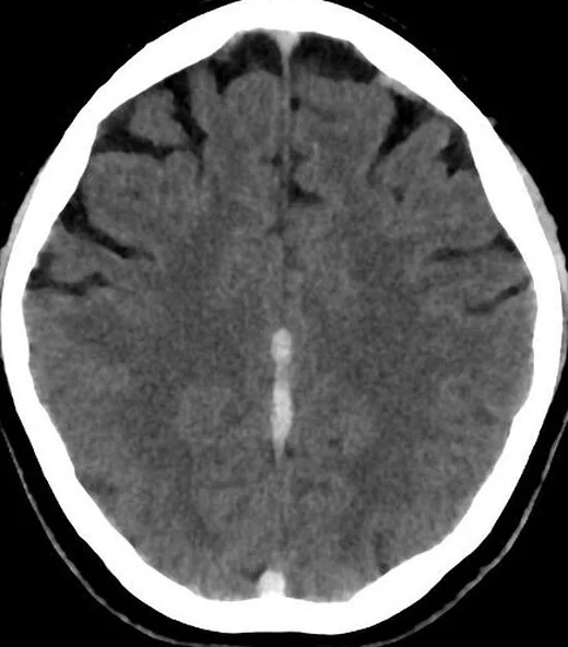
- Akut pıhtı superior sagittal, transvers veya sigmoid sinüs içinde normal venöz kana göre hiperdens görünebilir; ödem, sulkus silinmesi, hemorajik venöz infarkt veya konveksite subaraknoid kanaması eşlik edebilir. Bu kontrastsız seri başlangıç değerlendirmesidir; normal görünmesi venöz sinüs trombozunu tek başına dışlamaz.
- Gecikmiş tanısal
- Kontrastlı venografide venöz klotsun doğrudan tespitini sağlayan temel bulgu, sinüs lümeninde görülen dolum defektidir. Posterior superior sagittal sinüste üçgensel hiperdensite 'delta işareti' olarak adlandırılır ve bu bölgedeki trombozu temsil eder.
Önerilen pencereler: Beyin (G 80 / M 40), Anjiyo (G 600 / M 150).
BT bulguları
- Kontrastlı venografide superior sagittal sinüste doğrudan bulgu, klotsun yerini gösteren venöz dolum defektidir.
- Posterior superior sagittal sinüste üçgensel hiperdensite 'delta işareti' olarak adlandırılır ve bu bölgedeki trombozu temsil eder.
- Transvers-sigmoid sinüs opaklaşmaması — tek taraflı hipoplazi varyantını dışlamak için karşı sinüs kalibresi, sigmoid oluk ve juguler bulbus devamlılığıyla birlikte incelenir.
- Akut hiperdens sinüs — kontrastsız seride pıhtı sinüs lümenini çevre parankimden daha yoğun gösterir; hematokrit yüksekliği benzer yoğunluk yaratabilir.
- Kollateral venöz belirginleşme — falx, tentoryum veya kortikal venlerde belirgin kontrastlanma, çıkış engelinin eşlikçisi olabilir.
- Venöz ödem ve hemorajik infarkt — arteriyel sulama alanına uymayan kortikal-subkortikal hipodensite, şişme ve peteşiyel ya da parankimal kanama.
- Torcula ve düz sinüs uzanımı — birleşme bölgesi ile derin sistem çıkışındaki opaklaşma kusuru trombüsün yaygınlığını belirler.
Normalde
Normal kontrastlı venografide superior sagittal sinüs, torcula, iki transvers sinüs ve sigmoid sinüsler kesintisiz, lümen boyunca homojen opaklaşır; sağ-sol çap farkı tek başına tıkanıklık değildir. Kontrastsız seride sinüs yoğunluğunu komşu korteks ve hematokrit bağlamında, venografide ise karşı taraf, kemik oluk ve juguler bulbusa devamlılıkla kıyaslayın.
Normal BT ile kıyasla
Aynı bölgenin, kaynak veri setinde patoloji içermediği belirtilen bir BT incelemesini kesit kesit kaydırın; organ sınırlarını açarak anatomiyi hasta görüntüsüyle karşılaştırın. Küçük rastlantısal bulgular tümüyle dışlanmamıştır.
Kaynak: TotalSegmentator v2.01 veri seti (Wasserthal ve ark., Radiology: Artificial Intelligence 2023), CC BY 4.0, vaka s0103; organ sınırları veri setinin otomatik segmentasyonundan, birleştirilerek sadeleştirildi. Görüntüler 2,5 mm kesit aralığına yeniden örneklendi.
Ayırıcı tanı
- Konjenital transvers sinüs hipoplazisi
- küçük kemik oluğu ve juguler foramen/bulbus ile aynı tarafta gelişimsel küçüklük bulunur; karşı sinüs genellikle baskındır.
- Araknoid granülasyon
- çoğunlukla fokal, düzgün sınırlı, sinüs duvarına oturan dolum defektidir; sinüsü genişletmez ve çevresinde akım devam eder.
- Yüksek hematokrit
- kontrastsız BT'de birden fazla damar ve sinüs benzer biçimde yoğunlaşabilir; venografide gerçek intraluminal dolum kusuru oluşmaz.
- Akut arteriyel infarkt
- belirli arter alanına uyan erken hipodensite ve olası arter tıkanıklığı vardır; venöz sinüs defekti beklenmez.
- İntrakraniyal hipotansiyon
- yaygın dural kalınlaşma ve kontrastlanma yapabilir, fakat sinüs lümeninde trombüs dolum kusuru yoktur.
Tuzaklar
- Tek taraflı transvers sinüsün ince olması sık anatomik varyanttır; tromboz demeden önce ipsilateral sigmoid oluk ve juguler çıkışın küçük olup olmadığını kontrol edin.
- Arteriyel fazda sinüsün erken veya düzensiz opaklaşması yavaş akım gibi görünebilir; venöz zamanlı venografiye dayanmadan açıklık ya da tromboz kararı vermeyin.
- Yüksek hematokrit sinüsü yalancı hiperdens gösterebilir; kontrastsız dansite bulgusunu venografideki gerçek dolum kusuruyla doğrulayın.
- Normal kontrastsız BT venöz trombozu dışlamaz; doğrudan venografi veya MR venografi gerektiren klinik kuşkuyu görüntüde kapatmayın.
Kendini dene
Kontrastsız BT'de posterior superior sagittal sinüste üçgensel hiperdensite ('delta işareti') görünüyor. En olası bulgu nedir?
Cevabı göster
Dural sinüs trombozu. Üçgensel hiperdensite delta işareti olarak bilinir ve trombozu temsil eder. Araknoid granülasyon fokal ve düzgün sınırlıdır; meningiom dural tabanlı kitle olabilir; hematokrit artışı kontrastsız yoğunluğu etkiler.
Sol transvers sinüs ince; aynı tarafta sigmoid oluk ve juguler bulbus da küçük, lümen düzgün opaklaşıyor. En olası yorum nedir?
Cevabı göster
Gelişimsel hipoplazi. Sinüsle birlikte kemik oluğun ve juguler çıkışın küçük olması gelişimsel hipoplaziyi destekler. Akut trombozda lümen dolum kusuru veya opaklaşmama beklenir; diğer seçenekler bu bütün taraflı küçüklük ilişkisini açıklamaz.
İlgili konular
- Kortikal veya derin serebral ven trombozu
- Venöz konjesyon ve venöz infarkt
- Sinüs trombozu taklitçileri ve normal varyantlar
Kaynaklar
Bu sayfadaki 40 cümle ve 2 quiz sorusu aşağıdaki kaynaklardan alıntılarla tek tek karşılaştırıldı; 4 cümle bu karşılaştırmada düzeltildi (2026-10-08).
- Therapeutic Management of Septic Venous Thrombosis: A Narrative Reviewpmc.ncbi.nlm.nih.gov
- PHACE syndrome: a systematic literature review and illustrative case report of a patient with severe cerebrovascular and neurodevelopmental sequelaepmc.ncbi.nlm.nih.gov
- New Developments in the Pathophysiology, Workup, and Diagnosis of Dural Venous Sinus Thrombosis (DVST) and a Systematic Review of Endovascular Treatmentspmc.ncbi.nlm.nih.gov
- Disseminated Melioidosis Complicated by Dural Venous Sinus Thrombosis: A Case Reportpmc.ncbi.nlm.nih.gov
- Hyperthyroidism-Induced Dural Venous Sinus Thrombosis: A Case Reportpmc.ncbi.nlm.nih.gov
- Imaging of cerebral venous thrombosis.pubmed.ncbi.nlm.nih.gov
- Venous Thrombosis: Causes and Imaging Appearance.pubmed.ncbi.nlm.nih.gov
- Issues in the diagnosis and management of the papilledema shunt.pubmed.ncbi.nlm.nih.gov
- An uncommon disease included commonly in the differential diagnosis of neurological diseases: Neuro-Behçet's syndrome.pubmed.ncbi.nlm.nih.gov
- Dural Venous Sinus Thrombosis After Vestibular Schwannoma Surgery: Should We Anticoagulate?pubmed.ncbi.nlm.nih.gov
- Papilloedema associated with dural venous sinus thrombosis.pubmed.ncbi.nlm.nih.gov
- Dural Venous Sinus Thrombosis in Patients Presenting with Blunt Traumatic Brain Injuries and Skull Fractures: A Systematic Review and Meta-Analysis.pubmed.ncbi.nlm.nih.gov
- A sickle cell disease patient with dural venous sinus thrombosis: a case report and literature review.pubmed.ncbi.nlm.nih.gov
- Unusual neuroimaging findings in spontaneous intracranial hypotension.pubmed.ncbi.nlm.nih.gov
- Acute dural venous sinus injuries: Imaging and challenges.pubmed.ncbi.nlm.nih.gov
- Cranial involvement in sickle cell disease.pubmed.ncbi.nlm.nih.gov
- Imaging of Pregnancy-related Vascular Complications.pubmed.ncbi.nlm.nih.gov
- Torcular pseudomass in a 14-month-old child: illustrative case.pubmed.ncbi.nlm.nih.gov
- COVID-Related Leukoencephalopathy: Unusual MRI Features and Comparability to Delayed Post Hypoxic Ischemic Encephalopathy.pubmed.ncbi.nlm.nih.gov
- Cerebral venous sinus thrombosis in pregnancy and the postpartum period: epidemiology, management, and maternal mortality-a systematic review and meta-analysis.pubmed.ncbi.nlm.nih.gov
- Cytotoxic Edema and Intra-parenchymal Hemorrhage: A Mediated Pathway to Mortality and Functional Outcome in Cerebral Venous Sinus Thrombosis- A sub-analysis of the CLOT-VENUS Registry.pubmed.ncbi.nlm.nih.gov
- Hidden Culprit: Stroke-Like Presentation and Seizure Reveal Parasagittal Meningioma: A Case Report.pubmed.ncbi.nlm.nih.gov
- Intracranial venous thrombosis imaging by anatomical location: an educational review with mimics and diagnostic pitfalls.pubmed.ncbi.nlm.nih.gov
- Bilateral Vein of Trolard Thrombosis Presenting with Seizure and Minimal Deficits: A Rare Case Report.pubmed.ncbi.nlm.nih.gov
- Extensive cerebral venous sinus thrombosis with hemorrhagic venous infarction as the initial presentation of acute myeloid leukemia: A case report.pubmed.ncbi.nlm.nih.gov
- Balloon-assisted retrograde cerebral venography for enhanced visualization of the posterior and lateral dural venous sinuses: a feasibility study.pubmed.ncbi.nlm.nih.gov
- Beyond the Concussion: Cerebral Venous Thrombosis Following Mild-to-Moderate Traumatic Brain Injury.pubmed.ncbi.nlm.nih.gov
- Penetrating injuries of the posterior dural venous sinuses: a systematic review of injury mechanisms and repair techniques.pubmed.ncbi.nlm.nih.gov
- Venous-significant intracranial hemorrhage in neonatal HIE treated with therapeutic hypothermia: imaging phenotypes, diagnostic pitfalls, and prognostic implications.pubmed.ncbi.nlm.nih.gov
- Fulminant <i>Fusobacterium necrophorum</i> Meningoencephalitis Secondary to Frontal Sinusitis in a Previously Healthy Young Adult: A Case Report.pubmed.ncbi.nlm.nih.gov
- A Rare Case of Postpartum Cerebral Venous Thrombosis and Hemorrhagic Infarction From Somalia.pubmed.ncbi.nlm.nih.gov
- An Integrated Approach on the Diagnosis of Cerebral Veins and Dural Sinuses Thrombosis (a Narrative Review).pubmed.ncbi.nlm.nih.gov
- A Case-Based Review of Cerebral Venous Infarcts With Perfusion Imaging and Comparison to Arterial Ischemic Stroke.pubmed.ncbi.nlm.nih.gov
- Neuroradiology in women of childbearing age.pubmed.ncbi.nlm.nih.gov
- Overview of the utility of noncontrast CT brain in the diagnosis of cerebral vein thrombosis.pubmed.ncbi.nlm.nih.gov
- Diagnostic Sensitivity of Unenhanced CT for Cerebral Venous Thrombosis: Can Clot Density Measurement Replace CT Venogram?pubmed.ncbi.nlm.nih.gov
- Systematic review of commercial artificial intelligence tools for the detection and volume quantification in intracerebral hemorrhage.pubmed.ncbi.nlm.nih.gov
- Imaging and Management of Blunt Cerebrovascular Injury.pubmed.ncbi.nlm.nih.gov
- Spontaneous bacteremia and spontaneous bacterial peritonitis share similar prognosis in patients with cirrhosis: a cohort study.pubmed.ncbi.nlm.nih.gov
- Case Report: Mechanical thrombectomy of cerebral venous sinuses using the NeVa stent retriever.pubmed.ncbi.nlm.nih.gov
- Cerebral venous thrombosis revealed by psychiatric disorders: a diagnosis not to be missed.pubmed.ncbi.nlm.nih.gov
- Seizures as the initial presentation of cerebral venous sinus thrombosis in an elderly patient: a case report and literature review.pubmed.ncbi.nlm.nih.gov
- The Anticoagulation Dilemma: Concurrent Traumatic Brain Hemorrhage and Cerebral Venous Thrombosis in a Blast Injury Patient.pubmed.ncbi.nlm.nih.gov
- Diagnostic value of machine-learning using conventional magnetic resonance imaging markers for pediatric idiopathic intracranial hypertension: a retrospective study.pubmed.ncbi.nlm.nih.gov
- Mastoid emissary vein, mastoid emissary foramen, and mastoid emissary canal: anatomy, variability, imaging, and clinical implications.pubmed.ncbi.nlm.nih.gov
- Cerebral Venous Outflow Implications in Idiopathic Intracranial Hypertension-From Physiopathology to Treatment.pubmed.ncbi.nlm.nih.gov
- Polycythemia, a radiologic mimic in plain CT head: A case report.pubmed.ncbi.nlm.nih.gov
- Extensive Cerebral Venous Sinus Thrombosis With Obstructive Hydrocephalus in a Young Female With Underlying Essential Thrombocythemia: A Case Report.pubmed.ncbi.nlm.nih.gov
- Admission hyperdense sinus sign predicts poorer outcomes in cerebral venous sinus thrombosis.pubmed.ncbi.nlm.nih.gov
- The cuneus vein: initial anatomical description and preservation technique for posterior interhemispheric approaches.pubmed.ncbi.nlm.nih.gov
- A Practical Approach to Intracranial Dural Arteriovenous Fistulas: Pathogenesis, Classification and Management.pubmed.ncbi.nlm.nih.gov
- Neuroimaging for neurovascular complications of traumatic brain injury.pubmed.ncbi.nlm.nih.gov
- Quadriplegia Following Depressed Skull Fracture Over Superior Sagittal Sinus: A Rare Presentation of a Common Event.pubmed.ncbi.nlm.nih.gov
- Interesting neuroimaging findings in a child with hyperlipidemia: a case report.pubmed.ncbi.nlm.nih.gov
- An Unusual Case of Cerebral Venous Sinus Thrombosis With Negative D-dimer Mimicking Giant Cell Arteritis.pubmed.ncbi.nlm.nih.gov
- Cerebral Venous Sinus Thrombosis in Pediatric Critical Care.pubmed.ncbi.nlm.nih.gov
- Dural Venous Sinus Thrombosis: A Rare Cause of Intracranial Hemorrhage.pubmed.ncbi.nlm.nih.gov
- Cerebral venous disorders: clinical presentation, diagnostic strategy, and contemporary management.pubmed.ncbi.nlm.nih.gov
- Morphological variations of the dural venous sinuses: a cadaveric study.pubmed.ncbi.nlm.nih.gov
- An in-depth examination of variations in cerebral venous sinuses and the occurrence of sinovenous thrombosis.pubmed.ncbi.nlm.nih.gov
- Septic cerebral venous sinus thrombosis from maxillary sinusitis with concomitant right-sided sigmoid sinus hypoplasia: A case report and review of the literature.pubmed.ncbi.nlm.nih.gov
- Non-Thrombotic Filling Defects in Cerebral Veins and Sinuses: When Normal Structures Mimic a Disease.pubmed.ncbi.nlm.nih.gov
- Case Report: Intracranial hypertension occurring in association with subacute thyroiditis.pubmed.ncbi.nlm.nih.gov
- Bilateral Papilledema without Evident Intracranial Hypertension in Pediatric Chiari Malformation Type I: A Case Report.pubmed.ncbi.nlm.nih.gov
- An Overview of Vascular Compression Syndromes and Associations with Autonomic Dysfunction: A Review.pubmed.ncbi.nlm.nih.gov
- Neuroimaging of Vermiform Giant Arachnoid Granulations in Children.pubmed.ncbi.nlm.nih.gov
- When headache meets visual disturbance: An evidence-based review of common and critical entities.pubmed.ncbi.nlm.nih.gov
- Venous Thromboembolism in Neonates, Children, and Adolescents: A Comprehensive Narrative Review of Risk Factors, Diagnosis, Treatment, and Prevention.pubmed.ncbi.nlm.nih.gov
- Endovascular Embolization in Neurovascular Disease: Material Science, Multimodal Management, and Future Horizons.pubmed.ncbi.nlm.nih.gov
- Dengue-Associated Encephalopathy With Multiple Cranial Neuropathies and Multisystem Involvement in a Patient With Epilepsy: A Comprehensive Case Report With Longitudinal Multimodality Imaging.pubmed.ncbi.nlm.nih.gov
- Long-term safety and effectiveness of the "OptEase" vena cava filter.pubmed.ncbi.nlm.nih.gov
- Anatomy imaging and hemodynamics research on the cerebral vein and venous sinus among individuals without cranial sinus and jugular vein diseases.pubmed.ncbi.nlm.nih.gov
- Imaging Evaluation of Acute Traumatic Brain Injury.pubmed.ncbi.nlm.nih.gov
- Imaging the brain: diagnosis aided by structural features on neuroimaging studies.pubmed.ncbi.nlm.nih.gov
- The Role of Magnetic Resonance Imaging and Cardiac Computed Tomography in the Assessment of Left Atrial Anatomy, Size, and Function.pubmed.ncbi.nlm.nih.gov
- Patterns of injury and violence in Yaoundé Cameroon: an analysis of hospital data.pubmed.ncbi.nlm.nih.gov
- Anatomical Variability of the Transverse and Sigmoid Sinus Grooves: Implications for Skull Base Surgery.pubmed.ncbi.nlm.nih.gov
- Computed tomography perfusion stroke mimics on RAPID commercial software: A case-based review.pubmed.ncbi.nlm.nih.gov
- Imaging of Central Nervous System Involvement in Pediatric Hematologic Disorders.pubmed.ncbi.nlm.nih.gov
- Fetal intracranial hemorrhage and infarct: Main sonographic and MRI characteristics: A review article.pubmed.ncbi.nlm.nih.gov
- Efficacy and safety of a "radical" surgical strategy in the treatment of parasagittal sinus meningioma.pubmed.ncbi.nlm.nih.gov
- Anatomical Reasons for an Impaired Internal Jugular Flow.pubmed.ncbi.nlm.nih.gov
- Pulsatile Tinnitus: A Comprehensive Clinical Approach to Diagnosis and Management.pubmed.ncbi.nlm.nih.gov
- Management and Characteristics of Embolism and Thrombosis After COVID-19 Vaccination: Scoping Review.pubmed.ncbi.nlm.nih.gov
- Penetrating dural venous sinus injury: a scoping review of clinical presentation, operative management, and prognosis.pubmed.ncbi.nlm.nih.gov
- Disseminated Melioidosis Complicated by Dural Venous Sinus Thrombosis: A Case Report.pubmed.ncbi.nlm.nih.gov
- Cerebrospinal fluid venous fistula causing spontaneous intracranial hypotension resulting in venous sinus thrombosis and coma.pubmed.ncbi.nlm.nih.gov
- Surgical Management of Meningiomas Involving the Torcula, Transverse Sinus, and Jugular Bulb: A Technical Note and Review of the Literature.pubmed.ncbi.nlm.nih.gov
- Congenital and childhood atrioventricular blocks: pathophysiology and contemporary management.pubmed.ncbi.nlm.nih.gov
- Perfusion imaging abnormalities in acute cerebral venous sinus thrombosis: A case series.pubmed.ncbi.nlm.nih.gov
- A Case of Trousseau Syndrome: A Report Detailing Recurrent Ischaemic Strokes Associated With Metastatic Pancreatic Cancer and Pulmonary Embolisms Despite Treatment With Direct Oral Anticoagulation.pubmed.ncbi.nlm.nih.gov
- Non-contrast CT findings suggestive of secondary intracerebral haemorrhage.pubmed.ncbi.nlm.nih.gov
- Posterior Circulation Ischemic Stroke Occurring After ChAdOx1 nCoV-19/AZD1222 Vaccination Without Evidence of Vaccine-Induced Immune Thrombotic Thrombocytopenia: A Case Report and Focused Narrative Review.pubmed.ncbi.nlm.nih.gov
- Beyond plaque morphology: a multimodal imaging model combining structural vulnerability and functional reserve predicts stroke recurrence in symptomatic intracranial atherosclerosis.pubmed.ncbi.nlm.nih.gov
- Venous-phase Tan-based collateral assessment on CTP-derived CTA for outcome stratification after reperfusion therapy in acute ischemic stroke.pubmed.ncbi.nlm.nih.gov
- Baseline prolonged venous transit on CT perfusion predicts the post-treatment Brush Sign after successful reperfusion in acute ischemic stroke patients.pubmed.ncbi.nlm.nih.gov
Eğitim amaçlıdır; hasta başında tanı ve tedavi kararının yerine geçmez. İçerik yapay zekâ destekli bir süreçle güncel literatüre göre cümle cümle kaynaklarla karşılaştırıldı, ancak uzman radyolog incelemesinden geçmedi. Klinik kararlarda güncel kılavuzları, kurum protokollerini ve radyoloji raporunu esas alın.