Özet
Yeni başlayan baş ağrısı, nöbet veya fokal bulguyla gelen hastada trombüs yalnızca bir ya da birkaç kortikal veni tutabilir; dural sinüsler ve derin venöz sistem açık kalabilir. Kontrastsız BT tromboze kortikal veni hiperdens kord olarak gösterebilir; ancak kord işareti seyrektir ve BT’de belirgin bulgu görülmeyebilir. BT venografiyle kortikal ven trombüsü dolum kusuru olarak gösterilebilir; ancak noninvaziv görüntüleme her zaman tanısal olmayabilir ve bulgular belirsizse ek değerlendirme gerekebilir. Tanı gecikirse venöz konjesyon, kortikal kanama veya hemorajik infarkt arteriyel hastalık ya da primer kanama gibi yorumlanabilir.
Faz ve pencere
- Kontrastsız tanısal
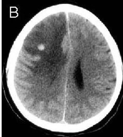
- Tromboze kortikal ven kontrastsız BT’de hiperdens kord işareti oluşturabilir; izole kortikal ven trombozuna fokal ödem, hemoraji veya subaraknoid kanama eşlik edebilir. Kontrastlı inceleme gerekmeden kontrastsız BT’de kord işareti görülebilir; ancak düşük duyarlılığı nedeniyle bu işaretin görülmemesi izole kortikal ven trombozunu dışlamaz.
- Venöz (BTV) tanısal
- BT venografide kortikal ven trombüsü dolum kusuru olarak doğrudan görülebilir. İzole kortikal ven trombozunda dural sinüs tutulumu bulunmaz.
Önerilen pencereler: Beyin (G 80 / M 40), Anjiyo (G 600 / M 150).
BT bulguları
- Kortikal ven kord işareti — kontrastsız seride sulkus boyunca uzanan tübüler/serpijinöz hiperdens damar görünümü.
- Sulkus içi trombüs odağı — kısa kesitlerde yuvarlak veya noktasal hiperdensite, ardışık kesitlerde kortikal ven seyriyle devam eder.
- Kortikal ven dolum kusuru — BT venografide şüpheli ven içinde kontrastla çevrili dolmayan bölüm veya kesintili opaklaşma.
- BT venografide kortikal venlerin bölgesel seyrekliği görülebilir; izole kortikal ven trombozunda kortikal venlerin bölgesel yokluğu ve subkortikal venöz opaklaşma azalması da tanımlanmıştır.
- Venöz konjesyon — ilgili girusta hacim artışı ve sulkus daralması, bir arter dalı sınırına uymayan dağılım gösterebilir.
- İzole kortikal ven trombozuna eşlik eden parankimal değişiklikler arasında ödem, venöz infarkt ve parankimal hematom bulunabilir.
- İzole kortikal ven trombozuna konveksite subaraknoid kanaması eşlik edebilir; kord işaretiyle ayırımı güç olabilir.
Normalde
Şüpheli tarafı karşı hemisferle karşılaştırmak, kortikal venlerin bölgesel yokluğu veya azlığını değerlendirmeye yardımcı olabilir. Şüpheli tarafı karşı hemisferle karşılaştırın; kortikal venlerin bölgesel yokluğunu ve venöz drenaj alanındaki değişiklikleri birlikte değerlendirin.
Normal BT ile kıyasla
Aynı bölgenin, kaynak veri setinde patoloji içermediği belirtilen bir BT incelemesini kesit kesit kaydırın; organ sınırlarını açarak anatomiyi hasta görüntüsüyle karşılaştırın. Küçük rastlantısal bulgular tümüyle dışlanmamıştır.
Kaynak: TotalSegmentator v2.01 veri seti (Wasserthal ve ark., Radiology: Artificial Intelligence 2023), CC BY 4.0, vaka s0103; organ sınırları veri setinin otomatik segmentasyonundan, birleştirilerek sadeleştirildi. Görüntüler 2,5 mm kesit aralığına yeniden örneklendi.
Ayırıcı tanı
- Konveksite subaraknoid kanaması kord işaretiyle karışabilir; izole kortikal ven trombozunda bu iki bulgunun ayrımı güç olabilir.
- Kortikal arter dalı trombozu
- damar tıkanıklığı arteriyel ağaçta izlenir ve parankim değişikliği ilgili arter dağılımına oturabilir.
- Kortikal arteriyel infarkt
- kama biçimli hipoattenüasyon bir arter havzasını izler; sulkusta tromboze ven gösterilmesi beklenmez.
- Kavernöz malformasyon kanaması, SWI/T2* görüntülerde hemosiderin halkalı dut benzeri bir lezyonla ilişkili olabilir.
Tuzaklar
- Yüksek hematokrit durumlarında kontrastsız BT’de venler görece hiperdens görünebilir; bu durumda arterler de benzer yoğunlukta olabilir.
- Konveksite subaraknoid kanaması kord işaretiyle karışabilir; bu iki bulgunun ayrımı güç olabilir.
- BT venografide kortikal venlerin bölgesel azlığı görülebilir; karşı hemisferle karşılaştırma tanıya yardımcı olabilir.
- Ana dural sinüslerin dolu olması kortikal ven pıhtısını dışlamaz; şüpheli yüzeyel venin tamamını ayrı bir damar hedefi olarak takip edin.
Kendini dene
Yeni nöbet geçiren hastada tek parietal sulkusta kıvrımlı hiperdens damar, komşu girusta hafif şişme var; venografide sinüsler açık. En olası tanı nedir?
Cevabı göster
İzole kortikal ven trombozu. İzole kortikal ven trombozu, dural sinüs veya derin ven tutulumu olmadan görülebilir; kaynakta kortikal ven dolum kusurları ve venöz çıkış bozukluğuna bağlı bulgular tanımlanmıştır. Anevrizma rüptürü sulkuslarda kan yapar, fistül anormal şant damarlarıyla değerlendirilir, kavernom ise parankim içi odaktır.
İzole kortikal ven trombozu şüphesinde noninvaziv görüntüleme tanısal değilse, tanıyı netleştirmede hangi inceleme yardımcı olabilir?
Cevabı göster
Kateter anjiyografi/venografi. Kaynak, izole kortikal ven trombozu tanısında, özellikle MR venografi belirsiz olduğunda kateter anjiyografi/venografinin önemli olabileceğini bildiriyor.
İlgili konular
- Dural venöz sinüs trombozu
- Venöz konjesyon ve venöz infarkt
- Sinüs trombozu taklitçileri ve normal varyantlar
Kaynaklar
Bu sayfadaki 37 cümle ve 2 quiz sorusu aşağıdaki kaynaklardan alıntılarla otomatik olarak karşılaştırıldı; 19 cümle bu karşılaştırmada düzeltildi (2026-10-09).
- Clinical and Angiographic Imaging Features of Isolated Cortical Venous Thrombosispmc.ncbi.nlm.nih.gov
- Cerebral venous thrombosis: imaging patternspmc.ncbi.nlm.nih.gov
- Isolated cortical vein thrombosis: systematic review of case reports and case series.pubmed.ncbi.nlm.nih.gov
- Isolated superficial sylvian vein thrombosis with long cord sign: case report and review of the literature.pubmed.ncbi.nlm.nih.gov
- Ipsilateral Dural Thickening and Enhancement: A Sign of Isolated Cortical Vein Thrombosis? A Case Report and Review of the Literature.pubmed.ncbi.nlm.nih.gov
- Bilateral Vein of Trolard Thrombosis.pubmed.ncbi.nlm.nih.gov
- Clinicoradiological Profile of Superficial Middle Cerebral Vein Thrombosis.pubmed.ncbi.nlm.nih.gov
- Isolated cortical vein thrombosis: case series.pubmed.ncbi.nlm.nih.gov
- Isolated cortical vein thrombosis in a young male adult.pubmed.ncbi.nlm.nih.gov
- Isolated Cortical Vein Thrombosis with Long Cord Sign.pubmed.ncbi.nlm.nih.gov
- Isolated cortical vein thrombosis: a widely variable clinicoradiological spectrum.pubmed.ncbi.nlm.nih.gov
- Isolated cortical vein thrombosis after epidural anesthesia: report of three cases.pubmed.ncbi.nlm.nih.gov
- COVID-19-associated Isolated Cortical Vein Thrombosis: Detection of SARS-CoV-2 in CSF.pubmed.ncbi.nlm.nih.gov
- Clinical and Angiographic Imaging Features of Isolated Cortical Venous Thrombosis.pubmed.ncbi.nlm.nih.gov
- Malignant isolated cortical vein thrombosis with type II protein S deficiency: a case report.pubmed.ncbi.nlm.nih.gov
- Incidental Diagnosis of Cerebral Cortical Venous Thrombosis in Postdural Puncture Headache on Brain Computed Tomography.pubmed.ncbi.nlm.nih.gov
- Bilateral Vein of Trolard Thrombosis Presenting with Seizure and Minimal Deficits: A Rare Case Report.pubmed.ncbi.nlm.nih.gov
- Isolated Psychiatric Presentation of Cerebral Venous Thrombosis in a Patient Without Risk Factors: A Diagnostic Challenge.pubmed.ncbi.nlm.nih.gov
- Seizures as the initial presentation of cerebral venous sinus thrombosis in an elderly patient: a case report and literature review.pubmed.ncbi.nlm.nih.gov
- Intracranial venous thrombosis imaging by anatomical location: an educational review with mimics and diagnostic pitfalls.pubmed.ncbi.nlm.nih.gov
- Admission hyperdense sinus sign predicts poorer outcomes in cerebral venous sinus thrombosis.pubmed.ncbi.nlm.nih.gov
- Clinical presentation domains and venous anatomical patterns in cerebral venous thrombosis: a retrospective cohort study.pubmed.ncbi.nlm.nih.gov
- Cerebral venous disorders: clinical presentation, diagnostic strategy, and contemporary management.pubmed.ncbi.nlm.nih.gov
- When the Brainstem Is the Target: Cryptogenic Paramedian Pontine Infarction Associated With Patent Foramen Ovale.pubmed.ncbi.nlm.nih.gov
- Cerebral venous thrombosis revealed by psychiatric disorders: a diagnosis not to be missed.pubmed.ncbi.nlm.nih.gov
- The laminar subarachnoid hemorrhage of infant venous stroke: Evidence of a 4<sup>th</sup> meningeal layer?pubmed.ncbi.nlm.nih.gov
- Homonymous hemianopia due to cerebral venous thrombosis: A case report.pubmed.ncbi.nlm.nih.gov
- A Case-Based Review of Cerebral Venous Infarcts With Perfusion Imaging and Comparison to Arterial Ischemic Stroke.pubmed.ncbi.nlm.nih.gov
- Perfusion imaging abnormalities in acute cerebral venous sinus thrombosis: A case series.pubmed.ncbi.nlm.nih.gov
- Non-Thrombotic Filling Defects in Cerebral Veins and Sinuses: When Normal Structures Mimic a Disease.pubmed.ncbi.nlm.nih.gov
- Case report: An illusive cortical venous infarction mimicking glioma hemorrhage.pubmed.ncbi.nlm.nih.gov
- Neuroimaging Modalities Used for Ischemic Stroke Diagnosis and Monitoring.pubmed.ncbi.nlm.nih.gov
- Reversible coma and elevated central venous pressure: warning signs of acute superior vena cava graft thrombosis following Warden procedure: a case report.pubmed.ncbi.nlm.nih.gov
- The expanding role of ultrasound in acute kidney injury: from B-mode to microcirculation.pubmed.ncbi.nlm.nih.gov
- Cerebral venous sinus thrombosis in pregnancy and the postpartum period: epidemiology, management, and maternal mortality-a systematic review and meta-analysis.pubmed.ncbi.nlm.nih.gov
- Conduction Aphasia in a Case of Left Cortical Veins and Left Lateral Sinus Thrombosis Due to Multiple Risk Factors: A Case Report and Review of the Literature.pubmed.ncbi.nlm.nih.gov
- Delayed Intracerebral Hemorrhage Following Transarterial Embolization for a Tentorial Dural Arteriovenous Fistula: Postoperative Venous Outflow Impairment Detected by Susceptibility-Weighted Imaging.pubmed.ncbi.nlm.nih.gov
- Intracranial dural arteriovenous fistulas with and without pial arterial supply: a case series of 227 patients.pubmed.ncbi.nlm.nih.gov
- Unruptured distal anterior cerebral artery branch pseudoaneurysm in the setting of intracranial pressure monitor placement: illustrative case.pubmed.ncbi.nlm.nih.gov
- Subarachnoid hemorrhage complicated by cerebral venous sinus thrombosis: a quantitative systematic review of cases.pubmed.ncbi.nlm.nih.gov
- Case Report: Mechanical thrombectomy of cerebral venous sinuses using the NeVa stent retriever.pubmed.ncbi.nlm.nih.gov
- Complex vasculitic overlap: temporal arteritis complicating suspected neuro-behçet disease with recurrent ischemic injury.pubmed.ncbi.nlm.nih.gov
- When headache meets visual disturbance: An evidence-based review of common and critical entities.pubmed.ncbi.nlm.nih.gov
- Hyperviscosity encephalopathy in suspected polycythemia vera at high altitude: a case report.pubmed.ncbi.nlm.nih.gov
- Cerebral Blood Transit in Sickle Cell Anemia.pubmed.ncbi.nlm.nih.gov
- Case Report: Structured MRI assessment of posterior thalamic infarction in a distribution compatible with posterior choroidal artery territory presenting as Déjerine-Roussy syndrome in an adolescent: differentiating arterial ischemia from venous thrombosis and thalamic neoplasm.pubmed.ncbi.nlm.nih.gov
- Reperfusion Injury and Infarct Size: Why Translation has Been Difficult, and How We Move Forward.pubmed.ncbi.nlm.nih.gov
- Developmental venous anomalies on fetal magnetic resonance imaging: prevalence and reproducible radiological phenotypes.pubmed.ncbi.nlm.nih.gov
- Association Between Pulse Wave Velocity and Cerebral Small Vessel Disease: A Scoping Review.pubmed.ncbi.nlm.nih.gov
- Predictors of seizure as presenting symptom of cerebral cavernomas.pubmed.ncbi.nlm.nih.gov
- Cognition in Cardiovascular Disease.pubmed.ncbi.nlm.nih.gov
- Cytomegalovirus meningoencephalitis in an immunocompetent host with evolution of cystic encephalomalacia.pubmed.ncbi.nlm.nih.gov
- Cryptogenic Multifocal Infarctions Involving Brain and Spinal Cord in a Patient With Patent Foramen Ovale: A Case Report.pubmed.ncbi.nlm.nih.gov
- Cerebral Venous Sinus Thrombosis Mimicking Tumor Hemorrhage: Successful Anticoagulation in a Resource-Limited Setting.pubmed.ncbi.nlm.nih.gov
- Exploring Central Venous Thrombosis as a Rare Postinfectious Complication of Dengue: A Case Report.pubmed.ncbi.nlm.nih.gov
- Reversible cerebral vasoconstriction syndrome: A narrative review.pubmed.ncbi.nlm.nih.gov
- Fetal intracranial hemorrhage and infarct: Main sonographic and MRI characteristics: A review article.pubmed.ncbi.nlm.nih.gov
- Venous-significant intracranial hemorrhage in neonatal HIE treated with therapeutic hypothermia: imaging phenotypes, diagnostic pitfalls, and prognostic implications.pubmed.ncbi.nlm.nih.gov
- Clinical management of isolated transverse-sigmoid sinus dural arteriovenous fistulas: a three-case illustrative series and narrative review.pubmed.ncbi.nlm.nih.gov
- Sensorineural Hearing Loss After Transvenous Embolization of a Transverse-Sigmoid Sinus Dural Arteriovenous Fistula.pubmed.ncbi.nlm.nih.gov
- Cerebral Venous Thrombosis: Pathophysiologic Insights, Clinical Evaluation Tools, and Novel Therapeutic Strategies.pubmed.ncbi.nlm.nih.gov
- Three-dimensional rotational angiography-based multimodal fusion imaging for preoperative neurovascular assessment in intracranial meningiomas: a retrospective technical feasibility study of 72 cases.pubmed.ncbi.nlm.nih.gov
- Penetrating Ballistic Trauma to the Popliteal Fossa Without Neurovascular Injury Despite High-Risk Trajectory: An Unusual Case Report.pubmed.ncbi.nlm.nih.gov
- When Cancer Clots: An Extensive Radiologic Analysis of Cancer-Associated Thromboembolism.pubmed.ncbi.nlm.nih.gov
- Cerebral Venous Sinus Thrombosis Revealing ALK-Positive Anaplastic Large-Cell Lymphoma in a Patient with Inherited Thrombophilia and Concomitant Infection: Case Report and Narrative Review.pubmed.ncbi.nlm.nih.gov
- Spectrum of neuroimaging mimics in children with COVID-19 infection.pubmed.ncbi.nlm.nih.gov
- Acute cerebral venous thrombosis - still an underdiagnosed pathology in emergency computed tomography of the brain.pubmed.ncbi.nlm.nih.gov
- Tumor-like cortical venous thrombosis: Case report and literature review.pubmed.ncbi.nlm.nih.gov
- Evaluating the diagnostic value of multi-detector brain CT angiography in diagnosing acute cerebral venous thrombosis.pubmed.ncbi.nlm.nih.gov
- An Integrated Approach on the Diagnosis of Cerebral Veins and Dural Sinuses Thrombosis (a Narrative Review).pubmed.ncbi.nlm.nih.gov
- Balloon-assisted retrograde cerebral venography for enhanced visualization of the posterior and lateral dural venous sinuses: a feasibility study.pubmed.ncbi.nlm.nih.gov
- Non-restrictive cerebral venous dysgenesis in an 11-year-old girl: a case report.pubmed.ncbi.nlm.nih.gov
- Adjunctive venous sinus stenting in transvenous embolization of vein of Galen malformations.pubmed.ncbi.nlm.nih.gov
- Mastoid emissary vein, mastoid emissary foramen, and mastoid emissary canal: anatomy, variability, imaging, and clinical implications.pubmed.ncbi.nlm.nih.gov
- Vascular lesions of the pediatric orbit: A radiological walkthrough.pubmed.ncbi.nlm.nih.gov
- Imaging and Management of Blunt Cerebrovascular Injury.pubmed.ncbi.nlm.nih.gov
- Non-contrast CT findings suggestive of secondary intracerebral haemorrhage.pubmed.ncbi.nlm.nih.gov
- Subarachnoid haemorrhage associated with septic cavernous sinus thrombosis secondary to odontogenic maxillary sinusitis.pubmed.ncbi.nlm.nih.gov
- Stroke-Mimic: An Intracranial Dural Arteriovenous Fistula Revealed by New-Onset Seizures and Post-Ictal Deficit.pubmed.ncbi.nlm.nih.gov
- Low-flow vascular malformations without arteriovenous shunting of the central nervous system: a pictorial review.pubmed.ncbi.nlm.nih.gov
- Functional and Hemodynamic Restoration After Microsurgical Resection of Compact High-Flow Temporo-Parieto-Occipital Arteriovenous Malformation.pubmed.ncbi.nlm.nih.gov
- Cerebral Superficial Siderosis : Etiology, Neuroradiological Features and Clinical Findings.pubmed.ncbi.nlm.nih.gov
- Imaging of inferior vena cava normal variants, anomalies and pathologies, Part 2: Acquired.pubmed.ncbi.nlm.nih.gov
- Cerebral venous thrombosis: Current management, recent advances and future directions.pubmed.ncbi.nlm.nih.gov
- Isolated cortical vein thrombosis with hemorrhagic infarction during the application of a transdermal estradiol patch: A case report.pubmed.ncbi.nlm.nih.gov
- Long-term safety and effectiveness of the "OptEase" vena cava filter.pubmed.ncbi.nlm.nih.gov
- Adrenocortical carcinoma.pubmed.ncbi.nlm.nih.gov
- Patterns of injury and violence in Yaoundé Cameroon: an analysis of hospital data.pubmed.ncbi.nlm.nih.gov
- Case Report of Cerebral Sinus Thrombosis Related to Immune Thrombotic Thrombocytopenia Following Administration of ChAdOx1 nCoV-19 for Vaccination against COVID-19.pubmed.ncbi.nlm.nih.gov
- Spontaneous Non-Aneurysmal Convexity Subarachnoid Hemorrhage: A Scoping Review of Different Etiologies beyond Cerebral Amyloid Angiopathy.pubmed.ncbi.nlm.nih.gov
- Malignant Isolated Cortical Vein Thrombosis as the Initial Manifestation of Primary Antiphospholipid Syndrome: Lessons on Diagnosis and Management From a Case Report.pubmed.ncbi.nlm.nih.gov
- Disease- and Treatment-related Complication on F-18-Fluorodeoxyglucose Positron Emission Tomography/Computed Tomography in Oncology Practice: A Pictorial Review.pubmed.ncbi.nlm.nih.gov
- Atypical manifestations of Dengue fever: case series in tertiary care hospital in Nepal.pubmed.ncbi.nlm.nih.gov
- Current state and advancements of imaging in acute ischemic stroke: a practical review.pubmed.ncbi.nlm.nih.gov
- Rogue AI: Cautionary Cases in Neuroradiology and What We Can Learn From Them.pubmed.ncbi.nlm.nih.gov
- Imaging of Cerebral Venous Thrombosis.pubmed.ncbi.nlm.nih.gov
Eğitim amaçlıdır; hasta başında tanı ve tedavi kararının yerine geçmez, tıbbi tavsiye değildir. Metinler yapay zekâ desteğiyle yazıldı ve otomatik bir adımda kaynak alıntılarıyla karşılaştırıldı; bu bir tıbbi doğrulama değildir ve içerik uzman radyolog incelemesinden geçmedi. Klinik kararlarda güncel kılavuzları, kurum protokollerini ve radyoloji raporunu esas alın.