
Özet
Yüksek enerjili künt kafa travmasında sfenoid kemik, sfenoid sinüs çevresi ve petröz kafa tabanından geçen kırıklar; damar, kraniyal sinir ve dura komşuluğundaki yaralanmaları haber verebilir. İlk inceleme kontrastsız ince kesit kafa BT'dir; kemik algoritması kırık hattını, beyin penceresi ise hemorajik sinüs içeriğini, pnömosefaliyi ve eşlik eden kanamayı gösterir. Orta kafa tabanı kırıklarında karotid kanal, kavernöz sinüs ve optik kanal tutulumu değerlendirilmeli; BT anjiyografi vasküler yaralanma tanısında birincil yöntemdir. Bu uzanımlar atlanırsa diseksiyon, psödoanevrizma, karotid-kavernöz fistül, görme kaybı veya BOS kaçağına yol açabilecek yaralanmalar fark edilmeyebilir.
Faz ve pencere
- Kontrastsız tanısal
- İnce kesit kemik algoritmalı BT'de sfenoid sinüs duvarı, basisfenoid, klivus, pterigoid çıkıntılar ve petröz temporal kemik boyunca kortikal kesinti, çizgisel açıklık veya yer değiştirmiş fragman aranır. Sinüs içinde kanla uyumlu yüksek dansiteli sıvı, kafa içinde hava ve eşlik eden akut kanama yumuşak doku/beyin pencerelerinde incelenir; kırık tanısı için kontrast gerekmez.
- BTA
- Kırık karotid kanalına ya da internal karotid arterin kavernöz segment komşuluğuna uzanmışsa BTA'da lümen daralması, intimal düzensizlik/flep, oklüzyon, psödoanevrizma veya kavernöz sinüste erken venöz dolum araştırılır. BTA kemik kırığını dışlamak ya da sınıflamak için kullanılan temel seri değildir.
Önerilen pencereler: Kemik (G 1800 / M 400), Beyin (G 80 / M 40), Yumuşak doku (G 400 / M 40), Anjiyo (G 600 / M 150).
BT bulguları
- Sfenoid sinüs duvarı kırığı — sinüsün ön, arka, lateral veya üst duvarında keskin kortikal basamaklanma ya da devamlılık kaybı.
- Basisfenoid veya klivus çatlağı — orta hatta uzanan çizgi sella tabanını, klivusu ya da karşı kafa tabanı yapılarını kesebilir.
- Karotid kanal tutulumu — kırık hattı petroz kanalın kemik halkasına ulaşır; bu bulgu tek başına arter lümen yaralanması demek değildir.
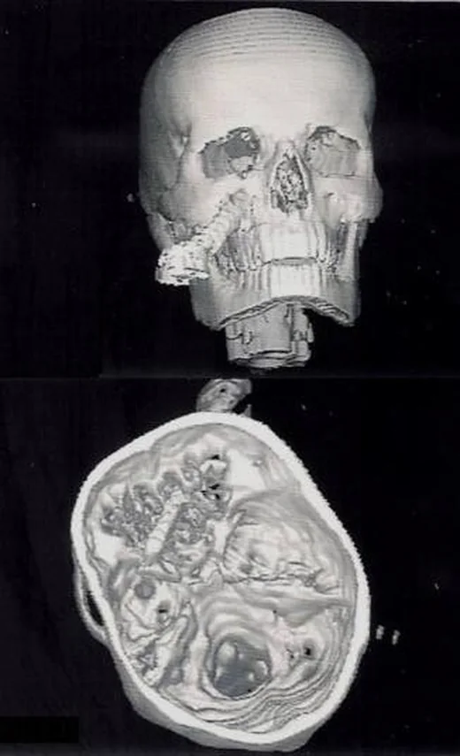
- Travmada sfenoid sinüs duvarı kırığı görülebilir.
- Pnömosefali — kırık hattıyla komşu bazal sisternlerde, ekstraaksiyel aralıkta veya intraventriküler alanda hava odakları.
- Kafa içi eşlikçi yaralanma — temporal kontüzyon, ekstraaksiyel hematom veya subaraknoid kanama kırıkla aynı travma incelemesinde aranır.
Normalde
Kırık şüphesinde karşı sfenoid sinüs duvarı ve ardışık aksiyel-koronal-sagittal kesitler kıyaslanarak gerçek kesinti, yer değiştirme ve kanal uzanımı ayrılır; yalnız tek kesitteki çizgisel lucensi kırık sayılmaz.
Normal BT ile kıyasla
Aynı bölgenin, kaynak veri setinde patoloji içermediği belirtilen bir BT incelemesini kesit kesit kaydırın; organ sınırlarını açarak anatomiyi hasta görüntüsüyle karşılaştırın. Küçük rastlantısal bulgular tümüyle dışlanmamıştır.
Kaynak: TotalSegmentator v2.01 veri seti (Wasserthal ve ark., Radiology: Artificial Intelligence 2023), CC BY 4.0, vaka s0103; organ sınırları veri setinin otomatik segmentasyonundan, birleştirilerek sadeleştirildi. Görüntüler 2,5 mm kesit aralığına yeniden örneklendi.
Ayırıcı tanı
- Normal sfenoid sinüs septumu
- sinüs içinde orta hatta uzanır ve dış kortekste travmatik basamak oluşturmaz.
- Klivus gelişimsel kanal veya sinkondroz
- düzgün sınırlı, beklenen anatomik hatta ve karşı tarafla uyumlu görünür.
Tuzaklar
- Sfenoid sinüsteki sıvı seviyesini kırık hattı sanmayın; tanı için duvar korteksindeki gerçek kesintiyi çok düzlemli kemik rekonstrüksiyonunda gösterin.
- Karotid kanal kırığı tek başına arter yaralanmasını garanti etmez; damar içi lezyonun doğrulanması için BT anjiyografi gereklidir.
- Yoğun bazal kemik artefaktı küçük kırık çizgisini gizleyebilir; sfenoid sinüs duvarlarını koronal ve sagittal reformasyonlarda yeniden izleyin.
- Sella ve kavernöz sinüs çevresindeki kırıklarda göz küresi/optik kanal ve karşı kafa tabanı uzanımını yalnız ilk belirgin çizgide bırakmayın.
Kendini dene
Travma BT'sinde sfenoid sinüs duvarından karotid kanal korteksine uzanan keskin çizgi var. Arter lümeninde diseksiyon olup olmadığını hangi inceleme gösterir?
Cevabı göster
BT anjiyografi. BT anjiyografi internal karotid arter lümenindeki flep, daralma veya psödoanevrizmayı gösterebilir. İlk travma görüntülemesinde kontrastsız kafa BT'si kullanılır.
Skull base travmasında internal karotid arter yaralanması (BCVI) şüphesi durumunda ilk tercih edilen görüntüleme yöntemi hangisidir?
Cevabı göster
BT anjiyografi (CTA). Kaynaklar, hızlı elde edilebilmesi ve yüksek tanısal doğruluğu nedeniyle BT anjiyografisinin (CTA) BCVI taraması ve tanısında birincil yöntem olduğunu belirtir.
İlgili konular
Kaynaklar
Bu sayfadaki 32 cümle ve 2 quiz sorusu aşağıdaki kaynaklardan alıntılarla otomatik olarak karşılaştırıldı; 3 cümle bu karşılaştırmada düzeltildi (2026-10-09).
- Imaging of cerebrovascular complications from blunt skull base traumapmc.ncbi.nlm.nih.gov
- Crumple zone effect of nasal cavity and paranasal sinuses on posterior cranial fossa.pubmed.ncbi.nlm.nih.gov
- Transoral Intracranial Injury via Middle Skull Base by a Blunt Chopstick in a Child.pubmed.ncbi.nlm.nih.gov
- [Skull and cervical spine fractures].pubmed.ncbi.nlm.nih.gov
- Traumatic ossicular disruption with isolated fracture of the stapes suprastructure: comparison with incudostapedial joint dislocation.pubmed.ncbi.nlm.nih.gov
- Pneumocephalus associated with massive cerebral air embolism.pubmed.ncbi.nlm.nih.gov
- Occipital condyle fractures revisited.pubmed.ncbi.nlm.nih.gov
- [Traumatology of the lateral skull base].pubmed.ncbi.nlm.nih.gov
- Misplaced incus: an unusual complication of a temporal bone fracture.pubmed.ncbi.nlm.nih.gov
- Defining the Zygomaticosphenoidal Angle as a Guide to Anatomic Zygomaticomaxillary Complex Fracture Reduction.pubmed.ncbi.nlm.nih.gov
- Blunt cerebrovascular injury screening guidelines: what are we willing to miss?pubmed.ncbi.nlm.nih.gov
- Cranial nerve injury after minor head trauma.pubmed.ncbi.nlm.nih.gov
- Blunt cerebrovascular injury: incidence and long-term follow-up.pubmed.ncbi.nlm.nih.gov
- [Endoscopic endonasal treatment of cerebrospinal fluid leak caused by clival fracture--a case report].pubmed.ncbi.nlm.nih.gov
- Frequency and prognostic factors of olfactory dysfunction after traumatic brain injury.pubmed.ncbi.nlm.nih.gov
- Delayed diagnosis and surgical repair of a chronic post-traumatic cerebrospinal fluid fistula with encephalocele in a child.pubmed.ncbi.nlm.nih.gov
- Right internal carotid artery C7 segment aneurysm with carotid occlusion following traumatic brain injury: illustrative case.pubmed.ncbi.nlm.nih.gov
- Application of 3D Slicer-based three-dimensional visual surgical planning in endoscopic optic canal decompression surgery: a prospective randomised controlled study.pubmed.ncbi.nlm.nih.gov
- Trauma Unmasking Pituitary Apoplexy: Coexisting Epidural Hematoma and Sellar Mass - A Case Report with Selected Cases from the Literature.pubmed.ncbi.nlm.nih.gov
- AI-Based Approaches for the Identification and Quantification of Traumatic Brain Injury in Computed Tomography Imaging: Systematic Review.pubmed.ncbi.nlm.nih.gov
- Traumatic Venous Sinus Thrombosis: Patient and Practice Patterns at a Major Trauma Center.pubmed.ncbi.nlm.nih.gov
- Morphological and Epidemiological Analysis of the Sphenoid Sinus Based on Computed Tomography and Magnetic Resonance Imaging: A Narrative Review.pubmed.ncbi.nlm.nih.gov
- Transarterial Embolization via the Occipital Artery for a Posterior Condylar Canal Dural Arteriovenous Fistula Following Head Trauma.pubmed.ncbi.nlm.nih.gov
- The infraorbital vein: embryology, anatomy, histology, variations, and clinical relevance.pubmed.ncbi.nlm.nih.gov
- Cerebrospinal fluid leak following traumatic skull base fractures: Predictors and outcomes.pubmed.ncbi.nlm.nih.gov
- A case report and literature review: Cerebral spinal fluid leak after COVID nasopharyngeal swab testing.pubmed.ncbi.nlm.nih.gov
- Translabyrinthine Resection of Posterior Petrous Face Meningiomas: Surgical, Functional, and Oncological Outcomes and Associations with Anatomical Extension and Tumor Burden.pubmed.ncbi.nlm.nih.gov
- Sphenoid sinus hyperpneumatization: anatomical variants, molecular blueprints, and AI-augmented roadmaps for skull base surgery.pubmed.ncbi.nlm.nih.gov
- The Unspoken Danger of the Mount Fuji Sign Leading to Sudden Death.pubmed.ncbi.nlm.nih.gov
- Neuroimaging for neurovascular complications of traumatic brain injury.pubmed.ncbi.nlm.nih.gov
- Transnasal Penetrating Intracranial Injury with a Retained Foreign Body: Diagnostic Challenges and Endoscopic Surgical Management-A Case Report.pubmed.ncbi.nlm.nih.gov
- A Case of Pneumocephalus on Valsalva Maneuver: A Case Report and Literature Review.pubmed.ncbi.nlm.nih.gov
- Clinical and Radiological Characteristics of Pediatric Skull Fractures and Their Association with Neurosurgical Intervention: A Retrospective Cohort Study.pubmed.ncbi.nlm.nih.gov
- Occult ascending pharyngeal artery pseudoaneurysm masked by CTA artifact after penetrating facial trauma: a case report.pubmed.ncbi.nlm.nih.gov
- Penetrating stab injury extending from the temporal region to the orbital apex and ethmoidal cells - A unique case report and review of the literature.pubmed.ncbi.nlm.nih.gov
- Occipital plate-assisted posterior reduction for tumor-related severe atlantoaxial dislocation: a single-case surgical technique.pubmed.ncbi.nlm.nih.gov
- Emergency radiology of severe acute injury in extreme alpine summer sports: a pictorial essay.pubmed.ncbi.nlm.nih.gov
- A review and commentary on congenital anomalies of the craniocervical junction.pubmed.ncbi.nlm.nih.gov
- Retroclival Epidural Hematoma in Pediatric Head Trauma: A Case Report and Comprehensive Literature Review.pubmed.ncbi.nlm.nih.gov
- Intrapetrous Internal Carotid Artery Pseudoaneurysm Secondary to Malignant Otitis Externa: A Case Report.pubmed.ncbi.nlm.nih.gov
- Spontaneous Resolution of a Bilateral Barrow Type D Indirect Carotid-Cavernous Fistula: A Rare Case Report and Literature Review.pubmed.ncbi.nlm.nih.gov
- Spontaneous Paraclinoid Carotid Artery Bleeding Presenting as Massive Epistaxis: A Case Report and Literature Review.pubmed.ncbi.nlm.nih.gov
- Anatomic, Radiographic, and Computed Tomographic Study of the Skull in the Persian Squirrel (Sciurus anomalus).pubmed.ncbi.nlm.nih.gov
- Specimen-level phylogenetic analysis and taxonomic revision of North American metoposaurids: a case for monospecific genera in vertebrate paleontology.pubmed.ncbi.nlm.nih.gov
- Delayed Expanding Intracerebral Pneumatocele Following Severe Craniofacial Trauma: A Case Report With Serial Imaging Correlation.pubmed.ncbi.nlm.nih.gov
- Predictors of Intracranial Hemorrhage on Initial Head Computed Tomography After Mild Traumatic Brain Injury.pubmed.ncbi.nlm.nih.gov
- Characteristics of head injuries in children at King Abdullah Specialized Children's Hospital, Jeddah: a cross-sectional study.pubmed.ncbi.nlm.nih.gov
- Impact of endoscopy and related technologies on indications for transsphenoidal pituitary surgery for growth hormone-producing pituitary neuroendocrine tumors in McCune-Albright syndrome: illustrative case.pubmed.ncbi.nlm.nih.gov
- Clinician-Guided Deep Learning Segmentation of Skull Base Pneumatization on Computed Tomography Using 3D Slicer and MONAI Label.pubmed.ncbi.nlm.nih.gov
- Optimizing Endo-DCR Outcomes in Traumatic Nasolacrimal Duct Obstruction.pubmed.ncbi.nlm.nih.gov
- Forensic Analysis of Head Traumas: Can Biomechanics Shed Light?-A Case Report.pubmed.ncbi.nlm.nih.gov
- Temporal Bone Fractures on High-Resolution CT: Bridging Radiologic Detail with Otologic Anatomy and Surgical Implications.pubmed.ncbi.nlm.nih.gov
- Sphenoid fungal rhinosinusitis across the disease spectrum: a case series of non-invasive fungal ball and fatal acute invasive fungal rhinosinusitis.pubmed.ncbi.nlm.nih.gov
- Chronic Sinonasal Mucocele Manifesting as a Distinctive Collapsed Fibrous Hyalinized Cyst Wall.pubmed.ncbi.nlm.nih.gov
- Giant Oncocytic Sinonasal Papilloma: A Rare Case.pubmed.ncbi.nlm.nih.gov
- Systematic anatomical validation of the endoscopic mononostril transethmoid-paraseptal approach to the central skull base.pubmed.ncbi.nlm.nih.gov
- Anatomical Blueprint of the Sphenoid Sinus in Saudis: A Radiological Observational Perspective.pubmed.ncbi.nlm.nih.gov
- Sellar, suprasellar, and parasellar masses: Imaging features and neurosurgical approaches.pubmed.ncbi.nlm.nih.gov
- Imaging and Management of Blunt Cerebrovascular Injury.pubmed.ncbi.nlm.nih.gov
- Advances and Challenges in Wearable Sensors for Health Monitoring.pubmed.ncbi.nlm.nih.gov
- Syndrome of Inappropriate Antidiuresis: From Pathophysiology to Management.pubmed.ncbi.nlm.nih.gov
- Occult clival chordoma initially unrecognized on MRI presenting with cerebrospinal fluid rhinorrhea: a case report.pubmed.ncbi.nlm.nih.gov
- Contribution of computed tomography in the postmortem diagnosis of drowning: a systematic review.pubmed.ncbi.nlm.nih.gov
- A Case Report of Growth Hormone-Secreting Pituitary Adenoma Complicated by Apoplexy With Atypical Clinical Presentation.pubmed.ncbi.nlm.nih.gov
- Early Experience of Endoscopic Endonasal Transphenoidal Surgery in the Democratic Republic of Congo: Expanding Access to Skull Base Oncology.pubmed.ncbi.nlm.nih.gov
- Imaging of skull vault tumors in adults.pubmed.ncbi.nlm.nih.gov
- Rare Diseases of the Orbit.pubmed.ncbi.nlm.nih.gov
- Trigeminal-Facial Nerve Anatomical Connections and Their Clinical Value: A Narrative Review.pubmed.ncbi.nlm.nih.gov
- Multimodal imaging in neuro-ophthalmology.pubmed.ncbi.nlm.nih.gov
- Intracranial venous thrombosis imaging by anatomical location: an educational review with mimics and diagnostic pitfalls.pubmed.ncbi.nlm.nih.gov
- Clinical applications of portable CT scanners.pubmed.ncbi.nlm.nih.gov
- Pituitary metastasis revisited: a retrospective single-center experience with diagnostic challenges and surgical nuances. Illustrative cases.pubmed.ncbi.nlm.nih.gov
- Cranial anatomy, palaeoneurology, palaeobiology and stratigraphic age of the large-bodied ornithopod, <i>Muttaburrasaurus langdoni</i> Bartholomai and Molnar, 1981, from the mid-Cretaceous of Australia.pubmed.ncbi.nlm.nih.gov
- A Comprehensive Review of Skull Base Osteomyelitis: Diagnostic and Therapeutic Challenges among Various Presentations.pubmed.ncbi.nlm.nih.gov
- New information on the braincase and inner ear of Euparkeria capensis Broom: implications for diapsid and archosaur evolution.pubmed.ncbi.nlm.nih.gov
- Application and prognostic analysis of endoscopic sinus surgery combined with multidisciplinary team management in rhino-orbito-cerebral mucormycosis.pubmed.ncbi.nlm.nih.gov
- Radiologic Characterization of Invasive Fungal Infections of the Paranasal Sinuses and Skull Base: A Prospective Analysis.pubmed.ncbi.nlm.nih.gov
- Imaging for Pediatric Head and Neck Emergencies: Critical Insights.pubmed.ncbi.nlm.nih.gov
- Fatal Injuries Caused by Falling Trees: Correlation Between Postmortem Computed Tomography and Forensic Autopsy Findings in Eight Cases.pubmed.ncbi.nlm.nih.gov
- Case Report and Literature Review: A Severe Case of Blast-Related Traumatic Brain Injury.pubmed.ncbi.nlm.nih.gov
- Neuroimaging Characterization of Acute Traumatic Brain Injury with Focus on Frontline Clinicians: Recommendations from the 2024 National Institute of Neurological Disorders and Stroke Traumatic Brain Injury Classification and Nomenclature Initiative Imaging Working Group.pubmed.ncbi.nlm.nih.gov
- Prevalence and Clinical Significance of Limb Arterial Variations: A Systematic Review and Proportional Meta-Analysis with an Evidence-Based Educational Framework.pubmed.ncbi.nlm.nih.gov
- Staged visceral revascularization prior to central aortic repair in acute type A aortic dissection complicated by mesenteric malperfusion: a case report.pubmed.ncbi.nlm.nih.gov
- AI-assisted detection of an initially missed infrarenal abdominal aortic injury in a polytrauma patient: A case report.pubmed.ncbi.nlm.nih.gov
- Calvarial hemangiomas: Series of 6 cases and review of literature.pubmed.ncbi.nlm.nih.gov
- Spectrum of Magnetic Resonance Imaging Findings in Acute Pediatric Traumatic Brain Injury - A Pictorial Essay.pubmed.ncbi.nlm.nih.gov
- A Comprehensive Review of the Role of the Latest Minimally Invasive Neurosurgery Techniques and Outcomes for Brain and Spinal Surgeries.pubmed.ncbi.nlm.nih.gov
- Traumatic pseudoaneurysm of a distal middle cerebral artery branch with findings suggestive of an interdural location: illustrative case.pubmed.ncbi.nlm.nih.gov
- Post-Traumatic Middle Cerebral Artery Pseudoaneurysm Following Depressed Skull Fracture: Case Report and PRISMA-Guided Literature Review.pubmed.ncbi.nlm.nih.gov
- Posttraumatic iatrogenic meningoencephalocele following tympanoplasty: illustrative case.pubmed.ncbi.nlm.nih.gov
- A Rare Case of Non-traumatic Tension Pneumocephalus Following a Lumbar Puncture: Unusual Complications of a Common Procedure.pubmed.ncbi.nlm.nih.gov
- ESR Essentials: imaging in nasal obstruction and epistaxis-practice recommendations by the European Society of Head and Neck Radiology.pubmed.ncbi.nlm.nih.gov
- Osteomyelitis of the Jaw Bones and Its Mimics: Resolving the Diagnostic Enigma.pubmed.ncbi.nlm.nih.gov
- Subtotal Petrosectomy (SP) in Cochlear Implantation (CI): A Report of 92 Cases.pubmed.ncbi.nlm.nih.gov
- Assessment of a novel patient-specific 3D printed multi-material simulator for endoscopic sinus surgery.pubmed.ncbi.nlm.nih.gov
- Slab maximum intensity projection images of rotational angiography are useful for evaluation of the perforating branch in a case of basilar artery incarceration secondary to clival fracture.pubmed.ncbi.nlm.nih.gov
- The preoperative temporal bone CT: a mnemonic for reducing surgical risk.pubmed.ncbi.nlm.nih.gov
- Key pre-operative CT parameters for cochlear implantation: a focused radiologic review with surgical relevance.pubmed.ncbi.nlm.nih.gov
- Robot-Assisted Placement of Thoracic Carbon-Fiber-Reinforced Polyetheretherketone (CFR-Peek) Pedicle Screws in the Cervical Spine for Giant Cell Tumor: Technical Note.pubmed.ncbi.nlm.nih.gov
- Review of imaging modalities and radiological findings of calvarial lesions.pubmed.ncbi.nlm.nih.gov
- Stepwise clinical and diagnostic strategy for coma of unknown origin.pubmed.ncbi.nlm.nih.gov
- Cytotoxic Edema and Intra-parenchymal Hemorrhage: A Mediated Pathway to Mortality and Functional Outcome in Cerebral Venous Sinus Thrombosis- A sub-analysis of the CLOT-VENUS Registry.pubmed.ncbi.nlm.nih.gov
- Penetrating maxillofacial trauma with transmaxillary migration of a metallic foreign body to the sphenoid sinus: A rare case.pubmed.ncbi.nlm.nih.gov
- A case series of 99mTc-MDP bone scintigraphy (planar and SPECT CT) in mucormycosis in the era of COVID 19.pubmed.ncbi.nlm.nih.gov
- Morphometric Analysis of the Maxillary Sinus Using CT-Based Air-Cavity Reconstruction: A Retrospective Study.pubmed.ncbi.nlm.nih.gov
- From anatomy to surgical safety: scenario-based learning for internal carotid artery patterns in undergraduate education.pubmed.ncbi.nlm.nih.gov
- Mandibular angle osteotomy in Asian patients: A narrative review on techniques, emerging technologies, and personalized surgical strategies.pubmed.ncbi.nlm.nih.gov
- Traumatic carotid artery dissection without head or neck trauma: a case report following thoracoabdominal crush injury.pubmed.ncbi.nlm.nih.gov
- The Impact of a Horse Kick: A Systematic Review of Horse-Related Maxillofacial Trauma.pubmed.ncbi.nlm.nih.gov
- Isolated sphenoid sinus foreign body following slingshot injury: a case report.pubmed.ncbi.nlm.nih.gov
- Successful Decompression and Reduction of Displaced Sphenoid Wing Fracture into the Superior Orbital Fissure Causing Complete Ophthalmoplegia Secondary to Compression of Cranial Nerves III, IV, and VI.pubmed.ncbi.nlm.nih.gov
Eğitim amaçlıdır; hasta başında tanı ve tedavi kararının yerine geçmez, tıbbi tavsiye değildir. Metinler yapay zekâ desteğiyle yazıldı ve otomatik bir adımda kaynak alıntılarıyla karşılaştırıldı; bu bir tıbbi doğrulama değildir ve içerik uzman radyolog incelemesinden geçmedi. Klinik kararlarda güncel kılavuzları, kurum protokollerini ve radyoloji raporunu esas alın.