
Özet
Düşük dereceli diffüz gliom, çoğunlukla genç veya orta yaşlı erişkinlerde nöbetle fark edilen, beyin parankimine yayılan glial tümör grubudur. BT, özellikle ilk nöbet incelemesinde kortikal-subkortikal hipodens alanı ve bazı tümörlerdeki kalsifikasyonu gösterebilir; küçük ya da infiltratif lezyonları dışlayamaz. MRG, tümör hacmini ve infiltratif bileşeni tanımlamada tercih edilen yöntemdir; kesin sınıflama doku ve moleküler inceleme gerektirir. BT’de belirsiz görünen lezyonun normal kabul edilmesi tanıyı ve ilerleme takibini geciktirebilir.
Faz ve pencere
- Kontrastsız tanısal
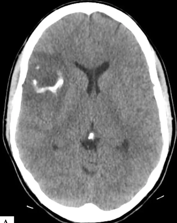
- Korteks ve subkortikal beyaz cevherde silik, düşük atenüasyonlu genişleme; oligodendroglial tümörde giriform veya nodüler kalsifikasyon görülebilir. Kontrastsız BT tümörün hipodensitesini ve kalsifikasyonunu gösterebilir, ancak infiltratif sınırlarını tam olarak belirlemez; WHO CNS5'e göre kesin sınıflama ve derece ataması histopatoloji ile entegre moleküler bulgulara dayanır.
Önerilen pencereler: Beyin (G 80 / M 40), Kemik (G 1800 / M 400).
BT bulguları
- Kortikomedüller hipodensite — normal gri-beyaz cevher ayrımını yerel olarak bulanıklaştırır.
- Kortikal genişleme — sulkusların yerel silinmesi ve girus konturunun dolgunlaşmasıyla fark edilir.
- Kalsifikasyon — özellikle oligodendrogliomlarda korteksi izleyen giriform ya da kümeleşmiş hiperdens odaklar olabilir.
- Hafif kitle etkisi — komşu sulkus daralır; ventrikül belirgin biçimde yer değiştirmeyebilir.
- Az ödemli görünüm tümörün derecesini tek başına belirlemez; moleküler özellikler gösteren düşük histolojik dereceli tümörlerde bile agresif seyre neden olunabilir, bu nedenle entegre tanı şarttır.
- Kontrastlanma olmaması veya sınırlı olması — düşük dereceyi tek başına doğrulamaz; dönüşüm veya yüksek dereceli bileşen MRG ile araştırılır.
Ölçütler ve sınıflamalar
- WHO CNS5’te diffüz gliomların sınıflanması
- WHO CNS5’te “düşük dereceli diffüz gliom” tek bir erişkin tümör tipi değildir. Erişkin tipi diffüz gliomlar arasında astrositom, IDH-mutant (CNS WHO derece 2–4) ve oligodendrogliom, IDH-mutant ve 1p/19q-kodelesyonlu (derece 2–3) ayrı tiplerdir; pediatrik tipi diffüz düşük dereceli gliomlar da ayrı gruptur. BT görünümü histomoleküler tipi veya dereceyi tek başına belirlemez.
Normalde
Karşılaştırma yapılırken aynı düzeydeki kontralateral insula ve frontal-operkulumun kesintisiz gri-beyaz cevher sınırını ve düzgün sulkus konturlarını referans alınız; centrum semiovale simetrik beyaz cevher atenüasyonu gösterir. Şüpheli tarafta korteksin kalınlaşmasını, gri-beyaz sınırın silinmesini ve insular/opercular sulkusların karşı tarafa göre daralmasını kıyasla.
Normal BT ile kıyasla
Aynı bölgenin, kaynak veri setinde patoloji içermediği belirtilen bir BT incelemesini kesit kesit kaydırın; organ sınırlarını açarak anatomiyi hasta görüntüsüyle karşılaştırın. Küçük rastlantısal bulgular tümüyle dışlanmamıştır.
Kaynak: TotalSegmentator v2.01 veri seti (Wasserthal ve ark., Radiology: Artificial Intelligence 2023), CC BY 4.0, vaka s0103; organ sınırları veri setinin otomatik segmentasyonundan, birleştirilerek sadeleştirildi. Görüntüler 2,5 mm kesit aralığına yeniden örneklendi.
Ayırıcı tanı
- Ensefalomalazi
- parankim hacim kaybına bağlı komşu sulkusların genişlemesi ve ex vacuo ventrikül büyümesi ile karakterizedir; kitle etkisi veya tümöral genişleme göstermez.
- Subakut enfarkt
- arteriyel dağılımı izler ve klinik başlangıçla uyumlu olabilir; seri görüntü ve MRG ayrımı sağlar.
- Dizembriyoplastik nöroepitelyal tümör (DNET)
- çoğunlukla kortikal yerleşimli glioneuronal tümördür; nöbetle ortaya çıkabilir ve görüntüleme özellikleri düşük dereceli gliomla örtüşebilir, bu nedenle MRG ve histomoleküler inceleme ayırıcıdır.
- Yüksek dereceli gliom
- daha belirgin heterojenite, nekroz, ödem ve kontrastlanma gösterebilir; BT’de bu özelliklerin yokluğu düşük dereceyi ispatlamaz.
Tuzaklar
- Kalsifikasyonu otomatik olarak oligodendrogliom sayma: kalsifikasyon özgül değildir; yerleşim ve MRG özellikleriyle birlikte yorumlanır.
- Hafif hipodensiteyi kronik iskemi ya da artefakt diye göz ardı etme: kortikal genişleme ve sulkus silinmesi tümör olasılığını artırır.
- Kontrastlanmayan lezyonu iyi huylu kabul etme: BT’de kontrastlanma tümör derecesini güvenilir biçimde dışlamaz veya belirlemez.
Kendini dene
Nöbetle başvuran erişkinde silik insular hipodensite, kortikal genişleme ve giriform kalsifikasyon var. Bu BT örüntüsü en çok hangi tümörü düşündürür?
Cevabı göster
Oligodendrogliom. Kortikal-subkortikal infiltrasyonla birlikte giriform kalsifikasyon oligodendrogliomu görüntüleme düzeyinde destekler; kalsifikasyon tek başına özgül değildir. Kesin tümör tipi ve derece histolojik-moleküler değerlendirmeyle belirlenir. Glioblastom genellikle daha heterojen ve ödemli görünür; menenjiyom ekstraaksiyeldir; kavernöz malformasyonda kan ürünü sinyali öne çıkar.
Şüpheli hemisferde sulkuslar silinmiş ve girus kalınlaşmış. Bu görünüm hangisini destekler?
Cevabı göster
İnfiltratif tümör. Kortikal genişleme ve sulkus silinmesi parankimin yer kaplayan infiltratif sürecini destekler. Ensefalomalazi ve atrofi hacim kaybı ile sulkus genişlemesi yapar; basit kist ise düzgün sınırlı sıvı yoğunluğundadır.
İlgili konular
Kaynaklar
Bu sayfadaki 39 cümle ve 2 quiz sorusu aşağıdaki kaynaklardan alıntılarla tek tek karşılaştırıldı; 4 cümle bu karşılaştırmada düzeltildi (2026-10-08).
- Artificial intelligence-based diagnostic model for identifying PTPRZ1-MET fusion in a clinically defined secondary glioblastoma cohortpmc.ncbi.nlm.nih.gov
- Imaging diagnosis of adult-type diffuse gliomas in the era of WHO CNS5 and cIMPACT-NOW updates 8–11: a narrative reviewpmc.ncbi.nlm.nih.gov
- Right Frontal Isocitrate Dehydrogenase (IDH)-Mutant WHO Grade 3 Oligodendroglioma Presenting With Morning Headache and Gait Imbalancepmc.ncbi.nlm.nih.gov
- Diffuse high-grade glioma with gliomatosis cerebri pattern mimicking symmetric white matter diseasepmc.ncbi.nlm.nih.gov
- Integrated Transcriptomic Analysis Reveals Coordinated Gliovascular Remodeling Across Diffuse Glioma Gradespmc.ncbi.nlm.nih.gov
- Pseudo-insular glioma syndrome: illustrative cases.pubmed.ncbi.nlm.nih.gov
- T2-FLAIR mismatch sign in dysembryoplasticneuroepithelial tumor.pubmed.ncbi.nlm.nih.gov
- Fractal analysis of 11 C-methionine PET in patients with newly diagnosed glioma.pubmed.ncbi.nlm.nih.gov
- CSF-based liquid biopsy pointing to a diagnosis of diffuse glioma in a patient with supposed neurodegenerative disorder.pubmed.ncbi.nlm.nih.gov
- MRI and CT Identify Isocitrate Dehydrogenase (IDH) -Mutant Lower-Grade Gliomas Misclassified to 1p/19q Codeletion Status with Fluorescence in Situ Hybridization.pubmed.ncbi.nlm.nih.gov
- Combining hyperintense FLAIR rim and radiological features in identifying IDH mutant 1p/19q non-codeleted lower-grade glioma.pubmed.ncbi.nlm.nih.gov
- The prognostic power of [ 11 C]methionine PET in IDH-wildtype diffuse gliomas with lower-grade histological features: venturing beyond WHO classification.pubmed.ncbi.nlm.nih.gov
- [<sup>18</sup>F]FET uptake in diffuse lower-grade IDH-mutant glioma correlates with glioma-related epilepsy.pubmed.ncbi.nlm.nih.gov
- Seizure-Related Deterioration in Treated Glioma: A Narrative Review of Nonconvulsive Status Epilepticus, Peri-Ictal Pseudoprogression, Treatment-Related Change, and True Progression.pubmed.ncbi.nlm.nih.gov
- The Incidence and Electroencephalographic Features in Brain Tumor-Related Epilepsy.pubmed.ncbi.nlm.nih.gov
- Giant Calcified Frontal Lobe Metastasis as the Initial Manifestation of Pulmonary Large-Cell Neuroendocrine Carcinoma: Diagnostic and Surgical Insights From Intraoperative Diagnosis.pubmed.ncbi.nlm.nih.gov
- Illuminating radiogenomic signatures in pediatric-type diffuse gliomas: insights into molecular, clinical, and imaging correlations. Part II: low-grade group.pubmed.ncbi.nlm.nih.gov
- White Epidermoid Cyst: An Illustrative Case.pubmed.ncbi.nlm.nih.gov
- Molecular tumor board: H3K27M diffuse midline glioma diagnosed during pregnancy by cerebrospinal fluid next-generation sequencing.pubmed.ncbi.nlm.nih.gov
- Diffuse high-grade glioma with gliomatosis cerebri pattern mimicking symmetric white matter disease.pubmed.ncbi.nlm.nih.gov
- Diagnostic Uncertainty in Rapidly Progressive Brainstem Lesions: A Radiological Diagnosis of Probable CNS Relapse of Diffuse Large B-cell Lymphoma.pubmed.ncbi.nlm.nih.gov
- Case Report: Basal ganglia brain abscess caused by <i>Nocardia farcinica</i>.pubmed.ncbi.nlm.nih.gov
- Brainstem Glioma Prognostication: Static FET PET/CT.pubmed.ncbi.nlm.nih.gov
- Spectral CT and Photon-counting Detector CT: Principles and Applications in Neuroradiology.pubmed.ncbi.nlm.nih.gov
- Deep learning in neurosurgery: a systematic literature review with a structured analysis of applications across subspecialties.pubmed.ncbi.nlm.nih.gov
- Safety, efficacy, and side effects of sodium fluorescein-aided resection of glioblastoma: a quasi-experimental study.pubmed.ncbi.nlm.nih.gov
- Conventional MRI/CT for differentiation of IDH-mutant adult-type diffuse gliomas: revisited with a new imaging feature "scFLAIR-D".pubmed.ncbi.nlm.nih.gov
- Rosette-forming glioneuronal tumor of the cerebellum with intratumoral hemorrhage: case report with radiologic-pathologic correlation in a resource-limited setting.pubmed.ncbi.nlm.nih.gov
- Diffuse glioma with <i>FGFR3</i>::<i>TACC3</i> gene fusion and prominent calcification: A case report.pubmed.ncbi.nlm.nih.gov
- Anatomy-Guided Microsurgical Resection of a Dominant Frontal Lobe Tumor Without Intraoperative Adjuncts: A Case Report from a Resource-Limited Context.pubmed.ncbi.nlm.nih.gov
- Paediatric cranial ultrasound: abnormalities of the brain in term neonates and young infants.pubmed.ncbi.nlm.nih.gov
- Temporoinsular Extraventricular Neurocytoma Initially Diagnosed as Glioblastoma in a 57-Year-Old Woman: A Case Report and Literature Review.pubmed.ncbi.nlm.nih.gov
- Diagnostic imaging and emerging image-guided neurointervention in brain tumors: a narrative review.pubmed.ncbi.nlm.nih.gov
- Purely cystic meningioma: extremely rare clinical and radiographic mimic of intra-axial glioblastoma multiforme with systematic review of the literature. Illustrative case.pubmed.ncbi.nlm.nih.gov
- Imaging diagnosis of adult-type diffuse gliomas in the era of WHO CNS5 and cIMPACT-NOW updates 8-11: a narrative review.pubmed.ncbi.nlm.nih.gov
- Prostate-specific membrane antigen in malignant brain tumors: vascular heterogeneity, imaging promise, and therapeutic challenges.pubmed.ncbi.nlm.nih.gov
- Bilateral lesions of the basal ganglia and thalami (central grey matter)-pictorial review.pubmed.ncbi.nlm.nih.gov
- Imaging of Non-atherosclerotic Vasculopathies.pubmed.ncbi.nlm.nih.gov
- Special Issue: EACR 2026 Congress: Innovative Cancer Science, 8-11 June 2026.pubmed.ncbi.nlm.nih.gov
- Neuroimaging of epilepsy.pubmed.ncbi.nlm.nih.gov
- Commentary: an eye on PET quantification.pubmed.ncbi.nlm.nih.gov
- AI-Powered Histology for Molecular Profiling in Brain Tumors: Toward Smart Diagnostics from Tissue.pubmed.ncbi.nlm.nih.gov
- Recent Advancements and Future Perspectives on Molecular Biomarkers in Adult Lower-Grade Gliomas.pubmed.ncbi.nlm.nih.gov
- Artificial-intelligence-based molecular classification of diffuse gliomas using rapid, label-free optical imaging.pubmed.ncbi.nlm.nih.gov
- Role of CXCR4 PET Imaging for the Evaluation of Gliomas: A Comprehensive Literature Review.pubmed.ncbi.nlm.nih.gov
- Integrating Multimodal MRI Habitat and Transformer-Based Pathomics to Predict High-Risk Molecular Subtypes and Explore Biological Mechanisms in Adult Diffuse Gliomas.pubmed.ncbi.nlm.nih.gov
- Intracranial venous thrombosis imaging by anatomical location: an educational review with mimics and diagnostic pitfalls.pubmed.ncbi.nlm.nih.gov
- Spectrum of Neuroimaging Findings in CNS Lymphoproliferative Disorders.pubmed.ncbi.nlm.nih.gov
- A Forgotten Rare Cause of Unilateral Basal Ganglia Calcinosis Due to Venous Angioma and Complicating Acute Stroke Management: A Case Report.pubmed.ncbi.nlm.nih.gov
- Disseminated Intracranial and Spinal Dysembryoplastic Neuroepithelial Tumor: A Case Report with a Systematic Review.pubmed.ncbi.nlm.nih.gov
- Imaging diagnosis of cystic intraparenchymal brain neoplasms.pubmed.ncbi.nlm.nih.gov
- Clinicoradiological differentiation of TERT promoter-mutant molecular glioblastoma versus histological glioblastoma: a single-center cohort study of 105 IDH-wildtype tumors.pubmed.ncbi.nlm.nih.gov
- A systematic approach to MR spectroscopy of the brain: Indications, protocol, metabolites, and spectral analysis in neurologic diseases.pubmed.ncbi.nlm.nih.gov
- MRI and PET Alterations in Adult Skull Base Tumors: A Narrative Review of Proton Versus Photon Radiotherapy.pubmed.ncbi.nlm.nih.gov
- Cystathionine Detection via Proton Magnetic Resonance Spectroscopy (¹H-MRS) for the Diagnosis of Oligodendroglioma: A Case Report.pubmed.ncbi.nlm.nih.gov
- Artificial intelligence in brain tumor diagnosis and surgical planning: Recent advances.pubmed.ncbi.nlm.nih.gov
- Telovelar Approach for Fourth-Ventricular Epidermoid Cyst: Anatomical Respect, Functional Recovery, and Long-Term Stability.pubmed.ncbi.nlm.nih.gov
- Added prognostic value of 3D deep learning-derived features from preoperative MRI for adult-type diffuse gliomas.pubmed.ncbi.nlm.nih.gov
- Pituitary neuroendocrine tumors: imaging features, clinical relevance, and a proposal for standardized MRI reporting.pubmed.ncbi.nlm.nih.gov
- Retinoblastoma and beyond: pediatric orbital mass lesions.pubmed.ncbi.nlm.nih.gov
- Retinoic acid in health and disease.pubmed.ncbi.nlm.nih.gov
- Report from the National Pediatric Cancer Foundation - infantile glioma and other non-embryonal central nervous system tumors: evolving molecular advances and current treatment landscape.pubmed.ncbi.nlm.nih.gov
- Illuminating radiogenomic signatures in pediatric-type diffuse gliomas: insights into molecular, clinical, and imaging correlations. Part I: high-grade group.pubmed.ncbi.nlm.nih.gov
- Pediatric Temporal Lobe Meningioma With Meningioangiomatosis Mimicking Invasive Meningioma.pubmed.ncbi.nlm.nih.gov
- Mesenchymal Nonmeningothelial Tumors of the CNS: Evolving Molecular Landscape and Implications for Neuroradiologists.pubmed.ncbi.nlm.nih.gov
- Health system learning enables generalist neuroimaging models.pubmed.ncbi.nlm.nih.gov
- Imaging of supratentorial intraventricular masses in children: a pictorial review-part 2.pubmed.ncbi.nlm.nih.gov
- Integrated Transcriptomic Analysis Reveals Coordinated Gliovascular Remodeling Across Diffuse Glioma Grades.pubmed.ncbi.nlm.nih.gov
- Cosmic Signs in Radiology: A Pictorial Review.pubmed.ncbi.nlm.nih.gov
- Vascular Cognitive Impairment (VCI).pubmed.ncbi.nlm.nih.gov
- A Review of the Effectiveness of Neuroimaging Modalities for the Detection of Traumatic Brain Injury.pubmed.ncbi.nlm.nih.gov
- An ABC for decision making.pubmed.ncbi.nlm.nih.gov
- Differential diagnoses of cerebral hemiatrophy in childhood: a review of literature with illustrative report of two cases.pubmed.ncbi.nlm.nih.gov
- Post-therapy imaging of adult brain tumours: patterns, pitfalls, and practical framework.pubmed.ncbi.nlm.nih.gov
- Imaging of the Primary Visual Pathway based on Visual Deficits.pubmed.ncbi.nlm.nih.gov
- An Atlas of Neonatal Neurovascular Imaging Anatomy as Depicted with Microvascular Imaging: The Intracranial Veins.pubmed.ncbi.nlm.nih.gov
- A Review of the Role of Neuroimaging in Neurotoxicity Monitoring in Children with Acute Lymphoblastic Leukemia.pubmed.ncbi.nlm.nih.gov
- Preoperative computed tomography perfusion and angiography predict the need for shunting in carotid endarterectomy: a multicenter study.pubmed.ncbi.nlm.nih.gov
- Multiterritory Acute Ischemic Stroke with Bilateral Middle Cerebellar Peduncle T2/FLAIR Hyperintensities in the Setting of a Fatal Systemic Arterial Thrombotic Process: A Case Report.pubmed.ncbi.nlm.nih.gov
- Case Report: Structured MRI assessment of posterior thalamic infarction in a distribution compatible with posterior choroidal artery territory presenting as Déjerine-Roussy syndrome in an adolescent: differentiating arterial ischemia from venous thrombosis and thalamic neoplasm.pubmed.ncbi.nlm.nih.gov
- Beyond acrodynia: infantile mercury vapor poisoning presenting with refractory hypoglycemia and persistent neurological damage.pubmed.ncbi.nlm.nih.gov
- Baseline radiological parameters associated with good neurological outcome following ventriculoperitoneal shunt in normal pressure hydrocephalus: A single-center study on 82 patients.pubmed.ncbi.nlm.nih.gov
- Stroke or Seizure? Diagnostic Role of Neuroimaging in Acute Neurologic Mimics.pubmed.ncbi.nlm.nih.gov
- Low-Field and Portable MRI for Acute Ischemic Stroke: A Systematic Review.pubmed.ncbi.nlm.nih.gov
- Characterizing the association between complement-mediated TMA and cognitive dysfunction using MRI and neurocognitive assessment.pubmed.ncbi.nlm.nih.gov
- Imaging of Ischemic Stroke.pubmed.ncbi.nlm.nih.gov
- Stroke secondary to cancer-associated coagulopathy: a systematic review.pubmed.ncbi.nlm.nih.gov
Eğitim amaçlıdır; hasta başında tanı ve tedavi kararının yerine geçmez. İçerik yapay zekâ destekli bir süreçle güncel literatüre göre cümle cümle kaynaklarla karşılaştırıldı, ancak uzman radyolog incelemesinden geçmedi. Klinik kararlarda güncel kılavuzları, kurum protokollerini ve radyoloji raporunu esas alın.