
Özet
Menenjiyom, araknoid kap hücrelerinden köken alan ve çoğunlukla dura komşuluğunda büyüyen ekstraaksiyel tümördür; rastlantısal bulunabileceği gibi çevre beyin dokusuna bası da yapabilir. Kontrastsız BT tümör kalsifikasyonunu ve komşu kafatasındaki hiperostozu iyi gösterir; kitle ile beyin arasındaki sınır için MRG daha duyarlıdır. Dural tabanlı yerleşim tanıyı desteklese de dural metastaz ve başka ekstraaksiyel kitleler benzer görünür. Komşu vazojenik ödem, korteks itilmesi ve kemik değişikliklerinin (hiperostoz veya osteoliz) gözden kaçırılması, kitenin gerçek etki boyutunun yanlış değerlendirilmesine yol açabilir.
Faz ve pencere
- Kontrastsız tanısal
- Dura komşuluğundaki kitle kortekse göre sıklıkla izo- ya da hiperdens olup yaklaşık dörtte birinde kalsifikasyon görülür; komşu kemikte hiperostoz veya nadiren osteoliz aranırken, kontrastsız BT aşamasında tümörün canlı dokusunun veya reaktif dural kalınlaşmanın kontrastlanması ile dural kuyruk işareti değerlendirilemez. Kontrastsız BT’de dural kuyruk ve tümör dokusunun belirgin kontrastlanması değerlendirilemez.
- Kontrastlı tanısal
- Dural tabanlı kitle çoğunlukla belirgin kontrastlanır; dural kuyruk görülebilir ancak menenjiyoma özgü değildir ve dural metastazlarda da bulunabilir. Kontrastlanma paterni tek başına histolojiyi veya dereceyi belirlemez.
Önerilen pencereler: Beyin (G 80 / M 40), Kemik (G 1800 / M 400).
BT bulguları
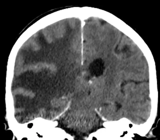
- Dural tabanlı ekstraaksiyel kitle — konveksite, falx, tentorium veya kafa tabanı boyunca geniş temas yüzeyi vardır.
- BOS yarığı — kitle ile komşu korteks arasında seçilebilen ince BOS aralığı ekstraaksiyel yerleşimi destekler; BT’de her zaman belirgin değildir.
- İç kalsifikasyon — noktasal ya da kaba hiperdens odaklar kitle içinde görülebilir.
- Komşu kemikte hiperostoz — kemik penceresinde iç tabula kalınlaşması ve yoğunluk artışı şeklindedir.
- Kemik destrüksiyonu — daha az tipiktir; dural metastaz veya daha agresif tümör olasılığını artırır.
- Peritümöral vazojenik ödem — komşu beyaz cevherde hipodensite ve sulkus basısı yapabilir.
- Kitle büyükse komşu korteksi içe iter ve parankimde geniş vazojenik ödem oluşturur.
Ölçütler ve sınıflamalar
- WHO CNS5 menenjiyom derecelendirmesi
- Menenjiyomlar CNS WHO derece 1, 2 veya 3 olarak sınıflandırılır. Derece histopatolojik ve uygun moleküler ölçütlerle belirlenir; BT görünümü tek başına derece atamaz.
Normalde
Aynı konveksite düzeyinde normal iç tabula ince ve düzgün, komşu korteks ise kafatasının iç yüzünü izleyen kesintisiz gri cevher konturudur. Şüpheli tarafta kemik kalınlığı ve yoğunluğunu karşı tarafla; korteksin itilmesini, aradaki BOS yarığını ve sulkusların açıklığını komşu normal giruslarla kıyasla.
Normal BT ile kıyasla
Aynı bölgenin, kaynak veri setinde patoloji içermediği belirtilen bir BT incelemesini kesit kesit kaydırın; organ sınırlarını açarak anatomiyi hasta görüntüsüyle karşılaştırın. Küçük rastlantısal bulgular tümüyle dışlanmamıştır.
Kaynak: TotalSegmentator v2.01 veri seti (Wasserthal ve ark., Radiology: Artificial Intelligence 2023), CC BY 4.0, vaka s0103; organ sınırları veri setinin otomatik segmentasyonundan, birleştirilerek sadeleştirildi. Görüntüler 2,5 mm kesit aralığına yeniden örneklendi.
Ayırıcı tanı
- Dural metastaz
- bilinen sistemik kanser, birden fazla dural odak ve kemik destrüksiyonu menenjiyomdan çok metastazı destekler.
- Soliter fibröz tümör
- dural tabanlı olabilir; menenjiyomdaki hiperostoz ve kalsifikasyon daha az karakteristiktir, kemik erozyonu eşlik edebilir.
- Primer santral sinir sistemi lenfoması
- sıklıkla parankimal/periventrikülerdir; dural yerleşim daha atipiktir.
- Kalsifiye ekstraaksiyel lezyon
- yerleşim, dural bağlantı ve kemik değişiklikleri birlikte değerlendirilmeden yalnız yoğunluğa göre menenjiyom denmemelidir.
Tuzaklar
- Dural kuyruk işaretini menenjiyoma özgü kabul etme: metastazlar ve diğer dural kitleler de benzer kontrastlanma yapabilir.
- Komşu kemikte hiperostoz sıklıkla görülür; WHO derecesi ve kemik invazyonu histopatoloji ile belirlenir, radyolojik bulgular tek başına kesin ayırım yapmaz.
- Kemik penceresini atlama: küçük konveksite veya sfenoid kanat menenjiyomunda kemik kalınlaşması yumuşak doku penceresinde geri planda kalabilir.
- Ödemi tümör sınırı sanma: ekstraaksiyel kitle komşu parankimde geniş vazojenik ödem oluşturabilir.
Kendini dene
Konveksitede dura ile geniş tabanlı temas eden, kalsifiye kitle ve komşu kemikte kalınlaşma izleniyor. En olası tanı nedir?
Cevabı göster
Menenjiyom. Dural taban, iç kalsifikasyon ve komşu hiperostoz menenjiyomu destekleyen BT özellikleridir. Glioblastom intraaksiyeldir; apse parankimal enfeksiyon odağıdır; ensefalomalazi doku kaybı gösterir.
Bilinen meme kanserli hastada birden çok dural kitle ve komşu kemikte erozyon var. En önemli taklitçi hangisidir?
Cevabı göster
Dural metastaz. Kanser öyküsü, çoklu dural odak ve litik kemik değişikliği dural metastazı düşündürür. Diğer seçenekler dural tabanlı çoklu kitle ve kalvaryal erozyon örüntüsünü açıklamaz.
İlgili konular
Kaynaklar
Bu sayfadaki 42 cümle ve 2 quiz sorusu aşağıdaki kaynaklardan alıntılarla tek tek karşılaştırıldı; 6 cümle bu karşılaştırmada düzeltildi (2026-10-08).
- Dural masses: meningiomas and their mimicspmc.ncbi.nlm.nih.gov
- EANO guideline on the diagnosis and management of meningiomas.pubmed.ncbi.nlm.nih.gov
- Cavernous sinus lesions.pubmed.ncbi.nlm.nih.gov
- Falcotentorial meningiomas.pubmed.ncbi.nlm.nih.gov
- Pathology of the carotid space.pubmed.ncbi.nlm.nih.gov
- Imaging of orbital disorders.pubmed.ncbi.nlm.nih.gov
- The optic chiasm.pubmed.ncbi.nlm.nih.gov
- 13 N-NH 3 PET/CT in oncological disease.pubmed.ncbi.nlm.nih.gov
- Visual loss and recovery in chiasmal compression.pubmed.ncbi.nlm.nih.gov
- REVIEW: MR elastography of brain tumors.pubmed.ncbi.nlm.nih.gov
- PET imaging for brain tumor diagnostics.pubmed.ncbi.nlm.nih.gov
- Meningiomas engaging major venous sinuses.pubmed.ncbi.nlm.nih.gov
- Clinical Aspects of Spinal Meningiomas: a Review.pubmed.ncbi.nlm.nih.gov
- MRI of intracranial intraventricular lesions.pubmed.ncbi.nlm.nih.gov
- Predicting Meningioma Consistency on Preoperative Neuroimaging Studies.pubmed.ncbi.nlm.nih.gov
- Dural-based metastasis mimicking meningioma: A rare case report.pubmed.ncbi.nlm.nih.gov
- Meningioma: A Review of Clinicopathological and Molecular Aspects.pubmed.ncbi.nlm.nih.gov
- Unusual coexistence of an epidermoid cyst with an atypical meningioma: Case report and review of the literature.pubmed.ncbi.nlm.nih.gov
- Diagnostic imaging and emerging image-guided neurointervention in brain tumors: a narrative review.pubmed.ncbi.nlm.nih.gov
- Magnetic resonance imaging of primary central nervous system neoplasms of dogs and cats: Basic concepts for diagnostic pathologists.pubmed.ncbi.nlm.nih.gov
- Malignant Meningiomas: From Diagnostics to Treatment.pubmed.ncbi.nlm.nih.gov
- Lung adenocarcinoma dural metastasis at the petroclival region mimicking meningioma: a frozen-section diagnostic pitfall. Illustrative case.pubmed.ncbi.nlm.nih.gov
- Tumor-to-tumor metastasis of small cell carcinoma of presumed pulmonary origin to a sphenoid ridge meningioma: A diagnostic pitfall in preoperative systemic evaluation.pubmed.ncbi.nlm.nih.gov
- Dural Metastasis of Prostatic Adenocarcinoma Mimicking Meningioma en Plaque: Case Report and Systematic Review.pubmed.ncbi.nlm.nih.gov
- Probabilistic MRI Maps for the Presentation and Management of Intracranial Meningiomas.pubmed.ncbi.nlm.nih.gov
- Ultra-high-field 7-Tesla MRI in neuro-oncology: a narrative review of current applications and future directions.pubmed.ncbi.nlm.nih.gov
- Increased intracranial pressure in NF2‑related schwannomatosis: an underestimated danger with serious consequences.pubmed.ncbi.nlm.nih.gov
- Case Report: Intracranial epidermoid cyst accompanied by bone hyperplasia forming a bone encapsulation.pubmed.ncbi.nlm.nih.gov
- Imaging of pituitary tumors: an update with the 5th WHO Classifications-part 2. Neoplasms other than PitNET and tumor-mimicking lesions.pubmed.ncbi.nlm.nih.gov
- Neoplasms and tumor-like lesions of the sellar region: imaging findings with correlation to pathology and 2021 WHO classification.pubmed.ncbi.nlm.nih.gov
- Spontaneous hemorrhage of cavernous malformation in the region of the cochlear nucleus: A case report.pubmed.ncbi.nlm.nih.gov
- Middle ear lymphoepithelioma-like carcinoma with cerebellopontine angle invasion misdiagnosed as meningioma: A case report and literature review.pubmed.ncbi.nlm.nih.gov
- Cinematic Rendering of Brain Magnetic Resonance Imaging: Photorealistic Visualization of Normal Neuroanatomy and Primary Brain Tumors.pubmed.ncbi.nlm.nih.gov
- A Case of Resection of Multiple Dural Cavernous Hemangiomas Mimicking a Convexity Meningioma.pubmed.ncbi.nlm.nih.gov
- Pituitary neuroendocrine tumors: imaging features, clinical relevance, and a proposal for standardized MRI reporting.pubmed.ncbi.nlm.nih.gov
- Imaging diagnosis of adult-type diffuse gliomas in the era of WHO CNS5 and cIMPACT-NOW updates 8-11: a narrative review.pubmed.ncbi.nlm.nih.gov
- Time-dependent Diffusion MRI-based Microstructural Mapping Value for Predicting Meningioma Grade, Subtype, and Proliferative Activity.pubmed.ncbi.nlm.nih.gov
- An intra-axial wolf in extra-axial clothing: A rare case of high-grade astrocytoma and literature review.pubmed.ncbi.nlm.nih.gov
- Extra-axial Desmoplastic Medulloblastoma in Adult Cerebellopontine Angle: Case Report and Noninvasive Molecular Subgrouping Utilizing Magnetic Resonance Imaging-Based Radiomics Nomogram.pubmed.ncbi.nlm.nih.gov
- Review of imaging modalities and radiological findings of calvarial lesions.pubmed.ncbi.nlm.nih.gov
- Sellar masses: diagnosis and treatment.pubmed.ncbi.nlm.nih.gov
- Intracranial solitary fibrous tumor with multiloculated cystic change: A case report.pubmed.ncbi.nlm.nih.gov
- A third ventricular floor-associated TTF-1-positive posterior pituitary tumor: illustrative case.pubmed.ncbi.nlm.nih.gov
- An Imitation Too Perfect: Isolated Neurosarcoidosis Mimicking Spheno-Orbital Meningioma With Hyperostosis.pubmed.ncbi.nlm.nih.gov
- Solitary Fibrous Tumours in the Central Nervous System and Their Clinical Mimickers: A Case Series.pubmed.ncbi.nlm.nih.gov
- Sacral benign notochordal cell tumor with atypical imaging features and 16-year spontaneous partial regression: A case report.pubmed.ncbi.nlm.nih.gov
- Metastatic Meningioma: Neuroradiologic and Molecular Imaging Perspectives.pubmed.ncbi.nlm.nih.gov
- Giant cell tumor of the orbital roof: A case report and brief review of literature.pubmed.ncbi.nlm.nih.gov
- Olfactory Groove Meningioma Mimicking Late-Life Depression: A Case Report.pubmed.ncbi.nlm.nih.gov
- Peritumoral edema in meningiomas: a review of influencing factors, mechanisms, and management.pubmed.ncbi.nlm.nih.gov
- Reversibility of cognitive and psychiatric impairments following surgical resection of frontotemporal meningiomas: a predictive factor analysis.pubmed.ncbi.nlm.nih.gov
- Hidden Hollows of the Fetal Cranium-Lacunar Skull in Isolation.pubmed.ncbi.nlm.nih.gov
- Computed Tomographic Grading of Ear Diseases in Guinea Pigs (Cavia porcellus).pubmed.ncbi.nlm.nih.gov
- Key pre-operative CT parameters for cochlear implantation: a focused radiologic review with surgical relevance.pubmed.ncbi.nlm.nih.gov
- Minimal Anterior and Posterior Combined Transpetrosal Approach for Posterior Clinoid Process Meningiomas.pubmed.ncbi.nlm.nih.gov
- Clear cell meningioma: Experience at a medical center.pubmed.ncbi.nlm.nih.gov
- "Updates on diagnostic and prognostic molecular biomarkers of CNS tumors".pubmed.ncbi.nlm.nih.gov
- Differential expression of TGF-β1, TGF-β2, and TGF-β3 across WHO grades in meningioma: convergent evidence from mRNA, methylation, miRNA, and protein analysis.pubmed.ncbi.nlm.nih.gov
- Clinical evaluation of the CrossNN DNA methylation classifier for central nervous system tumors.pubmed.ncbi.nlm.nih.gov
- Meningiomas classified as grade 2 due to brain invasion alone have better progression-free survival than other atypical meningiomas.pubmed.ncbi.nlm.nih.gov
- Radiation-induced cranial meningiomas are associated with earlier relapse: a propensity score-matched cohort study.pubmed.ncbi.nlm.nih.gov
- Human biomarker navigator.pubmed.ncbi.nlm.nih.gov
- Context aware machine learning techniques for brain tumor classification and detection - A review.pubmed.ncbi.nlm.nih.gov
- Molecular Testing for the World Health Organization Classification of Central Nervous System Tumors: A Review.pubmed.ncbi.nlm.nih.gov
- Decoding calvarial lesions: Contemporary neurosurgical management integrating radiological and histopathological features.pubmed.ncbi.nlm.nih.gov
- Operative diagnosis of a meningioma-like intraosseous skull lesion revealing multiple myeloma: Case report and literature review.pubmed.ncbi.nlm.nih.gov
- Spinal granulocytic sarcoma with both extradural and intradural involvement: a case report.pubmed.ncbi.nlm.nih.gov
- Solitary fibrous tumor of the posterior fossa: a rare and atypical case report.pubmed.ncbi.nlm.nih.gov
- Parieto-occipital interhemispheric transtentorial resection of a falcotentorial solitary fibrous tumor using a 3D-4K exoscope: Technical nuances and illustrative case.pubmed.ncbi.nlm.nih.gov
- Dural Lymphomas Unmasked: A Narrative Review of Extra-Axial Mimics and a Pragmatic Diagnostic Decision Algorithm.pubmed.ncbi.nlm.nih.gov
- Cerebral Venous Sinus Thrombosis Revealing ALK-Positive Anaplastic Large-Cell Lymphoma in a Patient with Inherited Thrombophilia and Concomitant Infection: Case Report and Narrative Review.pubmed.ncbi.nlm.nih.gov
- Challenges in diagnosing malignant lymphoma involving the spine: illustrative cases.pubmed.ncbi.nlm.nih.gov
- Pediatric Temporal Lobe Meningioma With Meningioangiomatosis Mimicking Invasive Meningioma.pubmed.ncbi.nlm.nih.gov
- Imaging of Abusive Head Trauma : A Radiologists' Perspective.pubmed.ncbi.nlm.nih.gov
- Neuroimaging of infections.pubmed.ncbi.nlm.nih.gov
- Immunoglobulin G4-Related Disease Mimicking Meningioma and Dural Metastasis: A Case Report.pubmed.ncbi.nlm.nih.gov
- Molecular Brain Tumor Imaging Beyond [<sup>18</sup>F]FDG: Somatostatin Receptor PET, Amino Acid PET, and Emerging Applications-From the <i>AJR</i> Special Series on Molecular Imaging.pubmed.ncbi.nlm.nih.gov
- Paradoxical evolution of spheno-orbital meningioma after cessation of progestin treatment.pubmed.ncbi.nlm.nih.gov
- An Unusual Therapy for an Atypical Case of Secondary Extranodal, Non-parenchymal Central Nervous System Marginal Zone Lymphoma.pubmed.ncbi.nlm.nih.gov
- Minimally invasive resection of a spheno-orbital tumor: Review of surgical techniques and a case report.pubmed.ncbi.nlm.nih.gov
- Incidentally discovered asymptomatic extrasinusal mucocele of the sphenoid greater wing: A case report and review of differential diagnoses.pubmed.ncbi.nlm.nih.gov
- Pediatric Maxillary Strut Anatomy: A CT-Based Analysis of Morphometry, Shape, and Pneumatization.pubmed.ncbi.nlm.nih.gov
- Spectrum of Neuroimaging Findings in CNS Lymphoproliferative Disorders.pubmed.ncbi.nlm.nih.gov
- Fluorescence-guided surgery combined with intraoperative photodynamic therapy for recurrent atypical and anaplastic intracranial meningiomas: a prospective feasibility study.pubmed.ncbi.nlm.nih.gov
- Transcranial ultrasonography imaging of a suprasellar meningioma: A case description and technical notes.pubmed.ncbi.nlm.nih.gov
- Pneumosinus dilatans frontalis: a cause of headaches mimicking chronic sinusitis-a case report and review of the literature.pubmed.ncbi.nlm.nih.gov
- Clival and Paraclival Lesions: A Pictorial Review.pubmed.ncbi.nlm.nih.gov
- Surgical strategy to minimize dominant hemisphere retraction in an asymmetric giant tuberculum sellae meningioma with contralateral anterior cerebral artery positioning: A case report.pubmed.ncbi.nlm.nih.gov
- Giant Right Sphenoid Wing Meningioma as a Reversible Frontal Network Lesion: A Pseudo-bvFTD Case with Venous-Sparing Skull-Base Resection.pubmed.ncbi.nlm.nih.gov
- Case of Langerhans Cell Histiocytosis That Mimics Meningioma in CT and MRI.pubmed.ncbi.nlm.nih.gov
- Surgical management of anterior clinoidal meningiomas: consensus statement on behalf of the EANS skull base section.pubmed.ncbi.nlm.nih.gov
- Magnetic resonance imaging of the orbit, Part 2: Characterization of orbital pathologies.pubmed.ncbi.nlm.nih.gov
- Giant Lumbosacral Meningioma Without Dural Attachment in an Adolescent: A Rare Diagnostic and Surgical Challenge.pubmed.ncbi.nlm.nih.gov
- Calcified Chronic Subdural Hematoma With Adjacent Cranial Thickening: A Case Report and Mechanistic Exploration.pubmed.ncbi.nlm.nih.gov
- Meningeal metastatic tumor with bone destruction from follicular thyroid carcinoma: a case report and literature review.pubmed.ncbi.nlm.nih.gov
- Lateral skull base metastasis from cervical adenocarcinoma: the masquerading trespasser - a case report.pubmed.ncbi.nlm.nih.gov
- Primary Extracranial Meningioma of the Sphenoid Bone Without Dural Attachment Presenting With Multicompartmental Extension: A Case Report.pubmed.ncbi.nlm.nih.gov
- The many faces of glioblastoma: Pictorial review of atypical imaging features.pubmed.ncbi.nlm.nih.gov
- Primary cerebellopontine angle glioblastoma in an adult.pubmed.ncbi.nlm.nih.gov
- A Mistaken Diagnosis of Secondary Glioblastoma as Parasitosis.pubmed.ncbi.nlm.nih.gov
- Imaging of Skull Base Tumors.pubmed.ncbi.nlm.nih.gov
- Imaging and diagnostic advances for intracranial meningiomas.pubmed.ncbi.nlm.nih.gov
- Commentary: an eye on PET quantification.pubmed.ncbi.nlm.nih.gov
- Skull Base Tumors and Tumor-Like Lesions: A Pictorial Review.pubmed.ncbi.nlm.nih.gov
- <sup>18</sup> F-FDG and <sup>68</sup> Ga-FAPI PET/CT Imaging in a Patient with High-Grade Meningioma: A Case Report.pubmed.ncbi.nlm.nih.gov
- Early Non-Contiguous Pachymeningeal Dissemination of Isocitrate Dehydrogenase (IDH)-Wildtype Giant Cell Glioblastoma: A Rare Finding During Adjuvant Therapy.pubmed.ncbi.nlm.nih.gov
- Rapid progression of recurrent intracranial solitary fibrous tumor following a previously reported TERT-wild-type primary tumor: a case report.pubmed.ncbi.nlm.nih.gov
- A Misleading Cystic Brain Lesion: An Uncommon Presentation of Meningioma.pubmed.ncbi.nlm.nih.gov
- Sphenoid Temporal Meningioma With Granular Cell Features and Extracranial Infratemporal Extension.pubmed.ncbi.nlm.nih.gov
- Atypical Foster Kennedy Syndrome With Orbital Apex Involvement Secondary to Intracranial Meningiomatosis: A Case Report.pubmed.ncbi.nlm.nih.gov
- FEBAIR microsurgical principles in meningiomas and recurrence: a long-term follow-up series.pubmed.ncbi.nlm.nih.gov
Eğitim amaçlıdır; hasta başında tanı ve tedavi kararının yerine geçmez. İçerik yapay zekâ destekli bir süreçle güncel literatüre göre cümle cümle kaynaklarla karşılaştırıldı, ancak uzman radyolog incelemesinden geçmedi. Klinik kararlarda güncel kılavuzları, kurum protokollerini ve radyoloji raporunu esas alın.