Özet
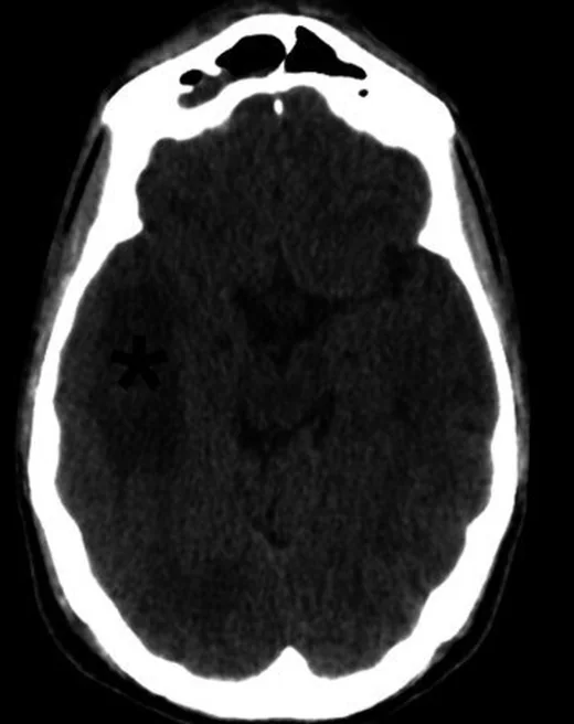
Serebrit, enfeksiyonun beyin parankiminde oluşturduğu, henüz olgun apse kapsülü gelişmemiş fokal inflamasyon evresidir ve çoğu kez komşu sinüs, mastoid, otit veya hematojen enfeksiyon kaynağıyla ilişkilidir. Kontrastsız BT'de enfekte parankimde sınırları belirsiz hipodensite görülebilir ve lezyon progresif olarak kapsül oluşturarak apse evresine ulaşabilir. BT acil durumda yaygınlığı, kitle etkisini ve gelişen nekroz/apseyi değerlendirmeye yardım eder; erken parankim enfeksiyonu için MR daha duyarlıdır. Serebrit, enfeksiyonun beyin parankiminde oluşturduğu ve henüz olgun apse kapsülü gelişmemiş fokal inflamasyon evresidir; ilerleyerek apse ve ekstraaksiyel yayılım gösterebilir.
Faz ve pencere
- Kontrastsız tanısal
- Enfekte parankimde sınırları belirsiz düşük atenüasyon izlenebilir ve olgun bir apse kapsülü henüz gelişmemiştir. Bu seride enfeksiyonun yayılımı ve komşu yapıların tutulumu değerlendirilir.
- Kontrastlı tanısal
- Kontrastlı görüntülerde lezyonun özellikleri incelenir ve enfeksiyonun ilerleyerek kapsül oluşturması takip edilir. Görüntüleme bulguları klinik bağlamla birlikte değerlendirilerek tanısal süreç desteklenir.
Önerilen pencereler: Beyin (G 80 / M 40), Kemik (G 1800 / M 400).
BT bulguları
- Sınırları belirsiz fokal hipodensite — inflamasyonlu beyin dokusu komşu normal parankime yumuşak geçiş gösterir.
- Kontrastlı fazda enfekte alanın boyanma paterni incelenir ve lezyonun yapısı değerlendirilir.
- Kapsül yokluğu — olgun apsedeki çevresel halka şeklinde belirgin duvar henüz seçilmez.
- Enfekte bölgede parankimal hipodensite ve ödem bulguları gözlenebilir.
- Lezyonun büyümesi ile çevre dokuda baskı etkileri izlenebilir.
- Kaynak odağa komşuluk — frontal serebritte sinüs, temporal/posterior fossada mastoid ve orta kulak değişiklikleri araştırılır.
- İlerleyen santral nekroz — takip görüntülemede odak daha belirginleşip kapsül boyanması ve sıvı merkez geliştirebilir.
Ölçütler ve sınıflamalar
- Evreli piyojenik beyin enfeksiyonunun BT görünümü
- Erken serebritte sınırları belirsiz düşük atenüasyon ve değişken boyanma görülür; geç serebritte nekroz ile halka boyanması gelişebilir; ardından erken ve olgun kapsül evreleri gelir.
Normalde
Normal aynı lobda kortikal gri cevher ile subkortikal beyaz cevher sınırı seçilir, sulkuslar açıktır ve parankimde yamalı kontrastlanma bulunmaz. Lezyonun karakteristiğini belirlemek için karşı hemisferdeki normal parankimle karşılaştırılmalıdır.
Normal BT ile kıyasla
Aynı bölgenin, kaynak veri setinde patoloji içermediği belirtilen bir BT incelemesini kesit kesit kaydırın; organ sınırlarını açarak anatomiyi hasta görüntüsüyle karşılaştırın. Küçük rastlantısal bulgular tümüyle dışlanmamıştır.
Kaynak: TotalSegmentator v2.01 veri seti (Wasserthal ve ark., Radiology: Artificial Intelligence 2023), CC BY 4.0, vaka s0103; organ sınırları veri setinin otomatik segmentasyonundan, birleştirilerek sadeleştirildi. Görüntüler 2,5 mm kesit aralığına yeniden örneklendi.
Ayırıcı tanı
- Vasküler hastalıklar ve diğer enfeksiyon dışı inflamatuvar süreçler benzer görüntüleme bulguları oluşturabilir.
- Tümör çevresi ödem
- ödem çoğunlukla kitle merkezinin çevresindedir; kontrastlanan solid/nodüler tümör bileşeni araştırılır.
- Farklı etkenlere bağlı ensefalitler de benzer parankimal tutulumlar gösterebilir.
- Demiyelinizan plak
- beyaz cevherde fokal hipodensite yapabilir; klinik ve MR paterni enfeksiyon yerine demiyelinizasyonu destekler.
- Travmatik lezyonlar ve hemorajik süreçler de ayırıcı tanıda değerlendirilmelidir.
Tuzaklar
- Tam halka kontrastlanmasının olmaması serebriti dışlamaz; erken evrede BT değişikliği silik veya yalnız hipodens olabilir.
- Fokal ödemi doğrudan serebrit diye adlandırmayın; komşu sinüs/otit kaynağı, seyir ve MR bulguları ayrımı güçlendirir.
- BT erken serebriti MR'dan daha az duyarlı gösterir; BT normal ya da belirsizken kuvvetli enfeksiyon şüphesi sürüyorsa MR ile inceleme gerekir.
- Serebrit ile kapsüllü apse arasında geçiş keskin değildir; kitle etkisi veya klinik durum değiştiğinde odak yeniden değerlendirilmelidir.
Kendini dene
Sinüzite komşu parankimde sınırları silik hipodens alan var, ancak halka kapsül henüz görünmüyor. En olası evre hangisidir?
Cevabı göster
Erken serebrit. Erken serebritte enfeksiyonlu parankim sınırları belirsiz düşük atenüasyon gösterebilir ve kapsül henüz oluşmamıştır. Olgun apse halka kapsüllü, metastaz nodüler/kitle biçimli, subdural hematom ise ekstraaksiyeldir.
Beyin apsesisinin gelişim sürecinde serebrit evresi nasıl tanımlanır?
Cevabı göster
Enfekte parankimde kapsül oluşmadan önceki fokal inflamasyon evresi. Beyin apsesisinin patogenezinde enfeksiyon parankimi tutar ve ilk evre olan serebritte henüz olgun apse kapsülü oluşmamıştır; ilerleyerek kapsül oluşum evresine geçer.
İlgili konular
Kaynaklar
Bu sayfadaki 39 cümle ve 2 quiz sorusu aşağıdaki kaynaklardan alıntılarla tek tek karşılaştırıldı; 15 cümle bu karşılaştırmada düzeltildi (2026-10-09).
- Fatal cerebral myiasis secondary to squamous cell carcinoma: case report and scoping reviewpmc.ncbi.nlm.nih.gov
- Crossing the Barrier: Eikenella corrodens Bacteremia Following CNS Infection in a Patient Treated with Nivolumab—A Case Report and Literature Reviewpmc.ncbi.nlm.nih.gov
- Tuberculosis revisted: classic imaging findings in childhoodpmc.ncbi.nlm.nih.gov
- Magnetic resonance imaging findings in central nervous system tuberculosis: A pictorial reviewpmc.ncbi.nlm.nih.gov
- Different causes and diverse outcomes of extremely rare septic cavernous sinus thrombosis complicated with internal carotid artery stenosispmc.ncbi.nlm.nih.gov
- [Brain abscess - overview].pubmed.ncbi.nlm.nih.gov
- Bacterial Brain Abscess: An Outline for Diagnosis and Management.pubmed.ncbi.nlm.nih.gov
- Role of imaging in the diagnosis of acute bacterial meningitis and its complications.pubmed.ncbi.nlm.nih.gov
- Imaging of cerebritis, encephalitis, and brain abscess.pubmed.ncbi.nlm.nih.gov
- Cocaine-induced encephalocele: case report and literature review.pubmed.ncbi.nlm.nih.gov
- A Brain Ring-Enhancing Lesion.pubmed.ncbi.nlm.nih.gov
- [Brain Abscess].pubmed.ncbi.nlm.nih.gov
- Neonatal cranial sonography: ultrasound findings in neonatal meningitis-a pictorial review.pubmed.ncbi.nlm.nih.gov
- Bacterial, Viral, and Prion Infectious Diseases of the Brain.pubmed.ncbi.nlm.nih.gov
- Neuroimaging of pediatric intracranial infection--part 1: techniques and bacterial infections.pubmed.ncbi.nlm.nih.gov
- Infectious Causes of Acute Encephalopathy.pubmed.ncbi.nlm.nih.gov
- Clinical, radiological, and microbiological profile of patients with autogenous cranioplasty infections.pubmed.ncbi.nlm.nih.gov
- Brain abscess caused by Salmonella Enteritidis following craniotomy for meningioma: A case report and literature review.pubmed.ncbi.nlm.nih.gov
- Multisystemic Imaging Features of Coccidioidomycosis.pubmed.ncbi.nlm.nih.gov
- Neonatal stress hyperglycemia mimicking diabetes mellitus secondary to massive intraparenchymal brain abscess due to escherichia coli sepsis: A case report.pubmed.ncbi.nlm.nih.gov
- Complications of Otitis Media: Mastoiditis and an Intracranial Abscess Presenting as Behavioral Changes.pubmed.ncbi.nlm.nih.gov
- Migratory Preseptal Cellulitis Secondary to Rhinosinusitis Complicated by Left Frontal Subdural Empyema in a Child: A Case Report.pubmed.ncbi.nlm.nih.gov
- Pathophysiology-driven neuroimaging: decoding brain injury in sepsis.pubmed.ncbi.nlm.nih.gov
- Neuroimaging of the Most Common Meningitis and Encephalitis of Adults: A Narrative Review.pubmed.ncbi.nlm.nih.gov
- Pediatric headache patient with cerebral abscesses: a brief review of the literature and case report.pubmed.ncbi.nlm.nih.gov
- Altered Mental Status in Emergency Department Patient with Cerebral Septic Emboli from Infective Endocarditis: Case Report.pubmed.ncbi.nlm.nih.gov
- Full recovery after severe bacterial encephalitis and brain abscess following deep brain stimulation surgery for Parkinson's disease.pubmed.ncbi.nlm.nih.gov
- Navigating complex presentations of central nervous system tuberculosis: A two-case analysis.pubmed.ncbi.nlm.nih.gov
- Disseminated and migratory sparganosis in the central nervous system: A case report and literature review of combined spinal and intracranial involvement.pubmed.ncbi.nlm.nih.gov
- Missed Cerebral Venous Thrombosis: A Diagnostic Challenge.pubmed.ncbi.nlm.nih.gov
- COVID-19 Co-Infection May Promote Development of Sinusitis Complication in Children.pubmed.ncbi.nlm.nih.gov
- Cerebral abscess secondary to post-esophageal dilation bacteremia: A case report with a pathophysiological association to consider.pubmed.ncbi.nlm.nih.gov
- Lumbar Puncture and Brain Herniation in Acute Bacterial Meningitis: An Updated Narrative Review.pubmed.ncbi.nlm.nih.gov
- Consecutive PRES and diffuse alveolar hemorrhage in a patient with active lupus nephritis and antiphospholipid antibodies: a case report and systematic literature review.pubmed.ncbi.nlm.nih.gov
- Potential infection foci in the oral cavity and their impact on the formation of central nervous system abscesses: A literature review.pubmed.ncbi.nlm.nih.gov
- Plasma Microbial Cell-Free DNA (CF-DNA) Next-Generation Sequencing in Diagnosing Intracranial Abscesses: Pathophysiology and a Scoping Review of the Literature.pubmed.ncbi.nlm.nih.gov
- Rhinosinusitis: Evidence and experience - 2024.pubmed.ncbi.nlm.nih.gov
- Emerging and re-emerging infectious disease in otorhinolaryngology.pubmed.ncbi.nlm.nih.gov
- Comprehensive review on endonasal endoscopic sinus surgery.pubmed.ncbi.nlm.nih.gov
- A systematic approach to detection of epileptogenic foci on imaging: protocols, entities, search pattern, and reporting tools.pubmed.ncbi.nlm.nih.gov
- Brain topography on adult ultrasound images: Techniques, interpretation, and image library.pubmed.ncbi.nlm.nih.gov
- Pediatric Meningococcal Meningitis Complicated by Cerebellar Abscess: A Case Report.pubmed.ncbi.nlm.nih.gov
- Bacterial Meningoencephalitis in Newborns.pubmed.ncbi.nlm.nih.gov
- A Twice-Vanishing Lesion: Recurrent Stroke Mimic Reveals Cerebral Amyloid Angiopathy-Related Inflammation.pubmed.ncbi.nlm.nih.gov
- Computed Tomography and Magnetic Resonance Imaging Findings in Early Glioblastoma (e-GBM): Spotting the Wolf in Sheep's Clothing.pubmed.ncbi.nlm.nih.gov
- Neuropathologic Autopsy Findings in Pediatric Sepsis: A Two-Center, Retrospective Study.pubmed.ncbi.nlm.nih.gov
- Blood-Brain Barrier Alterations and Edema Formation in Different Brain Mass Lesions.pubmed.ncbi.nlm.nih.gov
- The Management and Imaging of Vestibular Schwannomas.pubmed.ncbi.nlm.nih.gov
- Septic embolus resulting in M1 occlusion with separate, multiple distal mycotic aneurysms, complicated by surrounding concurrent cerebral abscess: illustrative case.pubmed.ncbi.nlm.nih.gov
- Polymicrobial brain abscesses: A complex condition with diagnostic and therapeutic challenges.pubmed.ncbi.nlm.nih.gov
- 후뇌염의 영상 소견 스펙트럼: 임상 화보.pubmed.ncbi.nlm.nih.gov
- Pediatric Meningeal Diseases: What Radiologists Need to Know.pubmed.ncbi.nlm.nih.gov
- Clinical applications of diffusion-weighted sequence in brain imaging: beyond stroke.pubmed.ncbi.nlm.nih.gov
- Delayed cerebellar abscess developed 38 years after craniocerebral gunshot wound: A case report.pubmed.ncbi.nlm.nih.gov
- Endoscopic endonasal drainage of a foreign body-associated brain abscess.pubmed.ncbi.nlm.nih.gov
- Iron rod-induced perforating head injury of the posterior cranial fossa: a case report and review of the literature.pubmed.ncbi.nlm.nih.gov
- Brain Abscess Mimicking Brain Tumors: A Systematic Review of Individual Patient's Data.pubmed.ncbi.nlm.nih.gov
- What every neuropathologist needs to know: Update on neuro infectious disease workups and consultation resources.pubmed.ncbi.nlm.nih.gov
- Post-craniotomy infections: A point-by-point approach.pubmed.ncbi.nlm.nih.gov
- Brain Abscess Likely Secondary to Infective Endocarditis Mimicking High-Grade Glioma in a Patient with Pyelonephritis.pubmed.ncbi.nlm.nih.gov
- From vesicles to ventricles: pediatric group a streptococcal meningitis post-varicella infection - a case report.pubmed.ncbi.nlm.nih.gov
- Atypical Presentation of an Odontogenic Brain Abscess Due to Eggerthella lenta and Bilophila wadsworthia Coinfection.pubmed.ncbi.nlm.nih.gov
- Neuroimaging Patterns of Intracranial Infections: Meningitis, Cerebritis, and Their Complications.pubmed.ncbi.nlm.nih.gov
- Magnetic resonance imaging findings in central nervous system tuberculosis: A pictorial review.pubmed.ncbi.nlm.nih.gov
- Community-Acquired Escherichia coli Meningoencephalitis and Brain Abscess in an Immunocompromised Patient: A Case Report and Literature Review.pubmed.ncbi.nlm.nih.gov
- Clinical infections in neurosurgical oncology: An overview.pubmed.ncbi.nlm.nih.gov
- Recurrent Pneumocephalus due to over Draining Thecoperitoneal Shunt in a Patient with Multiple Skull base Defects.pubmed.ncbi.nlm.nih.gov
- HIV Complicated with Talaromyces Marneffei Multisystem Infection: A Case Report and Literature Review.pubmed.ncbi.nlm.nih.gov
- Monoclonal Antibody Therapies in Alzheimer's Disease: A Guide for Emergency Physicians.pubmed.ncbi.nlm.nih.gov
- Fulminant <i>Fusobacterium necrophorum</i> Meningoencephalitis Secondary to Frontal Sinusitis in a Previously Healthy Young Adult: A Case Report.pubmed.ncbi.nlm.nih.gov
- Early secondary brain abscess formation caused by <i>Bacteroides fragilis</i> at the intracerebral hemorrhage site: A case report.pubmed.ncbi.nlm.nih.gov
- Multiple Brain Abscesses due to Streptococcus anginosus: Prediction of Mortality by an Imaging Severity Index Score.pubmed.ncbi.nlm.nih.gov
- Imaging of spinal metastatic disease.pubmed.ncbi.nlm.nih.gov
- A Guide to Utilization of the Microbiology Laboratory for Diagnosis of Infectious Diseases: 2018 Update by the Infectious Diseases Society of America and the American Society for Microbiology.pubmed.ncbi.nlm.nih.gov
- Insights into Accelerated MRI Protocols for Pediatric Brain Assessment in Emergency Cases.pubmed.ncbi.nlm.nih.gov
- Crossing the Barrier: <i>Eikenella corrodens</i> Bacteremia Following CNS Infection in a Patient Treated with Nivolumab-A Case Report and Literature Review.pubmed.ncbi.nlm.nih.gov
- Tuberculosis revisted: classic imaging findings in childhood.pubmed.ncbi.nlm.nih.gov
- Clinical Spectrum, Diagnosis, and Treatment Outcome in Individuals With Intravascular Large B-cell Lymphoma Affecting the Nervous System: A Case Series.pubmed.ncbi.nlm.nih.gov
- Autoimmune diseases of the brain, imaging and clinical review.pubmed.ncbi.nlm.nih.gov
- Neuroimaging of infections.pubmed.ncbi.nlm.nih.gov
- Encephalitis in Patients with COVID-19: A Systematic Evidence-Based Analysis.pubmed.ncbi.nlm.nih.gov
- A Diagnostic Challenge: From Suspected Ischemic Stroke to Confirmed Herpetic Encephalitis.pubmed.ncbi.nlm.nih.gov
- Neonatal herpes simplex virus encephalitis: a single-center retrospective study of 14 cases.pubmed.ncbi.nlm.nih.gov
- Etiological spectrum, risk factors, and outcomes of pediatric acute encephalitis: a 17-year pediatric intensive care unit retrospective cohort study.pubmed.ncbi.nlm.nih.gov
- A Review of the Role of Neuroimaging in Neurotoxicity Monitoring in Children with Acute Lymphoblastic Leukemia.pubmed.ncbi.nlm.nih.gov
- Conventional imaging-derived phenotypes of brain abscess and short-term clinical outcomes: a comparative study in children and adults.pubmed.ncbi.nlm.nih.gov
- Unleashing the Cytokine Storm: A Case of Macrophage Activation Syndrome in Systemic Lupus Erythematosus.pubmed.ncbi.nlm.nih.gov
- A Rare Battle: Multidisciplinary Care for a Child with Rhino-Orbital Cerebral Mucormycosis in Somali Region of Ethiopia.pubmed.ncbi.nlm.nih.gov
- <i>Citrobacter Sedlakii</i> as a Rare Cause of Brain Abscess in a Late-Preterm Omani Infant: <i>Case report and literature review</i>.pubmed.ncbi.nlm.nih.gov
- Integrating Brain Imaging Volumetrics and Quantitative Pupillometry for Predicting Neurologic Deterioration after Large Hemispheric Stroke.pubmed.ncbi.nlm.nih.gov
- Optic nerve sheath diameter for prediction of intracranial hypertension after ischemic sTrokE - The ONSITE study.pubmed.ncbi.nlm.nih.gov
- Optic nerve sheath diameter for prediction of intracranial hypertension after ischemic sTrokE - The ONSITE study.pubmed.ncbi.nlm.nih.gov
- Treatment of Herpes simplex encephalitis with the helicase-primase inhibitor pritelivir in an immunocompromised patient.pubmed.ncbi.nlm.nih.gov
- The Anticoagulation Dilemma: Concurrent Traumatic Brain Hemorrhage and Cerebral Venous Thrombosis in a Blast Injury Patient.pubmed.ncbi.nlm.nih.gov
- Persistent Post-Traumatic Brain Injury Hiccups Unresponsive to Standard Agents: Resolution Associated With Levetiracetam Therapy.pubmed.ncbi.nlm.nih.gov
- Post-Traumatic Middle Cerebral Artery Pseudoaneurysm Following Depressed Skull Fracture: Case Report and PRISMA-Guided Literature Review.pubmed.ncbi.nlm.nih.gov
- Co-VAN study: COVID-19 vaccine associated neurological diseases- an experience from an apex neurosciences centre and review of the literature.pubmed.ncbi.nlm.nih.gov
- Hemichorea-Hemiballismus as a Presentation of Cerebritis from Intracranial Toxoplasmosis and Tuberculosis.pubmed.ncbi.nlm.nih.gov
- HHV-6 Encephalitis in an Immunocompetent Child: A Steroid-Responsive Case Complicated by Cerebral Venous Sinus Thrombosis.pubmed.ncbi.nlm.nih.gov
- Cranial ultrasound for beginners.pubmed.ncbi.nlm.nih.gov
Eğitim amaçlıdır; hasta başında tanı ve tedavi kararının yerine geçmez. İçerik yapay zekâ destekli bir süreçle güncel literatüre göre cümle cümle kaynaklarla karşılaştırıldı, ancak uzman radyolog incelemesinden geçmedi. Klinik kararlarda güncel kılavuzları, kurum protokollerini ve radyoloji raporunu esas alın.