Özet
İntrakraniyal apse çocukta ateş, nöbet veya fokal nörolojik bulgularla başvurabilir; erken serebrit evresinde BT bulguları silik olabilir. Kontrastsız BT'de hipodens odak çevresinde ödem ve kitle etkisi izlenebilir; IV kontrastlı seride kapsüler boyanma ve hemorajik dönüşüm değerlendirilebilir. Halka kontrastlanması apseye özgü değildir; MR, özellikle difüzyon görüntüleme, karakterizasyonu güçlendirir. Ventriküle rüptür, ventrikülit veya hidrosefali fark edilmezse hızlı klinik kötüleşme gelişebilir.
Okuma sırası
- Serebral parankim. Aksiyel parankimde düşük atenüasyon odağını ve onun solid dokuya göre konumunu belirle.
- Koleksiyon duvarı. Kontrastlı seride odağın çevresinde kapsüler boyanma bulunup bulunmadığını ve duvarın düzgünlüğünü incele.
- Komşu beyaz cevher ve orta hat. Lezyon çevresinde ödem, sulkus silinmesi ve orta hat etkisini takip et.
- Ependim ve ventrikül lümeni. Ventriküler yüzü, bağımlı debris alanlarını ve hidrosefali işaretlerini apse yayılımı açısından kontrol et.
Faz ve pencere
- Kontrastsız tanısal
- Erken dönemde kontrastsız BT bulguları sınırlı kalabilir; olgun apsede hipodens santral içerik, çevresel ödem ve kitle etkisi izlenir.
- Kontrastlı tanısal
- IV kontrastlı seride olgun apsede hipodens merkez çevresinde halka şeklinde boyanma izlenir. Ventriküle yakın yerleşim (<5 mm) intraventriküler rüptür riskini artırır; ventrikül içinde debris veya ependimal tutulum ventrikülit açısından değerlendirilir.
Önerilen pencereler: Beyin (G 80 / M 40), Yumuşak doku (G 400 / M 40).
BT bulguları
- Erken dönemde kontrastsız BT'de odağın sınırları belirsiz kalabilir; çevresinde ödem izlenebilir.
- Olgun koleksiyonda hipodens santral içerik çevresel kapsülle ayrılır; IV kontrast sonrası kapsül halka şeklinde boyanabilir.
- Kapsül düzensiz veya nodüler olabilir; halka kontrastlanması apseye özgü değildir.
- Lezyon çevresindeki ödem ve komşu yapı basısı kitle etkisini yansıtır.
- Ventriküle yakın yerleşim (<5 mm) intraventriküler rüptür için önemli bir risk faktörüdür.
- Ventrikül içindeki pülanmatik içerik veya debris ventrikül/empiyem varlığını düşündürür.
- Artan intrakraniyal basınç bulguları ve hidrocefali işaretleri takip edilmelidir.
Ölçütler ve sınıflamalar
- Piyojenik enfeksiyonun görüntüleme evreleri
- Serebritin erken ve geç evreleri, kapsül oluşumunun erken dönemi ve olgun kapsüllü apse olarak tarif edilir; BT’de erken evre daha belirsiz, kapsüllü evre daha belirgin olabilir.
Normalde
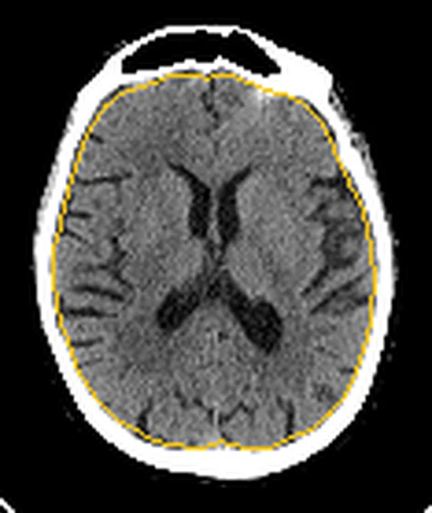
Aynı aksiyel düzeyde normal parankimde fokal düşük atenüasyon, halka kontrastlanması veya çevresel sulkus silinmesi beklenmez. Lateral ventrikül duvarı düzgün, ventrikül içi içerik homojen ve bazal sisternler açık görünür; şüpheli tarafı karşı hemisfer ve eş düzey normal referansla karşılaştır.
Normal BT ile kıyasla
Aynı bölgenin, kaynak veri setinde patoloji içermediği belirtilen bir BT incelemesini kesit kesit kaydırın; organ sınırlarını açarak anatomiyi hasta görüntüsüyle karşılaştırın. Küçük rastlantısal bulgular tümüyle dışlanmamıştır.
Kaynak: TotalSegmentator v2.01 veri seti (Wasserthal ve ark., Radiology: Artificial Intelligence 2023), CC BY 4.0, vaka s0103; organ sınırları veri setinin otomatik segmentasyonundan, birleştirilerek sadeleştirildi. Görüntüler 2,5 mm kesit aralığına yeniden örneklendi.
Ayırıcı tanı
- Nekrotik tümör
- kalın/nodüler kontrastlanan duvar oluşturabilir; BT görünümü apseyle örtüşür ve MR gerekir.
- Metastazlar da halka şeklinde kontrastlanan lezyonlar oluşturabilir ve ayırıcı tanıda değerlendirilir.
- Tümöral demiyelinizasyon
- açık halka biçimli kontrastlanma ve beyaz cevher yerleşimi apseyi taklit edebilir.
- Subdural ampiyem
- parankim içinde değil, dura boyunca ekstraaksiyel hilal biçimli koleksiyon şeklindedir.
Tuzaklar
- Erken serebritte kapsül henüz oluşmadığı için kontrastsız BT’de yalnız belirsiz ödem görülebilir; normal veya silik BT enfeksiyonu dışlamaz.
- Halka kontrastlanmasını tek başına apse kabul etme; nekrotik tümör ve diğer lezyonlarla örtüşür.
- Kontrastsız BT'de erken dönem bulgular sınırlı kalabileceğinden, şüphe durumunda MR ile tamamlama gerekebilir.
- Ventriküler sistemde pülanmatik deformite veya debris izlenirse ventrikülit/apsem açısından titizlikle değerlendirilmelidir.
Kendini dene
Kontrastsız BT’de fokal düşük atenüasyon ve çevresel ödem var; lezyon henüz kapsülsüz görünüyor. Hangi evre olasıdır?
Cevabı göster
Erken serebrit. Erken serebritte BT’de belirsiz düşük atenüasyon ve ödem görülebilir; belirgin kapsüler halka henüz gelişmemiş olabilir.
Apseye komşu ventrikülde yeni debris ve ependimal kontrastlanma saptanıyor. En olası komplikasyon nedir?
Cevabı göster
Ventrikülit. Ventrikül içi debris ve ependimal kontrastlanma enfeksiyonun ventrikül yüzeyine yayıldığını düşündürür. Diğer seçenekler ependim ve lümen bulgularını açıklamaz.
Kaynaklar
Bu sayfadaki 38 cümle ve 2 quiz sorusu aşağıdaki kaynaklardan alıntılarla tek tek karşılaştırıldı; 10 cümle bu karşılaştırmada düzeltildi (2026-10-08).
- Occult Bartonella Quintana Infective Endocarditis Incidentally Diagnosed During Pre-Transcatheter Aortic Valve Implantation (TAVI) Workup in a Patient Without Classical Risk Factorspmc.ncbi.nlm.nih.gov
- Acute otitis media complicated by mastoiditis and otogenic transverse-sigmoid sinus thrombosis in an adult: a case report on infectious pathogenesis and multidisciplinary therapypmc.ncbi.nlm.nih.gov
- Rapidly progressive subdural empyema secondary to acute pansinusitis complicated by superior sagittal sinus thrombosis in a young adult: a multidisciplinary case reportpmc.ncbi.nlm.nih.gov
- Enterococcus faecalis Aortic Valve Endocarditis Masquerading As Urinary Tract Infection: Diagnostic Delay, Septic Embolic Strokes, and a Fatal Therapeutic Impassepmc.ncbi.nlm.nih.gov
- Unusual Manifestations of Sinonasal Osteomas: A Narrative Reviewpmc.ncbi.nlm.nih.gov
- Bacterial orbital cellulitis - A review.pubmed.ncbi.nlm.nih.gov
- Orbital cellulitis.pubmed.ncbi.nlm.nih.gov
- [Orbital cellulitis].pubmed.ncbi.nlm.nih.gov
- Pott's puffy tumor: A comprehensive review of the literature.pubmed.ncbi.nlm.nih.gov
- Clostridium septicum Pneumocephalus.pubmed.ncbi.nlm.nih.gov
- Systematic review and case report: Intracranial complications of pediatric sinusitis.pubmed.ncbi.nlm.nih.gov
- Pott's puffy tumor in children: A review of the literature.pubmed.ncbi.nlm.nih.gov
- What is the optimal timing for surgery in infective endocarditis with cerebrovascular complications?pubmed.ncbi.nlm.nih.gov
- Role of imaging in the diagnosis of acute bacterial meningitis and its complications.pubmed.ncbi.nlm.nih.gov
- Too Much on Your "Plate"? Spectrum of Pathologies Involving the Tectal Plate.pubmed.ncbi.nlm.nih.gov
- Not quite Pott's puffy tumour.pubmed.ncbi.nlm.nih.gov
- Cranial hydatid abscess.pubmed.ncbi.nlm.nih.gov
- [Intracranial empyemas: Epidemiological, clinical, radiological and therapeutic aspects. Retrospective study of 100 observations].pubmed.ncbi.nlm.nih.gov
- Pediatric Meningococcal Meningitis Complicated by Cerebellar Abscess: A Case Report.pubmed.ncbi.nlm.nih.gov
- Brain abscess in Honduras: a five-year clinical and epidemiological study at Hospital Escuela.pubmed.ncbi.nlm.nih.gov
- From vesicles to ventricles: pediatric group a streptococcal meningitis post-varicella infection - a case report.pubmed.ncbi.nlm.nih.gov
- Brain Abscess Likely Secondary to Infective Endocarditis Mimicking High-Grade Glioma in a Patient with Pyelonephritis.pubmed.ncbi.nlm.nih.gov
- Navigating complex presentations of central nervous system tuberculosis: A two-case analysis.pubmed.ncbi.nlm.nih.gov
- Multifocal streptococcus intermedius abscess mimicking neurocysticercosis clinical and radiological findings: A case report and literature review.pubmed.ncbi.nlm.nih.gov
- Comparative Analysis of Magnetic Resonance Spectroscopy and Post-contrast Magnetic Resonance Imaging in the Evaluation of Intracranial Ring-Enhancing Lesions.pubmed.ncbi.nlm.nih.gov
- Gynecologic imaging errors and pitfalls: how to avoid misdiagnosis on pelvic MRI.pubmed.ncbi.nlm.nih.gov
- Intraventricular rupture of brain abscess: A two-center observational cohort study.pubmed.ncbi.nlm.nih.gov
- Suspected ruptured medulla oblongata abscess treated conservatively with a favorable outcome: A rare case report from Vietnam.pubmed.ncbi.nlm.nih.gov
- <i>Actinomyces urogenitalis</i> and <i>Peptoniphilus asaccharolyticus</i> brain abscess in an immunocompetent patient: A case report.pubmed.ncbi.nlm.nih.gov
- Neuroimaging of the Most Common Meningitis and Encephalitis of Adults: A Narrative Review.pubmed.ncbi.nlm.nih.gov
- Extrapulmonary tuberculosıs: an old but resurgent problem.pubmed.ncbi.nlm.nih.gov
- Multiple Brain Abscesses due to Streptococcus anginosus: Prediction of Mortality by an Imaging Severity Index Score.pubmed.ncbi.nlm.nih.gov
- Magnetic Resonance Imaging Features of Hepatic Hydatid Disease: A Pictorial Review with Emphasis on Atypical Presentations and Differential Diagnosis.pubmed.ncbi.nlm.nih.gov
- Collaborative Diagnostic Pathways for Unilateral Orbital Disease: A Clinicopathological Series.pubmed.ncbi.nlm.nih.gov
- Streptococcus anginosus group brain abscesses and subdural empyemas: exploratory compartment-specific phenotypes and discharge outcomes in a single-center retrospective cohort study.pubmed.ncbi.nlm.nih.gov
- Unveiling the Uncommon Ventriculitis in Tubercular Meningitis.pubmed.ncbi.nlm.nih.gov
- Complications of Otitis Media: Mastoiditis and an Intracranial Abscess Presenting as Behavioral Changes.pubmed.ncbi.nlm.nih.gov
- Listeria Cerebritis with Tumor Necrosis Factor Inhibition.pubmed.ncbi.nlm.nih.gov
- Encephalitis in Patients with COVID-19: A Systematic Evidence-Based Analysis.pubmed.ncbi.nlm.nih.gov
- Hemorrhagic brain abscess mimicking an intracranial neoplasm in invasive Klebsiella pneumoniae syndrome: illustrative case.pubmed.ncbi.nlm.nih.gov
- Spinal granulocytic sarcoma with both extradural and intradural involvement: a case report.pubmed.ncbi.nlm.nih.gov
- Evolution of neuroimaging findings in angioinvasive cerebral aspergillosis in a pediatric patient with leukemia during long-term observation.pubmed.ncbi.nlm.nih.gov
- Common and Uncommon Splenic Lesions: A Review.pubmed.ncbi.nlm.nih.gov
- Chronic expanding intracerebral hematoma: A case report.pubmed.ncbi.nlm.nih.gov
- Post-therapy imaging of adult brain tumours: patterns, pitfalls, and practical framework.pubmed.ncbi.nlm.nih.gov
- Central Nervous System Tuberculoma in Qatar: Clinical, Radiological, and Pathological Insights From a Rare Case With Literature Review.pubmed.ncbi.nlm.nih.gov
- Cerebral Abscess Masquerading as Tuberculoma in a Post-Renal Transplant Chronic Kidney Disease Patient: A Diagnostic and Therapeutic Challenge.pubmed.ncbi.nlm.nih.gov
- HIV Complicated with Talaromyces Marneffei Multisystem Infection: A Case Report and Literature Review.pubmed.ncbi.nlm.nih.gov
- Main Causes of Death in Piglets from Different Brazilian Nursery Farms Based on Clinical, Microbiological, and Pathological Aspects.pubmed.ncbi.nlm.nih.gov
- Magnetic resonance imaging of the orbit, Part 2: Characterization of orbital pathologies.pubmed.ncbi.nlm.nih.gov
- Advance in peptide-based drug development: delivery platforms, therapeutics and vaccines.pubmed.ncbi.nlm.nih.gov
- Ruptured intraventricular tuberculous brain abscess mimicking cystic neoplasm: a case report.pubmed.ncbi.nlm.nih.gov
- Smartphone Ocular Fundal Photography in the Diagnosis of Raised Intracranial Pressure: A Novel Adaptation to Neurosurgical Practice.pubmed.ncbi.nlm.nih.gov
- Rathke's cleft cyst: From history to molecular genetics.pubmed.ncbi.nlm.nih.gov
- Sellar, suprasellar, and parasellar masses: Imaging features and neurosurgical approaches.pubmed.ncbi.nlm.nih.gov
- Multimodality Imaging of Moyamoya Disease: A Practical Guide for Neuroradiologists Based on a Case Report.pubmed.ncbi.nlm.nih.gov
- All Three Supersystems-Nervous, Vascular, and Immune-Contribute to the Cortical Infarcts After Subarachnoid Hemorrhage.pubmed.ncbi.nlm.nih.gov
- Neurological manifestations and neuroimaging findings in patients with SARS-CoV2-a systematic review.pubmed.ncbi.nlm.nih.gov
- Polymicrobial brain abscesses: A complex condition with diagnostic and therapeutic challenges.pubmed.ncbi.nlm.nih.gov
- Multiple brain abscesses in a neonate: a rare case report along with review of literature.pubmed.ncbi.nlm.nih.gov
- Bacterial Meningoencephalitis in Newborns.pubmed.ncbi.nlm.nih.gov
- Candida parapsilosis intracerebral abscess and intralesional amphotericin B: a novel treatment approach to a rare infection. Illustrative case.pubmed.ncbi.nlm.nih.gov
- Spectrum of Neuroimaging Findings in CNS Lymphoproliferative Disorders.pubmed.ncbi.nlm.nih.gov
- Imaging diagnosis of cystic intraparenchymal brain neoplasms.pubmed.ncbi.nlm.nih.gov
- Aggressive metastatic parotid squamous cell carcinoma with pulmonary, cerebral, and hepatic metastases: A radiologic case report.pubmed.ncbi.nlm.nih.gov
- Predominantly cystic glioblastoma mimicking benign cystic lesions: A radiological diagnostic challenge.pubmed.ncbi.nlm.nih.gov
- A Case-Based Review of Cerebral Venous Infarcts With Perfusion Imaging and Comparison to Arterial Ischemic Stroke.pubmed.ncbi.nlm.nih.gov
- CT Imaging in Neurocritical Care.pubmed.ncbi.nlm.nih.gov
- Beyond the embolus: "do not miss" diffusion abnormalities of ischaemic and non-ischaemic neurological disease.pubmed.ncbi.nlm.nih.gov
- Multiplanar MRI approach to the differential diagnosis of central nervous system tumors and tumor-like lesions in patients with HIV: what radiologists need to know.pubmed.ncbi.nlm.nih.gov
- <sup>1</sup>H-MR Spectroscopy as a Decision-Support Tool in the Differential Diagnosis of Intracranial Lesions: A Real-World Bicentric Retrospective Cohort.pubmed.ncbi.nlm.nih.gov
- Pediatric Meningeal Diseases: What Radiologists Need to Know.pubmed.ncbi.nlm.nih.gov
- What every neuropathologist needs to know: Update on neuro infectious disease workups and consultation resources.pubmed.ncbi.nlm.nih.gov
- A systematic approach to MR spectroscopy of the brain: Indications, protocol, metabolites, and spectral analysis in neurologic diseases.pubmed.ncbi.nlm.nih.gov
- CT Angiography of the Upper Extremities: Review of Acute Arterial Entities.pubmed.ncbi.nlm.nih.gov
- Clinical Applications of Dual-Energy CT.pubmed.ncbi.nlm.nih.gov
- Patterns of injury and violence in Yaoundé Cameroon: an analysis of hospital data.pubmed.ncbi.nlm.nih.gov
- PCR-Confirmed Convexity Subarachnoid Neurocysticercosis with Negative Serology: A Case Report.pubmed.ncbi.nlm.nih.gov
- Ruptured intracranial dermoid cyst as an incidental finding: Case report.pubmed.ncbi.nlm.nih.gov
- A case report of bevacizumab-induced subcapsular renal hemorrhage in a patient with lung adenocarcinoma.pubmed.ncbi.nlm.nih.gov
- Invited review: Recommendations for reporting intervention studies on reproductive performance in dairy cattle: Improving design, analysis, and interpretation of research on reproduction.pubmed.ncbi.nlm.nih.gov
- Tumor-like Lesions in Primary Angiitis of the Central Nervous System: The Role of Magnetic Resonance Imaging in Differential Diagnosis.pubmed.ncbi.nlm.nih.gov
- Using non-invasive neuroimaging to enhance the care, well-being and experimental outcomes of laboratory non-human primates (monkeys).pubmed.ncbi.nlm.nih.gov
- Neuroimaging Patterns of Intracranial Infections: Meningitis, Cerebritis, and Their Complications.pubmed.ncbi.nlm.nih.gov
- Special Considerations in Pediatric Endoscopic Skull Base Surgery.pubmed.ncbi.nlm.nih.gov
- International Harmonization of Nomenclature and Diagnostic Criteria (INHAND): Nonproliferative and Proliferative Lesions of the Rabbit.pubmed.ncbi.nlm.nih.gov
- Tuberculoma mimicking en-plaque meningioma in a 45-year-old male: A case report.pubmed.ncbi.nlm.nih.gov
- Management of rare intraventricular empyema secondary to late onset neonatal meningitis in an infant: A case report.pubmed.ncbi.nlm.nih.gov
- Brain abscess mimicking a brain tumor only realized during surgery: A case report in a resource strained environment.pubmed.ncbi.nlm.nih.gov
- Acute otitis media complicated by mastoiditis and otogenic transverse-sigmoid sinus thrombosis in an adult: a case report on infectious pathogenesis and multidisciplinary therapy.pubmed.ncbi.nlm.nih.gov
- Occult Bartonella Quintana Infective Endocarditis Incidentally Diagnosed During Pre-Transcatheter Aortic Valve Implantation (TAVI) Workup in a Patient Without Classical Risk Factors.pubmed.ncbi.nlm.nih.gov
- Disseminated Melioidosis Complicated by Dural Venous Sinus Thrombosis: A Case Report.pubmed.ncbi.nlm.nih.gov
- Tumefactive Multiple Sclerosis Mimicking a High-Grade Glioma: A Case Report and Literature Review.pubmed.ncbi.nlm.nih.gov
- Tumefactive Demyelinating Lesion Mimicking Low-Grade Astrocytoma with a T2/FLAIR Mismatch Sign: A Case Report and Review of the Literature.pubmed.ncbi.nlm.nih.gov
- Vanishing brain space-occupying lesion in a child: illustrative case.pubmed.ncbi.nlm.nih.gov
- Tumefactive Multiple Sclerosis Mimicking an Intracranial Mass in a 34-Year-Old Man: A Case Report.pubmed.ncbi.nlm.nih.gov
- Posterior Fossa Ring-Enhancing Lesions in the Adult Immunocompetent Host: Illustrative Cases, Systematic Review, and Proposed Diagnostic Algorithm.pubmed.ncbi.nlm.nih.gov
- [⁶⁸Ga]Ga-FAPI-46 PET/CT imaging in neuromelioidosis: a case series demonstrating potential utility in diagnosis and response assessment.pubmed.ncbi.nlm.nih.gov
- Treatment of brain abscess rupturing into ventricle: a case report and literature review.pubmed.ncbi.nlm.nih.gov
- Influence of laboratory and radiographic parameters on the clinical presentation and outcome of surgically treated patients with primary brain abscesses.pubmed.ncbi.nlm.nih.gov
Eğitim amaçlıdır; hasta başında tanı ve tedavi kararının yerine geçmez. İçerik yapay zekâ destekli bir süreçle güncel literatüre göre cümle cümle kaynaklarla karşılaştırıldı, ancak uzman radyolog incelemesinden geçmedi. Klinik kararlarda güncel kılavuzları, kurum protokollerini ve radyoloji raporunu esas alın.