
Özet
İntrakraniyal kalsifikasyonlar kontrastsız BT’de belirginleşir; çocukta yaş, simetri, anatomik yerleşim ve dağılım fizyolojik odakları patolojiden ayırmaya yardım eder. Pineal bez, habenula, koroid pleksus ve dural katlantılar tipik fizyolojik yerlerdir. Parankim içi, damar boyunca veya yaygın kalsifikasyonlar enfeksiyöz, vasküler, metabolik ya da neoplastik nedenleri gündeme getirir. Beklenmedik dağılımı normal varyant saymak altta yatan hastalığın gözden kaçmasına neden olabilir.
Faz ve pencere
- Kontrastsız tanısal
- Kalsiyum yüksek atenüasyonlu odaklar olarak en iyi kontrastsız seride ayırt edilir; kontrastlı görüntü damar veya dura ile ilişkisini netleştirebilse de fizyolojik kalsifikasyon sorusunda rutin faz değildir.
Önerilen pencereler: Beyin (G 80 / M 40), Kemik (G 1800 / M 400).
BT bulguları
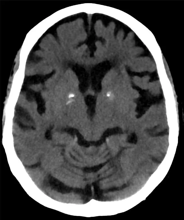
- Pineal bez ve habenula çevresindeki kompakt orta hat kalsifikasyonu yaşla birlikte görülebilen fizyolojik dağılımdır.
- Koroid pleksus kalsifikasyonu lateral ventriküllerin atriumlarında, çoğunlukla iki taraflı ve simetrik izlenir.
- Falks ve tentoryum boyunca ince çizgisel kalsifikasyonlar dural yerleşimlidir.
- Bazal gangliyonlarda iki taraflı, simetrik mineralizasyon parankimal dağılımdır; klinik/metabolik bağlamla birlikte yorumlanır.
- Girusları izleyen çizgisel veya giriform kalsifikasyonlar kortikal yüzey boyunca dağılım gösterir.
- Tek taraflı damar hattı boyunca serpijinöz kalsifikasyon kronik vasküler lezyon veya malformasyonla ilişkili olabilir.
- Kitle içinde kümeli, kaba veya düzensiz kalsifikasyon neoplastik ya da gelişimsel taklitçileri düşündürür.
Ölçütler ve sınıflamalar
- Dağılıma göre kalsifikasyon paterni
- Fizyolojik/yaşa bağlı; parankimal; vasküler; neoplastik veya enfeksiyöz dağılımlar anatomik yer ve morfolojiyle tarif edilir. Bu bir BT raporlama yaklaşımıdır, tek başına hastalık derecelemesi değildir.
Normalde
Aynı düzeyde normal BT’de pineal odak orta hatta, koroid pleksus odakları ventrikül atriumlarında ve dural mineralizasyon falks/tentoryum boyunca sınırlı görünür. Simetriyi, kalsifikasyonun anatomik kompartımanını ve çevresinde kitle, ödem ya da ventrikül deformasyonu olup olmadığını karşılaştır.
Normal BT ile kıyasla
Aynı bölgenin, kaynak veri setinde patoloji içermediği belirtilen bir BT incelemesini kesit kesit kaydırın; organ sınırlarını açarak anatomiyi hasta görüntüsüyle karşılaştırın. Küçük rastlantısal bulgular tümüyle dışlanmamıştır.
Kaynak: TotalSegmentator v2.01 veri seti (Wasserthal ve ark., Radiology: Artificial Intelligence 2023), CC BY 4.0, vaka s0103; organ sınırları veri setinin otomatik segmentasyonundan, birleştirilerek sadeleştirildi. Görüntüler 2,5 mm kesit aralığına yeniden örneklendi.
Ayırıcı tanı
- Konjenital enfeksiyon
- periventriküler kalsifikasyonlar ve eşlik eden beyin hacim/gelişim değişiklikleri tanıyı destekleyebilir.
- Sturge–Weber sendromu
- kortikal giriform kalsifikasyon, çoğunlukla tek hemisferde hacim kaybı ve sulkal belirginlikle birliktedir.
- Tüberoskleroz
- subependimal nodül ve kortikal tüberlerde kalsifikasyon görülebilir.
- Pineal bölge tümörü
- bezin beklenen küçük orta hat odağından farklı olarak kitle etkisi veya yer değiştirmiş kalsifikasyon olabilir.
- Arteriovenöz malformasyon
- serpijinöz damar yapıları ve ilişkili parankimal kalsifikasyonlar eşlik edebilir.
Tuzaklar
- Koroid pleksus kalsifikasyonunu intraventriküler kanama sanma; atriumlarda çift taraflı benzer odak ve kan ürünüyle uyumsuz dağılımı kontrol et.
- Pineal orta hat kalsifikasyonunun yer değiştirmesi kitle etkisi ipucu olabilir; odak çevresindeki pineal bölgeyi ve ventrikülleri izle.
- Bazal gangliyon mineralizasyonunu yalnız BT yoğunluğuna bakarak fizyolojik sayma; simetri, yaygınlık, yaş ve klinik bağlamla eşleştir.
- İntrakraniyal kalsifikasyonları BT’de parankim veya damar içindeki yerleşimlerine ve dağılımlarına göre değerlendir.
Kendini dene
İki taraflı atrial yerleşimli, koroid pleksus içinde sınırlı odakların yorumu en olası nedir?
Cevabı göster
Fizyolojik kalsifikasyon. Atriumlardaki simetrik koroid pleksus kalsifikasyonları tipik fizyolojik dağılımdır. Kanama genellikle pleksusla sınırlı, eşlenik mineralize odaklar şeklinde görünmez.
Tek hemisferde kortikal girusları izleyen kalsifikasyon hangi taklitçiyle uyumludur?
Cevabı göster
Sturge–Weber. Tek taraflı giriform kortikal kalsifikasyon Sturge–Weber paternini düşündürür. Tüberoskleroz daha çok kortikal tüberler/subependimal nodüllerle, konjenital enfeksiyon ise sıklıkla periventriküler dağılımla tanınır.
Kaynaklar
Bu sayfadaki 37 cümle ve 2 quiz sorusu aşağıdaki kaynaklardan alıntılarla tek tek karşılaştırıldı; 1 cümle bu karşılaştırmada düzeltildi (2026-10-08).
- Imaging and physiology across the high–low cerebrospinal fluid pressure spectrum: Navigating diagnostic uncertainty in headache practicepmc.ncbi.nlm.nih.gov
- Imaging findings of diabetic complications from head to toe: a comprehensive radiologic reviewpmc.ncbi.nlm.nih.gov
- Spontaneous Intracranial Hypotension and CSF-Venous Fistulae: from Diagnostic Imaging to Interventional Managementpmc.ncbi.nlm.nih.gov
- Monogenic Mimics of Neuroinflammatory Phenotypes in Children and Young Adultspmc.ncbi.nlm.nih.gov
- Advances of MR imaging in glioma: what the neurosurgeon needs to knowpmc.ncbi.nlm.nih.gov
- Intracranial calcifications in childhood: Part 2.pubmed.ncbi.nlm.nih.gov
- Ossifying fibroma of the ethmoid and sphenoid sinuses: A report of a rare case and literature review.pubmed.ncbi.nlm.nih.gov
- Guidelines for standard operation of imaging modalities in orbital diseases (2024).pubmed.ncbi.nlm.nih.gov
- Neuroimaging of pediatric tumors of the sellar region-A review in light of the 2021 WHO classification of tumors of the central nervous system.pubmed.ncbi.nlm.nih.gov
- Physiological Intracranial Calcifications in Children: A computed tomography-based study.pubmed.ncbi.nlm.nih.gov
- Intracranial calcifications in childhood: Part 1.pubmed.ncbi.nlm.nih.gov
- Intracranial Calcification Associated with 3-Methylcrotonyl-CoA Carboxylase Deficiency.pubmed.ncbi.nlm.nih.gov
- Paediatric cranial ultrasound: abnormalities of the brain in term neonates and young infants.pubmed.ncbi.nlm.nih.gov
- Pediatric CLIPPERS: Expanding the Neuroimaging Spectrum of a Rare Steroid-Responsive Neuroinflammatory Syndrome-A Systematic Review.pubmed.ncbi.nlm.nih.gov
- Insights from a novel monogenic autoinflammatory disease: overview of a multicentric European cohort of 38 patients with COPA syndrome.pubmed.ncbi.nlm.nih.gov
- Multimodal imaging in neuro-ophthalmology.pubmed.ncbi.nlm.nih.gov
- Study of Intraoperative Squash Cytology of Intracranial and Spinal Cord Lesions with Histopathological Correlation.pubmed.ncbi.nlm.nih.gov
- Sinonasal and skull base phosphaturic mesenchymal tumours: a case series and narrative review.pubmed.ncbi.nlm.nih.gov
- Hyperthyroidism-Induced Dural Venous Sinus Thrombosis: A Case Report.pubmed.ncbi.nlm.nih.gov
- Change in Emergency Department Length of Stay following Routine Adoption of Dual-Energy CT to Differentiate Intracranial Hemorrhage from Calcification.pubmed.ncbi.nlm.nih.gov
- Spot Sign in Intracerebral Hemorrhage: Critical Reappraisal and Future Clinical Implications.pubmed.ncbi.nlm.nih.gov
- Fetal intracranial hemorrhage and infarct: Main sonographic and MRI characteristics: A review article.pubmed.ncbi.nlm.nih.gov
- Intracranial calcifications on CT: an updated review.pubmed.ncbi.nlm.nih.gov
- Cranial ultrasound for beginners.pubmed.ncbi.nlm.nih.gov
- Artificial Intelligence-Assisted Segmentation of a Falx Cerebri Calcification on Cone-Beam Computed Tomography: A Case Report.pubmed.ncbi.nlm.nih.gov
- Meningioma: Molecular Updates from the 2021 World Health Organization Classification of CNS Tumors and Imaging Correlates.pubmed.ncbi.nlm.nih.gov
- Primary central nervous system lymphoma: Imaging features and differential diagnosis.pubmed.ncbi.nlm.nih.gov
- Rare Presentation of Homozygous S<i>LC20A2</i> Mutations Causing Intra-Arterial Cerebral Vasculopathy and Stroke in Infancy: Case Report and Review of the Literature.pubmed.ncbi.nlm.nih.gov
- Prosthodontic rehabilitation in Fahr's syndrome: clinical challenges and oral manifestations.pubmed.ncbi.nlm.nih.gov
- Basal Ganglia Calcifications for Nephrologists: Fahr/Primary Familial Brain Calcification (PFBC) at the Interface of Chronic Kidney Disease-Mineral and Bone Disorder (CKD-MBD) and Hypoparathyroidism.pubmed.ncbi.nlm.nih.gov
- A double threat: CNS toxoplasmosis and CMV encephalitis in a heart transplant recipientpubmed.ncbi.nlm.nih.gov
- Longitudinal Neuroimaging of a Pediatric Patient With Sturge-Weber Syndrome: From Birth to Adolescence.pubmed.ncbi.nlm.nih.gov
- Accidental finding of Sturge-Weber syndrome presented to the emergency department with persistent seizures: an unusual case report from Somalia.pubmed.ncbi.nlm.nih.gov
- Diagnosis and Management of Parotid Gland Arteriovenous Malformation: A Case Report.pubmed.ncbi.nlm.nih.gov
- A compendium of male breast imaging: The road less traveled.pubmed.ncbi.nlm.nih.gov
- Intracranial cerebrovascular lesions on T2-weighted magnetic resonance imaging.pubmed.ncbi.nlm.nih.gov
- Contemporary Clinical Definitions, Differential Diagnosis, and Novel Predictive Tools for Renal Cell Carcinoma.pubmed.ncbi.nlm.nih.gov
- Heart Disease and Stroke Statistics-2021 Update: A Report From the American Heart Association.pubmed.ncbi.nlm.nih.gov
- Spontaneous bacteremia and spontaneous bacterial peritonitis share similar prognosis in patients with cirrhosis: a cohort study.pubmed.ncbi.nlm.nih.gov
- Head Trauma Mechanisms and Neuroimaging Findings in Psychiatric Inpatients Referred for Neurosurgical Consultation.pubmed.ncbi.nlm.nih.gov
- Unique presentation of superficial siderosis of the central nervous system following pituitary tumor surgery: a case report and literature review.pubmed.ncbi.nlm.nih.gov
- Telovelar Approach for Fourth-Ventricular Epidermoid Cyst: Anatomical Respect, Functional Recovery, and Long-Term Stability.pubmed.ncbi.nlm.nih.gov
- Commentary: an eye on PET quantification.pubmed.ncbi.nlm.nih.gov
- High-affinity alphavbeta3 integrin targeted optical probe as a new imaging biomarker for early atherosclerosis: initial studies in Watanabe rabbits.pubmed.ncbi.nlm.nih.gov
- Prenatal diagnosis and outcomes of fetal cardiac tumors: a 10-year single-center study of 81 cases.pubmed.ncbi.nlm.nih.gov
- Carotid plaque vulnerability and circle of Willis anatomy predict ipsilateral brain infarcts and long-term mortality in carotid endarterectomy patients.pubmed.ncbi.nlm.nih.gov
- Non-Atherosclerotic and Genetic Intracranial Stenoses: Diagnostic Challenges and Emerging Genetic Insights.pubmed.ncbi.nlm.nih.gov
- Case Report: The cystic masquerade in type 1 Von Hippel-Lindau disease: coexisting clear cell renal cell carcinoma and an imaging-suspected pancreatic neuroendocrine tumor.pubmed.ncbi.nlm.nih.gov
- Comparative Analysis of Magnetic Resonance Spectroscopy and Post-contrast Magnetic Resonance Imaging in the Evaluation of Intracranial Ring-Enhancing Lesions.pubmed.ncbi.nlm.nih.gov
- Low-flow vascular malformations without arteriovenous shunting of the central nervous system: a pictorial review.pubmed.ncbi.nlm.nih.gov
- Congenital Toxoplasmosis With Atypical Neuroimaging: Hydrocephalus and Bilateral Chorioretinitis Without Intracranial Calcifications.pubmed.ncbi.nlm.nih.gov
- Neonatal Aicardi-Goutières syndrome presenting with macrophage activation syndrome-like hyperinflammation and severe congenital glaucoma: a case report.pubmed.ncbi.nlm.nih.gov
- Sturge-Weber Syndrome: A Narrative Review of Clinical Presentation and Updates on Management.pubmed.ncbi.nlm.nih.gov
- GNAQ/GNA11 Mosaicism Is Associated with Abnormal Serum Calcium Indices and Microvascular Neurocalcification.pubmed.ncbi.nlm.nih.gov
- Prenatal magnetic resonance imaging findings of tuberous sclerosis complex in fetuses.pubmed.ncbi.nlm.nih.gov
- Delayed Diagnosis of Tuberous Sclerosis Complex: A Case Report of Abdominal Distension, Pain, and Severe Anemia.pubmed.ncbi.nlm.nih.gov
- Transmantle sign-like calcified radial lesion on CT serves as a potential imaging feature for epileptogenic foci in tuberous sclerosis complex: a Case Report.pubmed.ncbi.nlm.nih.gov
- Spinal granulocytic sarcoma with both extradural and intradural involvement: a case report.pubmed.ncbi.nlm.nih.gov
- Primary intracranial adenocarcinoma associated with an epidermoid cyst: a case report and literature review.pubmed.ncbi.nlm.nih.gov
- Case Report: A case series of Lhermitte-Duclos disease with surgical intervention.pubmed.ncbi.nlm.nih.gov
- Vascular Malformations and Tumors: A Review of Classification and Imaging Features for Cardiothoracic Radiologists.pubmed.ncbi.nlm.nih.gov
- Unruptured Arteriovenous Malformations in the Multidetector Computed Tomography Era: Frequency of Detection and Predictable Failures.pubmed.ncbi.nlm.nih.gov
- AOSNP-ADAPTR resource level-based recommendations on practical diagnostic strategies for medulloblastomas and other embryonal tumors.pubmed.ncbi.nlm.nih.gov
- Hydrocephalus: historical analysis and considerations for treatment.pubmed.ncbi.nlm.nih.gov
- Calcified Pilocytic Astrocytomas and Calcifying Pseudoneoplasms of the Neuraxis: A Diagnostic Challenge.pubmed.ncbi.nlm.nih.gov
- Primary adrenal mature cystic teratoma: A rare retroperitoneal tumor with two decades of indolent growth.pubmed.ncbi.nlm.nih.gov
- Combined with chemotherapy and radiotherapy is effective in improving prognosis: a case of primary mediastinal embryonal carcinoma.pubmed.ncbi.nlm.nih.gov
- Basal Ganglia Calcification With Treatment-Resistant Hypocalcaemia and Persistent Psychosis in an 18-Year-Old: A Case Report.pubmed.ncbi.nlm.nih.gov
- Amyloid-induced mineralization: From biological systems to biomimetic materials.pubmed.ncbi.nlm.nih.gov
- Case Report: Developmental-like skeletal deformities and transient osteosclerosis as rare presentations of primary hyperparathyroidism.pubmed.ncbi.nlm.nih.gov
- Sphenoid fungal rhinosinusitis across the disease spectrum: a case series of non-invasive fungal ball and fatal acute invasive fungal rhinosinusitis.pubmed.ncbi.nlm.nih.gov
- Cardiovascular Outcomes Associated with Romosozumab Versus Denosumab in Chronic Kidney Disease.pubmed.ncbi.nlm.nih.gov
- Diagnostic imaging and emerging image-guided neurointervention in brain tumors: a narrative review.pubmed.ncbi.nlm.nih.gov
- Cerebrospinal Fluid Shunts: An Updated Radiologic Review of Devices, Malfunctions, and Complications.pubmed.ncbi.nlm.nih.gov
- Society for Cardiovascular Magnetic Resonance 2024 Cases of SCMR case series.pubmed.ncbi.nlm.nih.gov
- Pediatric Meningeal Diseases: What Radiologists Need to Know.pubmed.ncbi.nlm.nih.gov
- Storage diseases with hypertrophic cardiomyopathy phenotype.pubmed.ncbi.nlm.nih.gov
- Long-term safety and effectiveness of the "OptEase" vena cava filter.pubmed.ncbi.nlm.nih.gov
- Leptomeningeal metastasis with sturge-weber syndrome-like gyriform calcification on imaging: a case report.pubmed.ncbi.nlm.nih.gov
- Undiagnosed Epileptic Case Since Childhood of Sturge-Weber Syndrome: First Case Report from Somalia.pubmed.ncbi.nlm.nih.gov
- Imaging and physiology across the high-low cerebrospinal fluid pressure spectrum: Navigating diagnostic uncertainty in headache practice.pubmed.ncbi.nlm.nih.gov
- Spontaneous Intracranial Hypotension and CSF-Venous Fistulae: from Diagnostic Imaging to Interventional Management.pubmed.ncbi.nlm.nih.gov
- Monogenic Mimics of Neuroinflammatory Phenotypes in Children and Young Adults: An Evolving Landscape.pubmed.ncbi.nlm.nih.gov
- Artifact-Controlled Multi-Vertebral Transfer Learning Ensemble for Patient-Level Three-Class Osteoporosis Screening Using Thoracoabdominal CT-Derived Lumbar Images.pubmed.ncbi.nlm.nih.gov
- Extended Transoral Resection of Anterior Commissure Tumors in a Canine Model: Impact on Laryngeal Framework, Glottis, and Vocal Function.pubmed.ncbi.nlm.nih.gov
- Voxel-wise deep learning segmentation of hydroxyapatite and iodine in spectral photon-counting CT: A quantitative phantom study.pubmed.ncbi.nlm.nih.gov
- ESR Essentials: imaging in nasal obstruction and epistaxis-practice recommendations by the European Society of Head and Neck Radiology.pubmed.ncbi.nlm.nih.gov
- Simultaneous Burr hole drainage and middle meningeal artery occlusion through a single incision for chronic subdural hematoma guided by 3D reconstruction.pubmed.ncbi.nlm.nih.gov
- Sphenoid Temporal Meningioma With Granular Cell Features and Extracranial Infratemporal Extension.pubmed.ncbi.nlm.nih.gov
- Fatal Injuries Caused by Falling Trees: Correlation Between Postmortem Computed Tomography and Forensic Autopsy Findings in Eight Cases.pubmed.ncbi.nlm.nih.gov
- Theragnostic Use of Radiolabelled Dota-Peptides in Meningioma: From Clinical Demand to Future Applications.pubmed.ncbi.nlm.nih.gov
- Orbital tumours and tumour-like lesions: exploring the armamentarium of multiparametric imaging.pubmed.ncbi.nlm.nih.gov
- Rare anatomical localizations of hydatid cysts (2020-2025): Narrative review of presentation, imaging, and management.pubmed.ncbi.nlm.nih.gov
Eğitim amaçlıdır; hasta başında tanı ve tedavi kararının yerine geçmez. İçerik yapay zekâ destekli bir süreçle güncel literatüre göre cümle cümle kaynaklarla karşılaştırıldı, ancak uzman radyolog incelemesinden geçmedi. Klinik kararlarda güncel kılavuzları, kurum protokollerini ve radyoloji raporunu esas alın.