Özet
Şantlı çocukta kusma, baş ağrısı, irritabilite veya bilinç değişikliği, BOS drenajının bozulduğunu gösterebilir. Kontrastsız BT, ventrikül kalibresini, kateter ucunun yerini ve akut kitle etkisini hızlıca gösterir; önceki inceleme ile kıyas tanısal değeri artırır. Normal boyutlu ventriküller şant işlev bozukluğunu tek başına dışlamaz. Tıkanma veya ayrışma gözden kaçarsa hidrosefali ve basınç artışı ilerleyebilir.
Faz ve pencere
- Kontrastsız tanısal
- BT’de ventrikül boyutu, şantın konumu ve bileşenlerinin bütünlüğü değerlendirilir; eşlik edebilen transependimal ödem, subgaleal sıvı ve kateter boyunca ödem de aranır. Şantın mekanik değerlendirmesinde BT, ventrikül boyutunu, şantın konumunu ve bileşenlerinin bütünlüğünü değerlendirmede kullanılır.
Önerilen pencereler: Beyin (G 80 / M 40), Kemik (G 1800 / M 400), Yumuşak doku (G 400 / M 40).
BT bulguları
- Ventriküler kateter ucu BOS içeren ventrikül lümeninde değilse malpozisyon veya migrasyon düşünülür.
- Yeni ventrikül genişlemesi, temporal hornların belirginleşmesi ve sulkusların silinmesi önceki incelemeye göre tıkanmayı destekler.
- Periventriküler düşük atenüasyon transependimal BOS geçişiyle uyumlu olabilir.
- Ventriküllerin slit benzeri kollapsı aşırı drenajla ilişkili olabilir; klinik ve önceki görüntülerle eşleştirilir.
- Kateterin delik taşıyan segmentinin BOS boşluğunda bulunması açıklığı için önemlidir; koroid pleksusa yakınlık tıkanmaya yol açabilir.
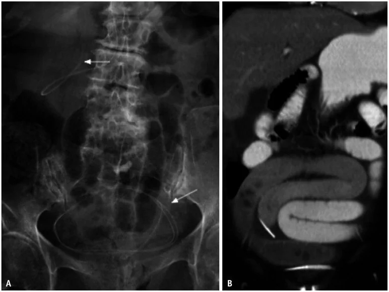
- Şant hattında kesinti, kıvrılma, bağlantı ayrışması veya cilt altına geri çekilme aranır.
- Kateter yolu boyunca subgaleal sıvı, şant ayrışması ya da BOS kaçağına eşlik edebilir.
Normalde
Aynı düzeydeki önceki BT’de frontal hornlar, üçüncü ventrikül, temporal hornlar ve sulkusların görünümüyle yan yana kıyasla. Normal çalışan şantta kateter ucu ventrikül içinde izlenebilir; ventrikül kalibresindeki değişim hastanın kendi bazal incelemesine göre yorumlanır.
Normal BT ile kıyasla
Aynı bölgenin, kaynak veri setinde patoloji içermediği belirtilen bir BT incelemesini kesit kesit kaydırın; organ sınırlarını açarak anatomiyi hasta görüntüsüyle karşılaştırın. Küçük rastlantısal bulgular tümüyle dışlanmamıştır.
Kaynak: TotalSegmentator v2.01 veri seti (Wasserthal ve ark., Radiology: Artificial Intelligence 2023), CC BY 4.0, vaka s0103; organ sınırları veri setinin otomatik segmentasyonundan, birleştirilerek sadeleştirildi. Görüntüler 2,5 mm kesit aralığına yeniden örneklendi.
Ayırıcı tanı
- Şant tıkanması
- önceki incelemeye göre ventrikül genişlemesi ve transependimal ödem eşlik edebilir.
- Şant aşırı drenajı slit ventriküller, ekstraaksiyel sıvı koleksiyonları veya subdural higromalarla ilişkili olabilir.
- Şant enfeksiyonunda kontrastlı BT veya MR tanısal değildir; ventrikülit varsa düzensiz ependimal kontrastlanma görülebilir.
- Şantlı hastada ventrikül boyutlarındaki artış, önceki görüntüleme ile karşılaştırıldığında şant arızasını düşündürür; ventriküllerde belirgin büyüme yoksa altta yatan hidrosefali progresyonu gibi alternatif tanılar değerlendirilir.
Tuzaklar
- Önceki görüntülemeye göre ventrikül boyutlarında artış olmaması şant arızasını tek başına dışlamaz; slit ventrikül sendromu da göz önünde bulundurulmalıdır.
- Önceki görüntülemeler, ventrikül boyutlarındaki değişimi değerlendirmek için önemlidir; karşılaştırmayı aynı düzeydeki görüntüler üzerinden yap.
- Kateter ucu ventrikül içinde görünse bile açıklığı BT ile kanıtlanamaz; yalnız konum ve ikincil işaretler değerlendirilir.
Kendini dene
Şantlı çocukta klinik kötüleşme var; ventriküller bazal BT’ye göre aynı. Bu bulgu neyi gösterir?
Cevabı göster
Arıza dışlanmaz. Bazal ventrikül boyutu değişmeden şant tıkanması görülebilir; kateter konumu ve ikincil işaretler incelenir. Diğer seçenekler tek başına ventrikül karşılaştırmasından çıkarılamaz.
Ventriküler şantın delik taşıyan segmentlerinin uzun dönem açıklığını koruması için nerede bulunması gerekir?
Cevabı göster
BOS boşluğunda. İşlevini sürdüren şantta kateterin delik taşıyan segmentlerinin BOS boşluğunda bulunması gerekir.
İlgili konular
- Posterior fossa tümörleri: medulloblastom, ependimom ve pilositik astrositom
- Pediatrik beyin tümörleri: BT’de kitle ve kalsifikasyon
Kaynaklar
Bu sayfadaki 33 cümle ve 2 quiz sorusu aşağıdaki kaynaklardan alıntılarla otomatik olarak karşılaştırıldı; 8 cümle bu karşılaştırmada düzeltildi (2026-10-09).
- Cerebrospinal Fluid Shunts: An Updated Radiologic Review of Devices, Malfunctions, and Complicationspmc.ncbi.nlm.nih.gov
- Management of Ventriculo-Peritoneal Shunts in the Paediatric Populationpmc.ncbi.nlm.nih.gov
- Papilledema: A review of etiology, pathophysiology, diagnosis, and management.pubmed.ncbi.nlm.nih.gov
- Cerebral Venous Thrombosis Due to Overdrainage in a Patient With Normal Pressure Hydrocephalus: A Case Report.pubmed.ncbi.nlm.nih.gov
- Obstruction of Ventriculoperitoneal Shunt After Myelography-Report of a Unique Case and Its Treatment.pubmed.ncbi.nlm.nih.gov
- A Case of Discontinued Proximal Limb of a Ventriculoperitoneal Shunt With Patent Fibrous Tract.pubmed.ncbi.nlm.nih.gov
- Preoperative risk and postoperative outcome from subdural fluid collections in African infants with postinfectious hydrocephalus.pubmed.ncbi.nlm.nih.gov
- From shunt to gut: managing a ventriculoperitoneal shunt-associated abdominal pseudocyst causing bowel obstruction - a case report.pubmed.ncbi.nlm.nih.gov
- Cerebrospinal Fluid Shunts: An Updated Radiologic Review of Devices, Malfunctions, and Complications.pubmed.ncbi.nlm.nih.gov
- Trapped Fourth Ventricle in a Pediatric Patient With a History of Post-Traumatic Hydrocephalus and Ventriculoperitoneal Shunt: A Case Report and Literature Review.pubmed.ncbi.nlm.nih.gov
- Incidence and management of overdrainage in pediatric hydrocephalus patients exclusively treated with modern gravitational VP shunt valves.pubmed.ncbi.nlm.nih.gov
- Radiologic Assessment of Malpositioned Indwelling Catheters and Medical Devices: A Retrospective Case Series With Pictorial Review.pubmed.ncbi.nlm.nih.gov
- Transcaval Access and Closure Best Practices.pubmed.ncbi.nlm.nih.gov
- Pseudocyst in Neck: A Case Report on Rare Complication of Ventriculoperitoneal Shunt.pubmed.ncbi.nlm.nih.gov
- Lumbar puncture-induced negative-pressure hydrocephalus in a child with a ventriculoperitoneal shunt and ventricular obstruction: illustrative case.pubmed.ncbi.nlm.nih.gov
- Occipital shuntalgia: Rethinking post-shunt occipital headache etiology and care.pubmed.ncbi.nlm.nih.gov
- α-Synuclein Pathology in Idiopathic Normal Pressure Hydrocephalus: Evidence for Concomitant Synucleinopathy and Clinical Implications.pubmed.ncbi.nlm.nih.gov
- Amenorrhea Induced by Hydrocephalus Due to Tuberculous Meningitis: A Rare Case Report and Literature.pubmed.ncbi.nlm.nih.gov
- Continuum of Irreversible and Potentially Reversible Traumatic Brain Injury Sequelae: Axonal Injury and Hydrocephalus.pubmed.ncbi.nlm.nih.gov
- Point-of-Care Ultrasound Assessment of Ventriculoperitoneal Shunt Patency: A Case Report.pubmed.ncbi.nlm.nih.gov
- Life-threatening exacerbation of a chronic porencephalic cyst in an adult: A case report and literature review.pubmed.ncbi.nlm.nih.gov
- Inferior phrenic artery-to-pulmonary artery fistula after surgery for catamenial pneumothorax: a case report.pubmed.ncbi.nlm.nih.gov
- Predominance of ventricular catheter misplacement with anterior approach in neonates and infants.pubmed.ncbi.nlm.nih.gov
- Etiology and Risk Factors for Shunt Revision in Adult Hydrocephalus: A Single-Center Retrospective Cohort Study.pubmed.ncbi.nlm.nih.gov
- The Wire Twisting/Locking Technique to Facilitate Precise PDA Stent Delivery in Neonates with Ductal-Dependent Pulmonary Blood Flow.pubmed.ncbi.nlm.nih.gov
- Paediatric cranial ultrasound: abnormalities of the brain in term neonates and young infants.pubmed.ncbi.nlm.nih.gov
- Persistent neurological symptoms and elevated intracranial pressures in a previously healthy host with cryptococcal meningitis.pubmed.ncbi.nlm.nih.gov
- Imaging Characteristics of Colloid Cysts: A Case Series Correlating Cyst Size and Location With Associated Acute Hydrocephalus.pubmed.ncbi.nlm.nih.gov
- Magnetic resonance imaging of primary central nervous system neoplasms of dogs and cats: Basic concepts for diagnostic pathologists.pubmed.ncbi.nlm.nih.gov
- Lipomatous Dural Metaplasia Associated With Chronic Ventricular Shunting: A Report of Two Cases.pubmed.ncbi.nlm.nih.gov
- Shunt-related cerebrospinal fluid overdrainage - a multicentre consensus definition.pubmed.ncbi.nlm.nih.gov
- Diagnostic Challenges and Surgical Outcomes of Miyazaki Syndrome: A Report of Two Cases and a Systematic Review.pubmed.ncbi.nlm.nih.gov
- Non-invasive intracranial pressure monitoring in management of slit ventricle syndrome-case report.pubmed.ncbi.nlm.nih.gov
- Imaging of Cerebrospinal Fluid Shunts and Their Complications.pubmed.ncbi.nlm.nih.gov
- Case Report: Endoscopic-assisted cistern ostomy with Ommaya reservoir insertion for trapped temporal horn: surgical technique and case illustration.pubmed.ncbi.nlm.nih.gov
- Flexible endoscopy for removal of a migrated fractured ventricular catheter in an adult: A case report and technical note.pubmed.ncbi.nlm.nih.gov
- Ultra-low dose protocol on photon-counting computed tomography as an alternative to radiographic shunt series in the diagnosis of mechanical ventriculoperitoneal shunt complications - an ex vivo phantom study for children and adults.pubmed.ncbi.nlm.nih.gov
- Ventriculoperitoneal shunt distal dysfunction due to peritoneal malabsorption: etiologies and management strategies-An institutional review.pubmed.ncbi.nlm.nih.gov
- A rare complication of autologous cranioplasty: symptomatic epidural fluid collection causing mass effect requiring removal of bone flap.pubmed.ncbi.nlm.nih.gov
- Ultrasound-guided needle technique as a relaxation strategy for early cranioplasty: illustrative case.pubmed.ncbi.nlm.nih.gov
- Lumboperitoneal shunting as a rescue strategy in the management of complex cranial wound conditions.pubmed.ncbi.nlm.nih.gov
- <i>Streptococcus suis</i> meningitis complicated with communicating hydrocephalus: complete clinical and radiological resolution with non-neurosurgical management-A case report.pubmed.ncbi.nlm.nih.gov
- Inflammation Meets Ventricular Dynamics: Combined Hematologic and Radiologic Predictors of Shunt-Dependent Hydrocephalus in Endovascularly Treated Patients with Aneurysmal Subarachnoid Hemorrhage.pubmed.ncbi.nlm.nih.gov
- Atrial volume reduction correlates with early improvement in hemorrhage-associated normal pressure hydrocephalus-a 3D computed tomography volumetric study.pubmed.ncbi.nlm.nih.gov
- Acute Neurological Deterioration in a Child with Shunt-Dependent Post-Hemorrhagic Hydrocephalus: A Case Report.pubmed.ncbi.nlm.nih.gov
- Transcatheter Mitral Valve Replacement in Failed Mitral Bioprosthesis: Case Report.pubmed.ncbi.nlm.nih.gov
- Jugular Venous Compression via Soft Cervical Collar in Shunt-Refractory Low-Pressure Hydrocephalus: A Report of Two Cases.pubmed.ncbi.nlm.nih.gov
- Basilar artery dolichoectasia presenting as obstructive hydrocephalus mimicking normal pressure hydrocephalus.pubmed.ncbi.nlm.nih.gov
- TachoSil<sup>®</sup> as a ventricular sealant after glioma resection: A single-center retrospective cohort study.pubmed.ncbi.nlm.nih.gov
- <i>Cryptococcus gattii</i> CNS infection presenting as a periaqueductal "soap-bubble" lesion causing obstructive hydrocephalus with refractory CSF diversion.pubmed.ncbi.nlm.nih.gov
- Post-Hemorrhagic Hydrocephalus with Secondary Cerebrospinal Fluid Pathway Obstruction in an Extremely Premature Infant: A Case Report.pubmed.ncbi.nlm.nih.gov
- Subarachnoid Hemorrhage Causing Hydrocephalus: Narrative Review of a Neurosurgical Emergency.pubmed.ncbi.nlm.nih.gov
- Persistent refractory hypercapnia following tracheostomy decannulation in a patient with brainstem hemorrhage: a case report.pubmed.ncbi.nlm.nih.gov
- Coarctation-like features from skeletal anomalies: recognising extrinsic compression in fetal echocardiography.pubmed.ncbi.nlm.nih.gov
- Left Ventricular Metastasis from Hepatocellular Carcinoma Presenting With Systemic Embolization and Stroke.pubmed.ncbi.nlm.nih.gov
- Diagnostic and management dilemmas in pulmonary hypertension.pubmed.ncbi.nlm.nih.gov
- Ultrasound Assessment of the Third Ventricle Diameter: Agreement With CT Results in the Pediatric Population.pubmed.ncbi.nlm.nih.gov
- SGLT2 inhibitors are associated with reductions in epicardial adipose tissue volume and thickness: a meta-analysis.pubmed.ncbi.nlm.nih.gov
- Automatic coarse-to-fine AC-PC localization on CT using registration-guided 3D-UNets.pubmed.ncbi.nlm.nih.gov
- From shunt dependency to shunt freedom: Endoscopic third ventriculostomy in ventriculoperitoneal shunt-dependent communicating tetraventricular hydrocephalus: A case report.pubmed.ncbi.nlm.nih.gov
- Neuroendoscopic-assisted abdominal wall puncture technique in ventriculoperitoneal shunt surgery: implication for clinical treatment.pubmed.ncbi.nlm.nih.gov
- Pathophysiological management of gastric varices: From hemodynamics to targeted therapies.pubmed.ncbi.nlm.nih.gov
- Third ventriculostomy and endoscopic aqueductoplasty in a child with isolated fourth ventricle after intraventricular hemorrhage of prematurity.pubmed.ncbi.nlm.nih.gov
- External ventricular drainage in modern neurosurgical practice: optimization, standardization, and emerging guidance technologies.pubmed.ncbi.nlm.nih.gov
- Delayed posterior tilting of a giant basilar tip aneurysm following stent-assisted coiling with therapeutic P1 occlusion: A case report.pubmed.ncbi.nlm.nih.gov
- Clinical manifestations, progression and treatment of post-traumatic hydrocephalus accompanied by contralateral subdural effusion after decompressive craniectomy: a single-center retrospective study.pubmed.ncbi.nlm.nih.gov
- Advances in temporizing treatment strategies and associated complications for posthemorrhagic hydrocephalus in preterm infants.pubmed.ncbi.nlm.nih.gov
- Post-hemorrhagic ventricular dilatation: Comparison of management pathways among North American level IV NICUs.pubmed.ncbi.nlm.nih.gov
- Clinicopathologic Features and Magnetic Resonance Imaging Findings in 24 Cats With Histopathologically Confirmed Neurologic Feline Infectious Peritonitis.pubmed.ncbi.nlm.nih.gov
- Clinically relevant imaging in tuberous sclerosis.pubmed.ncbi.nlm.nih.gov
- Fluid transport in the brain.pubmed.ncbi.nlm.nih.gov
- How do I do it? Laparoscopic extraperitoneal reverse puncture technique for distal catheter placement in ventriculoperitoneal shunting.pubmed.ncbi.nlm.nih.gov
- Unveiling Portal Vein Anatomy: Clinical Impact and Significance of Variants in Transjugular Intrahepatic Portosystemic Shunt Creation.pubmed.ncbi.nlm.nih.gov
- Chest X-ray Interpretation: Detecting Devices and Device-Related Complications.pubmed.ncbi.nlm.nih.gov
- Extensive Calcification of a Ventriculoperitoneal Shunt Causing Late Mechanical Failure in a Child.pubmed.ncbi.nlm.nih.gov
- Intraoperative cone-beam CT for real-time quality control in CSF shunt surgery: a prospective workflow study.pubmed.ncbi.nlm.nih.gov
- A hybrid omental-parietal peritoneal fibrous sheath: a case report of an unreported cause of recurrent ventriculoperitoneal shunt failure.pubmed.ncbi.nlm.nih.gov
- Case series: the right parasternal view and its prognostic value in detecting pathological abnormalities on transthoracic echocardiography.pubmed.ncbi.nlm.nih.gov
- Ebstein Anomaly: Imaging, Pathophysiology, and Therapeutics.pubmed.ncbi.nlm.nih.gov
- Role of choroid plexus cauterization in augmenting the success rate of endoscopic third ventriculostomy in Chiari II-associated hydrocephalus.pubmed.ncbi.nlm.nih.gov
- Progress in the diagnosis and treatment of post-traumatic hydrocephalus.pubmed.ncbi.nlm.nih.gov
- Ventricular volumetry in relation to clinical response and overdrainage after shunt surgery in idiopathic normal-pressure hydrocephalus: a three-year prospective study.pubmed.ncbi.nlm.nih.gov
- Low-Pressure Hydrocephalus After Traumatic Brain Injury: A Case Series and Scoping Review.pubmed.ncbi.nlm.nih.gov
- Transanal Extrusion of a Cystoperitoneal Shunt Catheter in a Child: A Case Report and Review of the Literature.pubmed.ncbi.nlm.nih.gov
- Revisiting the disabling potential of calcified ventriculoperitoneal shunt tubing causing intractable neck pain with a novel technique for operative excision: illustrative case.pubmed.ncbi.nlm.nih.gov
- Left Atrial Appendage: Mere Spectator or Protagonist? A Narrative Review of Its Multimodal Imaging Assessment and Its Closure Techniques.pubmed.ncbi.nlm.nih.gov
- Impact of Left Ventricular Mass Index and Geometric Pattern on Right Ventricular Dimensions and Function in Hypertensive Patients Using Dual-Source Computed Tomography.pubmed.ncbi.nlm.nih.gov
- Catheter architecture as a determinant of cerebrospinal fluid flow in pediatric hydrocephalus.pubmed.ncbi.nlm.nih.gov
- Comparative accuracy evaluation of patient-specific 3D-printed guide and neuronavigation for ventriculoperitoneal shunt in dogs: a dual-phase <i>ex vivo</i> and <i>in vivo</i> study.pubmed.ncbi.nlm.nih.gov
- Pediatric emergency medicine point-of-care ultrasound: summary of the evidence.pubmed.ncbi.nlm.nih.gov
- Neuroradiological Correlation of the Lateral Third Periventricular, Pituitary Gland and Stalk, Pineal Gland, Cerebral Aqueduct, and Foramen Magendie and Luschka With Intraventricular Neuroendoscopic Images: A Case Series.pubmed.ncbi.nlm.nih.gov
Eğitim amaçlıdır; hasta başında tanı ve tedavi kararının yerine geçmez, tıbbi tavsiye değildir. Metinler yapay zekâ desteğiyle yazıldı ve otomatik bir adımda kaynak alıntılarıyla karşılaştırıldı; bu bir tıbbi doğrulama değildir ve içerik uzman radyolog incelemesinden geçmedi. Klinik kararlarda güncel kılavuzları, kurum protokollerini ve radyoloji raporunu esas alın.