Özet
Yüksek enerjili mekanizma, çoklu yaralanma kuşkusu veya güvenilir fizik muayenenin mümkün olmadığı ağır travma hastasında tüm vücut BT, gözden kaçabilecek yaralanmaları aynı değerlendirmede haritalar. Beyin ve servikal omurga genellikle kontrastsız; gövde ise kurumsal protokole ve klinik soruya göre IV kontrastlı tek ya da çok fazlı incelenir. Tarama sınırlarının, kontrast zamanlamasının ve rekonstrüksiyonların kaydı bulguların doğru anatomik bağlama oturmasını sağlar. Görüntülenen alan dışındaki yaralanma veya artefakt altında saklı kanama atlanırsa hızlı tanı ve güvenli triyaj gecikebilir.
Okuma sırası
- Scout ve kafa serisi sınırları. Scout görüntüsünde kafa tepesinden başlayıp son kafa kesitine kadar anatomik kapsamın kesintisiz olup olmadığını gösterin.
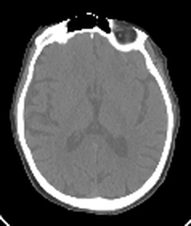
- Serebral hemisferler. Kontrastsız kafa BT’sinde ekstraaksiyel kanama ve parankim dansitesini değerlendirirken aksiyel görüntülere ek olarak koronal reformasyonları da inceleyin.
- Foramen magnum ve kafa tabanı. Kafa tabanı kesitinde kontrastlı gövde serisine geçişi ve beyin-kemik rekonstrüksiyonlarının kapsama devamını belirtin.
- Artefakt ve tarama alanı. Son olarak görüntü alanına giren metal, hareket çizgileri veya kesilmiş ekstremiteyi bulun; bunların hangi anatomik değerlendirmeyi sınırladığını açıklayın.
Faz ve pencere
- Kontrastsız tanısal
- Kafa serisi kontrastsız alınır; akut intrakraniyal kan, kafa kırığı ve pnömosefali bu seride değerlendirilir.
- Arteriyel
- Klinik soru ve yerel protokol gerektiriyorsa boyun-gövde arteriyel edinimi damar yaralanması, aktif ekstravazasyon ve aort hasarı için kullanılır; tüm merkezlerde zorunlu ayrı faz değildir.
- Portal tanısal
- IV kontrastlı gövde serisinde solid organ parankim hasarı, hemoperitoneum ve birçok damar dışı yaralanma görünür; çekim kapsamı merkez protokolüne göre değişir.
Önerilen pencereler: Beyin (G 80 / M 40), Kemik (G 1800 / M 400), Yumuşak doku (G 400 / M 40), Anjiyo (G 600 / M 150), Akciğer (G 1500 / M -600), Mediasten (G 350 / M 50), Batın (G 400 / M 50).
BT bulguları
- Tarama sınırı — kafa tepesi, servikal omurga, göğüs, abdomen ve pelvisin gerçekten kapsanıp kapsanmadığını scout görüntüsü ve seri son kesitlerinden kontrol et.
- Başlangıçta kontrastsız kafa serisi alınır; bu seri akut intrakraniyal kanama ve kemik yaralanmalarının değerlendirmesi için kullanılır.
- Kontrast bolusu ve faz zamanlaması — Damar lümenindeki kontrastlanma düzeyini, gövde çekiminin arteriyel veya portal faz zamanlamasını ve olası tekrar çekimleri değerlendir.
- Hareket ve metal artefaktı — kafatası tabanı, omuz kuşağı ve pelviste çizgilenmenin kanama ya da kırık hattını örtüp örtmediğini belirle.
- Reformat uyumu — aksiyel kesitleri koronal-sagittal yumuşak doku ve kemik rekonstrüksiyonlarıyla eşleştir; ince kemik yaralanmasını yalnız kalın yumuşak doku serisinde arama.
- Görüntü alanına giren ekstremite ve yabancı cisim — kesilmiş anatomiyi, mermi/metal parçalarını ve bunların oluşturduğu yerel değerlendirme kısıtını raporda belirt.
Normalde
Normal incelemede kafa kemikleri kesintisiz, beyin parankimi simetrik ve ekstraaksiyel aralıklar belirgin kitle etkisi olmadan izlenir; gövdede damar ve organlar bekendiği şekilde kontrastlanır. Kıyasın ilk noktası yalnız organ görünümü değil, scouttan son kesite kadar tüm planlanan anatomik kapsamın tamamlanması ve faza uygun opaklaşmanın bulunmasıdır. Hareket veya metal çizgileri normal anatomiyi bozuyorsa bu alanı tanısal güven düzeyinden ayrı kaydet.
Normal BT ile kıyasla
Aynı bölgenin, kaynak veri setinde patoloji içermediği belirtilen bir BT incelemesini kesit kesit kaydırın; organ sınırlarını açarak anatomiyi hasta görüntüsüyle karşılaştırın. Küçük rastlantısal bulgular tümüyle dışlanmamıştır.
Kaynak: TotalSegmentator v2.01 veri seti (Wasserthal ve ark., Radiology: Artificial Intelligence 2023), CC BY 4.0, vaka s0103; organ sınırları veri setinin otomatik segmentasyonundan, birleştirilerek sadeleştirildi. Görüntüler 2,5 mm kesit aralığına yeniden örneklendi.
Ayırıcı tanı
- Gerçek intrakraniyal kanama
- Kontrastsız kafa serisinde damar yolunu takip etmeyen, sulkus, sisterna veya parankim içinde izlenen hiperdensitedir.
- Hareket artefaktı
- Travma BT’sinde solunum hareketi, diyafram yapılarının değerlendirilmesini güçleştirebilir.
- Metal artefaktı
- cismin yakınında ışın sertleşmesi ve çizgilenme yapar; şüpheli alanın karşı düzlem ve komşu kesitlerde devam edip etmediğine bakılır.
Tuzaklar
- Tüm vücut BT'yi her ağır travma hastasına otomatik uygulanan tek reçete gibi sunma; hemodinamik durum, hemen müdahale gereksinimi ve yerel protokol görüntüleme kararını değiştirir.
- Tek fazlı gövde BT'sini arteriyel ve portal fazların tüm sorularını yanıtlıyor gibi yorumlama; damar yaralanması kuşkusunda amaçlı anjiyografik edinim gerekebilir.
- Kafa-boyun geçişini veya ekstremiteyi tarama alanı dışında bırakıp normalmiş gibi sonuçlandırma; scout ve son görüntü kapsamını raporla.
Kendini dene
Kafa travması sonrası hiperdansite yalnız kortikal sulkus lümenini dolduruyor. En olası tanı nedir?
Cevabı göster
Travmatik subaraknoid kanama. Sulkus içindeki kan travmatik subaraknoid kanamayı destekler. Subdural hematom ekstraaksiyel hilal biçimindedir; epidural hematom çoğunlukla bikonvekstir; kontüzyon kortikal parankim içinde yer alır.
Intrakraniyal kanama sınıflamasında 'intraserebral kanama' (ICH) terimi hangi yapıları kapsar?
Cevabı göster
Parankimal (IPH) ve/veya intraventriküler (IVH) kanamalar. S75'e göre ICH, parankimal (IPH) ve/veya intraventriküler (IVH) kanamaları kapsar; daha geniş kapsam ICrH'dir.
Bu konunun yer aldığı turlar
İlgili konular
Kaynaklar
Bu sayfadaki 33 cümle ve 2 quiz sorusu aşağıdaki kaynaklardan alıntılarla tek tek karşılaştırıldı; 7 cümle bu karşılaştırmada düzeltildi (2026-10-08).
- Current Standards for and Clinical Impact of Emergency Radiology in Major Traumapmc.ncbi.nlm.nih.gov
- Artificial intelligence for abdominopelvic trauma imaging: trends, gaps, and future directions.pubmed.ncbi.nlm.nih.gov
- The value of precontrast thoraco-abdominopelvic CT in polytrauma patients.pubmed.ncbi.nlm.nih.gov
- Dual-source CT in blunt trauma patients: elimination of diaphragmatic motion using high-pitch spiral technique.pubmed.ncbi.nlm.nih.gov
- Improving trauma imaging in Wales through Kotter's theory of change.pubmed.ncbi.nlm.nih.gov
- The "Innsbruck Emergency Algorithm" avoids the underdiagnosis of blunt cervical vascular injuries.pubmed.ncbi.nlm.nih.gov
- Maintaining immobilisation devices on trauma patients during CT: a feasibility study.pubmed.ncbi.nlm.nih.gov
- Multidetector computed tomography (MDCT): simple CT protocol for trauma patient.pubmed.ncbi.nlm.nih.gov
- Trauma Computed Tomography in the Modern Era: Not Always Quick and Safe.pubmed.ncbi.nlm.nih.gov
- Improving the protocol for whole-body CT scans in trauma patients.pubmed.ncbi.nlm.nih.gov
- [Update polytrauma and computed tomography in ongoing resuscitation : ABCDE and "diagnose first what kills first"].pubmed.ncbi.nlm.nih.gov
- Rapid imaging protocol in trauma: a whole-body dual-source CT scan.pubmed.ncbi.nlm.nih.gov
- Development of a user-friendly calculator for a pediatric split-bolus polytrauma computed tomography protocol.pubmed.ncbi.nlm.nih.gov
- Quality and Dose Optimized CT Trauma Protocol - Recommendation from a University Level-I Trauma Center.pubmed.ncbi.nlm.nih.gov
- Major trauma CT scanning: the experience of a regional trauma centre in the UK.pubmed.ncbi.nlm.nih.gov
- Emergency radiology of severe acute injury in extreme alpine summer sports: a pictorial essay.pubmed.ncbi.nlm.nih.gov
- Traumatic complete pyloric transection following blunt abdominal trauma: A case report and literature review.pubmed.ncbi.nlm.nih.gov
- Two cases of traumatic aortic isthmus pseudoaneurysm successfully managed with endovascular repair: Multidisciplinary critical care and anesthetic perspective.pubmed.ncbi.nlm.nih.gov
- Methodological standards for body composition assessment-an expert-endorsed guide for research and clinical applications: bioimpedance, dual-energy X-ray absorptiometry, computerized tomography, and ultrasound methods.pubmed.ncbi.nlm.nih.gov
- Spontaneous Intracranial Hypotension: A Review of Neuroimaging and Current Concepts.pubmed.ncbi.nlm.nih.gov
- Clinical Applications of a CT Window Blending Algorithm: RADIO (Relative Attenuation-Dependent Image Overlay).pubmed.ncbi.nlm.nih.gov
- Understanding Photon-Counting CT: Physics, Detector Technology, and Image Reconstructions.pubmed.ncbi.nlm.nih.gov
- Radiation dose optimization in coronary CT angiography clinical application of absolute phase gating in patients with atrial fibrillation.pubmed.ncbi.nlm.nih.gov
- Emergency endovascular repair of blunt thoracic aortic injury-related pseudoaneurysm using a castor single-branched stent graft: a case report and literature review.pubmed.ncbi.nlm.nih.gov
- Aortic Injury and Hemopericardium: Key Findings and Evaluation.pubmed.ncbi.nlm.nih.gov
- The Arrow and the Atlas-A Narrow Escape in the Cervical Spine: A Case Report About Penetrating Injury With an Arrow Impacted in the Neck.pubmed.ncbi.nlm.nih.gov
- Radiocapitellar Alignment in Suspected Pediatric Monteggia Lesions: Narrative Review and Imaging Interpretation Framework.pubmed.ncbi.nlm.nih.gov
- Traumatic basilar artery incarceration following longitudinal clival fracture: illustrative case.pubmed.ncbi.nlm.nih.gov
- Value of coronal reformations in the CT evaluation of acute head trauma.pubmed.ncbi.nlm.nih.gov
- Duodenal Obstruction and Grade III Acute Cholangitis Following Pancreaticoduodenal Artery Aneurysm Rupture During Conservative Management of Acute Type B Aortic Dissection: A Case Report.pubmed.ncbi.nlm.nih.gov
- Guidelines for Enhanced Recovery After Trauma and Intensive Care (ERATIC): Enhanced Recovery After Surgery (ERAS) Society and International Association of Trauma Surgery and Intensive Care (IATSIC) Recommendations: Paper 1: Initial Care-Pre and Intraoperative Care Until ICU, Including Non-Operative Management.pubmed.ncbi.nlm.nih.gov
- Pelvic Trauma: Anatomy and Interventions.pubmed.ncbi.nlm.nih.gov
- Point-of-care ultrasound in trauma: a systematic review of recent literature.pubmed.ncbi.nlm.nih.gov
- Relevance of intra-abdominal pressure monitoring in non-operative management of patients with blunt liver and splenic injuries.pubmed.ncbi.nlm.nih.gov
- Posttraumatic Corpus Luteal Cyst Rupture: A Diagnostic Enigma for Massive Hemoperitoneum.pubmed.ncbi.nlm.nih.gov
- Thoracic Inlet in Cervical Spine CT of Blunt Trauma Patients: Prevalence of Pathologies and Importance of CT Interpretation.pubmed.ncbi.nlm.nih.gov
- Patterns of injury and violence in Yaoundé Cameroon: an analysis of hospital data.pubmed.ncbi.nlm.nih.gov
- Commentary: an eye on PET quantification.pubmed.ncbi.nlm.nih.gov
- Diagnostic Value of the Split-Bolus CT Protocol in Blunt Abdominal Trauma: Comparison With Standard Portal Venous Phase Imaging.pubmed.ncbi.nlm.nih.gov
- Contrast-enhanced ultrasound (CEUS) in urgent and emergent pediatric conditions: a comprehensive review.pubmed.ncbi.nlm.nih.gov
- Blunt Traumatic Isolated Left External Iliac Vein Injury Without Pelvic Fracture.pubmed.ncbi.nlm.nih.gov
- Automatic identification of anatomical landmarks in three-dimensional computed tomography/cone-beam computed tomography: a scoping review.pubmed.ncbi.nlm.nih.gov
- Classification and correlation analysis of orbital medial wall fractures based on computed tomography data for prediction of surgical planning.pubmed.ncbi.nlm.nih.gov
- Role of Multidetector Computed Tomography in the Evaluation of Maxillofacial Trauma.pubmed.ncbi.nlm.nih.gov
- Evaluating the Effectiveness of Yttrium-90 Glass Microspheres in the Treatment of Hepatocellular Carcinoma, Intrahepatic Cholangiocarcinoma, and Metastatic Colorectal Cancer in Practice: Protocol for the Prospective PROACTIF Phase IV Registry Study in France.pubmed.ncbi.nlm.nih.gov
- Acute Traumatic Aortic Injury: What the Radiologist Needs to Know.pubmed.ncbi.nlm.nih.gov
- Do Artifacts From Dental Implants Impair the Diagnosis of Simulated Internal Root Resorption in Cone-Beam CT?pubmed.ncbi.nlm.nih.gov
- An Integrated Approach on the Diagnosis of Cerebral Veins and Dural Sinuses Thrombosis (a Narrative Review).pubmed.ncbi.nlm.nih.gov
- MRI and PET Alterations in Adult Skull Base Tumors: A Narrative Review of Proton Versus Photon Radiotherapy.pubmed.ncbi.nlm.nih.gov
- Spontaneous Intracranial Vertebral Artery Dissection: A Rare Cause of Ischemic Stroke.pubmed.ncbi.nlm.nih.gov
- Minimal Imaging Requirements.pubmed.ncbi.nlm.nih.gov
- Imaging of pediatric brain tumors: A COG Diagnostic Imaging Committee/SPR Oncology Committee/ASPNR White Paper.pubmed.ncbi.nlm.nih.gov
- Neuroimaging standards for research into small vessel disease and its contribution to ageing and neurodegeneration.pubmed.ncbi.nlm.nih.gov
- Dual-energy computed tomography in children: Technique and clinical applications.pubmed.ncbi.nlm.nih.gov
- Advances in Bone Joint Imaging-Metal Artifact Reduction.pubmed.ncbi.nlm.nih.gov
- The Role of Computed Tomography in Imaging Non-neurologic Disorders of the Head in Equine Patients.pubmed.ncbi.nlm.nih.gov
- Transarterial embolization in musculoskeletal care: an indication-stratified narrative review.pubmed.ncbi.nlm.nih.gov
- ESR Essentials: trauma team and the role of Interventional Radiology-practice recommendations by the Cardiovascular and Interventional Radiological Society of Europe.pubmed.ncbi.nlm.nih.gov
- Transcatheter arterial embolization for traumatic adrenal hemorrhage with contrast extravasation: case series and literature review.pubmed.ncbi.nlm.nih.gov
- Endovascular Treatment of an Iatrogenic Inferior Mesenteric Arteriovenous Fistula Presenting as Massive Lower Gastrointestinal Hemorrhage: A Case Report.pubmed.ncbi.nlm.nih.gov
- Rapid Interval Development of a Left Hepatic Artery Pseudoaneurysm During Acute Interstitial Edematous Pancreatitis.pubmed.ncbi.nlm.nih.gov
- "No Touch to Touch": Percutaneous Sclerotherapy of a Giant Hepatic Cavernous Hemangioma.pubmed.ncbi.nlm.nih.gov
- Guidelines on the Use of Therapeutic Apheresis in Clinical Practice-Evidence-Based Approach From the Writing Committee of the American Society for Apheresis: The Tenth Special Issue.pubmed.ncbi.nlm.nih.gov
- Physical exercise for people with Parkinson's disease: a systematic review and network meta-analysis.pubmed.ncbi.nlm.nih.gov
- Physical exercise for people with Parkinson's disease: a systematic review and network meta-analysis.pubmed.ncbi.nlm.nih.gov
- 2.5-mm articulated endoluminal forceps using a compliant mechanism.pubmed.ncbi.nlm.nih.gov
- Visualization-driven digital technologies in oncology: current applications, technological advances, and future directions.pubmed.ncbi.nlm.nih.gov
- Guided Limited Maxillectomy and Staged Septal-Palatal Reconstruction for Low-Grade Chondrosarcoma of the Hard Palate: A Case Report and Literature Review.pubmed.ncbi.nlm.nih.gov
- Management Protocol for Ballistic and Other High-Energy Avulsive Facial Injuries-An Update for the 21st Century.pubmed.ncbi.nlm.nih.gov
- AI-based synthetic CT attenuation correction enables reliable quantitative SPECT in unilateral condylar hyperplasia.pubmed.ncbi.nlm.nih.gov
- Whole-body magnetic resonance imaging (WBMRI) versus whole-body computed tomography (WBCT) for myeloma imaging and staging.pubmed.ncbi.nlm.nih.gov
- Neuroimaging for neurovascular complications of traumatic brain injury.pubmed.ncbi.nlm.nih.gov
- 'Carotid spells' - transient focal neurological episodes associated with convexity subarachnoid haemorrhage in severe ipsilateral extracranial internal carotid artery stenosis: a case report and concise literature review.pubmed.ncbi.nlm.nih.gov
- Middle cerebral artery dissection: pathophysiology, diagnosis, and therapeutic options.pubmed.ncbi.nlm.nih.gov
- A Standardized Anatomical Classification of Intracranial Hemorrhage.pubmed.ncbi.nlm.nih.gov
- Duropathies as Unifying Concept-Part Two: A Narrative Overview of Clinical and Neuroradiological Features.pubmed.ncbi.nlm.nih.gov
- Spinal lesions: a comprehensive radiologic overview.pubmed.ncbi.nlm.nih.gov
- Spectrum of Magnetic Resonance Imaging Findings in Acute Pediatric Traumatic Brain Injury - A Pictorial Essay.pubmed.ncbi.nlm.nih.gov
- CT Imaging in Neurocritical Care.pubmed.ncbi.nlm.nih.gov
- Terminal spreading depolarization and electrical silence in death of human cerebral cortex.pubmed.ncbi.nlm.nih.gov
- Incorporating algorithmic uncertainty into a clinical machine deep learning algorithm for urgent head CTs.pubmed.ncbi.nlm.nih.gov
- Cross-Subject Registration-Based Augmentation: Alleviating Anatomical Misalignment in Trauma CT for Robust Hemorrhage Segmentation.pubmed.ncbi.nlm.nih.gov
- Individualized treatment for pediatric intracranial aneurysms in China: case series and literature review.pubmed.ncbi.nlm.nih.gov
- AI-assisted detection of an initially missed infrarenal abdominal aortic injury in a polytrauma patient: A case report.pubmed.ncbi.nlm.nih.gov
- Isolated Parotitis Following a Cheek Slap: A Case Report and Clinical Implications.pubmed.ncbi.nlm.nih.gov
- Development of a novel clinical decision support tool for determining the need for whole-body computed tomography in pediatric trauma patients: the WHOLE score.pubmed.ncbi.nlm.nih.gov
- Dose-reduction strategies in whole-body CT for injured patients in the emergency department: a scoping review.pubmed.ncbi.nlm.nih.gov
- Liver Imaging Reporting and Data System (LI-RADS): A Comprehensive Review.pubmed.ncbi.nlm.nih.gov
- A new in silico model to precisely design focused ultrasound brain therapies.pubmed.ncbi.nlm.nih.gov
- New insights into enlarged parietal foramina: an anatomical, radiological, and histological study.pubmed.ncbi.nlm.nih.gov
- TomoRay cranial: synthesis of cranial CT imaging from biplanar radiographs using a generative adversarial network.pubmed.ncbi.nlm.nih.gov
- Subarachnoid Hemorrhage Confirmed by Magnetic Resonance Imaging in a Patient with Brain Death owing to Hypoxic Encephalopathy Following Suicide by Hanging.pubmed.ncbi.nlm.nih.gov
- Anoxic Brain Injury Presenting as Pseudosubarachnoid Hemorrhage in the Medical Intensive Care Unit.pubmed.ncbi.nlm.nih.gov
- Radon-Guided Wavelet-Domain Attention U-Net for Periodic Artifact Suppression in Brain MRI.pubmed.ncbi.nlm.nih.gov
- Regional and network patterns of sleep-depth-associated brain diffusion changes: an EEG-informed multishell diffusion MRI study.pubmed.ncbi.nlm.nih.gov
- A survey of transformer-based architectures in medical image analysis: models, applications, and challenges.pubmed.ncbi.nlm.nih.gov
- Late surgical site hemorrhage is a possible radiologic indicator of early tumor recurrence in grossly totally resected high-grade meningioma: A case report.pubmed.ncbi.nlm.nih.gov
- AI-Based Approaches for the Identification and Quantification of Traumatic Brain Injury in Computed Tomography Imaging: Systematic Review.pubmed.ncbi.nlm.nih.gov
- Admission hyperdense sinus sign predicts poorer outcomes in cerebral venous sinus thrombosis.pubmed.ncbi.nlm.nih.gov
- Current Practice: Rationale for Screening Children with Hereditary Hemorrhagic Telangiectasia for Brain Vascular Malformations.pubmed.ncbi.nlm.nih.gov
- Parenchymal Insults in Abuse-A Potential Key to Diagnosis.pubmed.ncbi.nlm.nih.gov
- Deep learning versus iterative image reconstruction algorithm for head CT in trauma.pubmed.ncbi.nlm.nih.gov
- Imaging Evaluation of Acute Traumatic Brain Injury.pubmed.ncbi.nlm.nih.gov
- Rib Reverberation: An important New Artifact in Lung Ultrasound.pubmed.ncbi.nlm.nih.gov
- Comparison of Ultrasonography, Contrast Radiographic Tenography, Cone-Beam Computed Tomographic Tenography, and Tenoscopy for Lesion Detection Within the Digital Flexor Tendon Sheath of Horses-A Prospective Clinical Trial.pubmed.ncbi.nlm.nih.gov
- Intelligence Architectures and Machine Learning Applications in Contemporary Spine Care.pubmed.ncbi.nlm.nih.gov
- The Role of Novel Imaging and Biofluid Biomarkers in Traumatic Axonal Injury: An Updated Review.pubmed.ncbi.nlm.nih.gov
- Cranial ultrasound for beginners.pubmed.ncbi.nlm.nih.gov
- Focal subarachnoid haemorrhage mimicking transient ischaemic attack--do we really need MRI in the acute stage?pubmed.ncbi.nlm.nih.gov
- Seizure and aspiration complicating acute expanding epidural hematoma in a tuberous sclerosis adolescent: a case report of clinical and surgical emergency.pubmed.ncbi.nlm.nih.gov
- Severe Craniocerebral Injury in a Child Caused by Repetitive Cleaver Strikes: Case Report and Review of the Literature.pubmed.ncbi.nlm.nih.gov
Eğitim amaçlıdır; hasta başında tanı ve tedavi kararının yerine geçmez. İçerik yapay zekâ destekli bir süreçle güncel literatüre göre cümle cümle kaynaklarla karşılaştırıldı, ancak uzman radyolog incelemesinden geçmedi. Klinik kararlarda güncel kılavuzları, kurum protokollerini ve radyoloji raporunu esas alın.