Özet
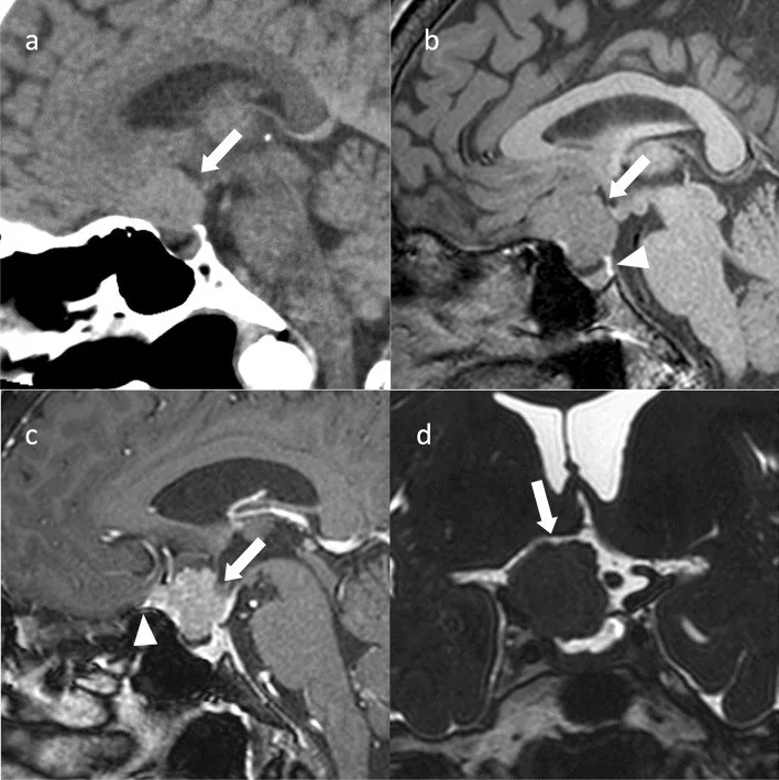
Tuberkulum sellae, planum sphenoidale veya kavernöz sinüs dura materinden gelişen meningiom, hipofiz kaynaklı kitleyle karışabilir; görme yolları ve kavernöz sinüs içindeki kraniyal sinirlerle yakınlığı klinik önem taşır. BT, özellikle kontrastsız incelemede tümörün yumuşak doku sınırını sınırlı gösterse de kalsifikasyonu ve kafa tabanı kemiğindeki hiperostoz ya da erozyonu ortaya koyar. Sella genişlemesinin olmaması, dural taban yerleşimi ve komşu kemik reaksiyonu PitNET’ten ayrımda ipucu verir; yumuşak doku yayılımı için sellaya yönelik MRI daha duyarlıdır. Kitle uzanımı ve optik kanal/kiazma basısı gözden kaçarsa görme yolu etkilenimi ve cerrahi açıdan önemli damar-sinir ilişkisi eksik değerlendirilir.
Faz ve pencere
- Kontrastsız
- Kitle beyin dokusuna benzer veya hafif yüksek atenüsyonda ekstraaksiyel yumuşak doku olarak seçilebilir; iç kalsifikasyonlar ile tuberkulum sellae, klinoid çıkıntı veya sfenoid planumda fokal kemik kalınlaşması kemik algoritmasında belirginleşir. Bu incelemede dural kuyruk ya da tümörün kavernöz sinüs içindeki sınırı güvenle gösterilemez.
Önerilen pencereler: Yumuşak doku (G 400 / M 40), Kemik (G 1800 / M 400).
BT bulguları
- Tuberkulum sellae, planum sphenoidale, klinoid çıkıntı veya kavernöz sinüs duvarına oturan; hipofiz yatağından ayrı izlenebilen ekstraaksiyel kitle
- Kitle tabanına komşu sfenoid kemikte kortikal kalınlaşma ve artmış dansiteyle görülen fokal hiperostoz; yokluğu meningiomu dışlamaz
- Tümör içinde noktasal ya da kaba kalsifikasyon; kemik algoritmasında yumuşak doku kitlesinden daha yoğun odaklar
- Kavernöz sinüs meningiomunda internal karotid arterin kitle tarafından sarılması ve lümeninde daralma; makroadenomda daralma tipik değildir
- Sella çapında belirgin artış olmadan suprasellar veya parasellar kitlenin tuberkulum sellae boyunca yerleşmesi
- En plaque meningiomlarda plak biçimli yayılım görülebilir; temporal kemikte yaygın hiperostotik değişiklikler eşlik edebilir.
- Tuberkulum sellae yerleşiminde suprasellar kitle etkisi; optik kanal ve kiazma basısı kontrastsız BT’de sınırlı seçilebileceğinden MRI ile tamamlanır
- Kontrastlı MRG’de meningiom genellikle homojen ve belirgin kontrastlanır; dural kuyruk eşlik edebilir.
Normalde
Normal sella turcica çevresinde tuberkulum sellae, planum sphenoidale ve klinoid çıkıntıların kemik konturu ince ve süreklidir; kavernöz karotid kanallarının çapı iki tarafta benzerdir. Aynı kesitlerde sellar boşluğun genişleyip genişlemediğini, dural yüzeye komşu yumuşak doku olup olmadığını ve fokal kemik yoğunluğu artışının kitle tabanıyla örtüşüp örtüşmediğini karşılaştır.
Normal BT ile kıyasla
Aynı bölgenin, kaynak veri setinde patoloji içermediği belirtilen bir BT incelemesini kesit kesit kaydırın; organ sınırlarını açarak anatomiyi hasta görüntüsüyle karşılaştırın. Küçük rastlantısal bulgular tümüyle dışlanmamıştır.
Kaynak: TotalSegmentator v2.01 veri seti (Wasserthal ve ark., Radiology: Artificial Intelligence 2023), CC BY 4.0, vaka s0103; organ sınırları veri setinin otomatik segmentasyonundan, birleştirilerek sadeleştirildi. Görüntüler 2,5 mm kesit aralığına yeniden örneklendi.
Ayırıcı tanı
- PitNET makroadenomu
- sella merkezinden genişleme ve suprasellar uzanım daha tipiktir; meningiomda dural taban, hiperostoz ve sarılmış arterde daralma ayrımı destekler.
- Parasellar anevrizma
- kısmen tromboze dev kavernöz internal karotid arter anevrizması kitle etkisi oluşturabilir ve menenjiyomu taklit edebilir.
- Kraniyofaringiom
- suprasellar yerleşim ve kistik bileşenle birlikte kaba kalsifikasyon görülebilir; tuberkulum sellae boyunca hiperostotik dural taban beklenen örüntü değildir.
- Kavernöz sinüs schwannomu
- kraniyal sinir boyunca uzanabilir ve kafa tabanı forameninde genişleme yapabilir; menenjiyomda komşu kemik hiperostozu ve internal karotid arter daralması görülebilir.
- Hipofiz metastazı
- hızlı gelişen infiltratif kitle, kemik destrüksiyonu veya infundibulum/arka lob tutulumu öykü ve MRI ile birlikte ayrımı gündeme getirir.
Tuzaklar
- Hiperostoz, reaktif değişikliklerle doğrudan tümör invazyonunu ayırt etmeyi güçleştirebilir; yalnız kemik kalınlaşması invazyon kanıtı sayılmamalı ve yumuşak doku uzanımıyla birlikte değerlendirilmelidir.
- Kavernöz internal karotid arter düzeyindeki kısmen tromboze dev anevrizma kitle etkisi oluşturabilir ve menenjiyomu taklit edebilir; ayırıcı tanıda göz önünde bulundur.
- Kontrastsız seride dural kuyruk ve homojen kontrastlanma değerlendirilemez; bunların yokluğunu raporlamak yerine bu fazın sınırını belirt.
- Tuberkulum sellae meningiomu komşu optik kanala uzanabilir; rutin kalın BT kesitlerinde kanal içi yayılım ve kiazma basısı olduğundan az görünebilir.
Kendini dene
Kavernöz sinüs menenjiyomlarında komşu kemikte hiperostoz ve internal karotid arter (ICA) lümeninde daralma görülebilir. En olası tanı hangisidir?
Cevabı göster
Kavernöz sinüs menenjiyomu. Komşu kemikte hiperostoz ve kavernöz ICA lümeninde daralma meningiomda görülebilen görüntüleme özellikleridir; tek başlarına tanıyı kesinleştirmezler.
Tuberkulum sellae yerleşimli ve komşu sfenoid kemikte fokal hiperostozu bulunan ekstraaksiyel kitle, en çok hangi tanıyı destekler?
Cevabı göster
Tuberkulum sellae menenjiyomu. Tuberkulum sellae meningiomu bu bölgede merkezlenebilir; meningiomlarda komşu kemik hiperostozu tanıyı destekleyen bir bulgudur.
İlgili konular
Kaynaklar
Bu sayfadaki 36 cümle ve 2 quiz sorusu aşağıdaki kaynaklardan alıntılarla otomatik olarak karşılaştırıldı; 15 cümle bu karşılaştırmada düzeltildi (2026-10-09).
- Pneumocephalus and delayed hydrocephalus following endoscopic endonasal biopsy of skull base lesion: diagnostic pitfalls and the role of immunohistochemistrypmc.ncbi.nlm.nih.gov
- Delayed Diagnosis and Multimodality Management of a Primary Myxoid Chondrosarcoma of the Skull Base: A Case Reportpmc.ncbi.nlm.nih.gov
- The vascular mimic: successful surgical management of cavernous sinus hemangioma via adapted approach: Case reportpmc.ncbi.nlm.nih.gov
- Pituitary neuroendocrine tumors: imaging features, clinical relevance, and a proposal for standardized MRI reportingpmc.ncbi.nlm.nih.gov
- Case Report: A case of craniopharyngioma in the cerebellopontine anglepmc.ncbi.nlm.nih.gov
- Usefulness of dual-energy computed tomography imaging in the differential diagnosis of sellar meningiomas and pituitary adenomas: preliminary report.pubmed.ncbi.nlm.nih.gov
- Endoscopic endonasal resection of ectopic suprasellar pituitary neuroendocrine tumors: comparison of intra-stalk and extra-stalk tumors. Illustrative cases.pubmed.ncbi.nlm.nih.gov
- Case Report: A case of craniopharyngioma in the cerebellopontine angle.pubmed.ncbi.nlm.nih.gov
- Compressive Optic Neuropathypubmed.ncbi.nlm.nih.gov
- Sphenoid Temporal Meningioma With Granular Cell Features and Extracranial Infratemporal Extension.pubmed.ncbi.nlm.nih.gov
- Three-dimensional rotational angiography-based multimodal fusion imaging for preoperative neurovascular assessment in intracranial meningiomas: a retrospective technical feasibility study of 72 cases.pubmed.ncbi.nlm.nih.gov
- Jugular Paraganglioma Presenting As Isolated Hypoglossal Nerve Palsy: An Anatomical Enigma.pubmed.ncbi.nlm.nih.gov
- Intrasellar meningioma versus pituitary apoplexy in the setting of postpartum hemorrhage: a case report.pubmed.ncbi.nlm.nih.gov
- An Illustrated Scoping Review of the Magnetic Resonance Imaging Characteristics of Canine and Feline Brain Tumors.pubmed.ncbi.nlm.nih.gov
- What to report in sellar tumor MRI? A nationwide survey among German pituitary surgeons, radiation oncologists, and endocrinologists.pubmed.ncbi.nlm.nih.gov
- Surgical strategy to minimize dominant hemisphere retraction in an asymmetric giant tuberculum sellae meningioma with contralateral anterior cerebral artery positioning: A case report.pubmed.ncbi.nlm.nih.gov
- Multimodal imaging in neuro-ophthalmology.pubmed.ncbi.nlm.nih.gov
- Precision in Complexity: A Protocol-Driven Quantitative Anatomic Strategy for Giant Olfactory Groove Meningioma Resection in a High-Risk Geriatric Patient.pubmed.ncbi.nlm.nih.gov
- Imaging of pituitary tumors: an update with the 5th WHO Classifications-part 2. Neoplasms other than PitNET and tumor-mimicking lesions.pubmed.ncbi.nlm.nih.gov
- Sclerosing Meningioma : Radiological and Clinical Characteristics of 21 Cases.pubmed.ncbi.nlm.nih.gov
- Nuances in the treatment of malignant tumors of the clival and petroclival region.pubmed.ncbi.nlm.nih.gov
- Proptosis Revealing a Neurological Form of IgG4-Related Disease: A Case Report and Literature Review.pubmed.ncbi.nlm.nih.gov
- Dural Lymphomas Unmasked: A Narrative Review of Extra-Axial Mimics and a Pragmatic Diagnostic Decision Algorithm.pubmed.ncbi.nlm.nih.gov
- Anterior basal meningiomas: Can different approaches make a difference?pubmed.ncbi.nlm.nih.gov
- The Endoscopic-Assisted Supraorbital Approach for Resection of Anterior Skull Base Meningiomas: A Large Single-Center Retrospective Surgical Study.pubmed.ncbi.nlm.nih.gov
- Atypical Foster Kennedy Syndrome With Orbital Apex Involvement Secondary to Intracranial Meningiomatosis: A Case Report.pubmed.ncbi.nlm.nih.gov
- Intravascular large B-cell lymphoma mimicking an orbital mass lesion and bilateral cavernous sinus syndrome: A case report.pubmed.ncbi.nlm.nih.gov
- Melanotic neuroectodermal tumor of infancy (MNTI): A report of two cases from Pakistan.pubmed.ncbi.nlm.nih.gov
- Magnetic resonance imaging of primary central nervous system neoplasms of dogs and cats: Basic concepts for diagnostic pathologists.pubmed.ncbi.nlm.nih.gov
- Spontaneous regression of a petroclival meningioma: illustrative case.pubmed.ncbi.nlm.nih.gov
- Radiogenomics: Current Understandings and Future Perspectives.pubmed.ncbi.nlm.nih.gov
- Complex skull base brain tumor resection: the role of microvascular doppler in surgical precision and outcomes.pubmed.ncbi.nlm.nih.gov
- Risk of new tumor, carotid stenosis, and stroke after stereotactic radiosurgery for pituitary tumor: A multicenter study of 2254 patients with imaging follow-up.pubmed.ncbi.nlm.nih.gov
- Isolated Intracranial Rosai-Dorfman Disease Mimicking Sellar Pituitary Adenoma: a case report and literature review.pubmed.ncbi.nlm.nih.gov
- Infiltrative and inflammatory systemic disorders involving the pituitary gland.pubmed.ncbi.nlm.nih.gov
- Pediatric endocrine disorders: a review of intracranial findings and appropriate imaging.pubmed.ncbi.nlm.nih.gov
- Middle meningeal artery embolization of meningioma en plaque misdiagnosed as subdural hematoma: illustrative cases.pubmed.ncbi.nlm.nih.gov
- Use of three-dimensional-printed models for preoperative planning in endoscopic endonasal surgery for tuberculum sellae meningioma: A case report and literature review.pubmed.ncbi.nlm.nih.gov
- Endoscopic endonasal approach for tuberculum sellae meningioma with optic canal involvement: standard technique for optimal visual outcomes.pubmed.ncbi.nlm.nih.gov
- Morphological and Epidemiological Analysis of the Sphenoid Sinus Based on Computed Tomography and Magnetic Resonance Imaging: A Narrative Review.pubmed.ncbi.nlm.nih.gov
- CT evaluation of the relationship between optic canal, anterior clinoid process, optic strut, caroticoclinoid foramen, and dimensions of sella turcica based on sphenoid sinus pneumatization patterns.pubmed.ncbi.nlm.nih.gov
- The Developing Human Sphenoid Bone: Linking Embryological Development to Adult Morphology.pubmed.ncbi.nlm.nih.gov
- Radiological Differentiation of Pituitary Adenomas From Other Sellar Masses: A Systematic Review.pubmed.ncbi.nlm.nih.gov
- Hybrid CNN-GCN framework for brain tumor MRI classification: A graph-based approach to smart healthcare diagnostics.pubmed.ncbi.nlm.nih.gov
- Endovascular Embolization in Neurovascular Disease: Material Science, Multimodal Management, and Future Horizons.pubmed.ncbi.nlm.nih.gov
- Three-Dimensional Constructive Interference in Steady State (3D CISS) Imaging and Clinical Applications in Brain Pathology.pubmed.ncbi.nlm.nih.gov
- Pheochromocytomas and Paragangliomas-Current Management.pubmed.ncbi.nlm.nih.gov
- Suprasellar Desmoplastic Infantile Ganglioglioma in a 7-Month-Old Infant: A Rare Diagnostic Consideration in Infant Suprasellar Masses.pubmed.ncbi.nlm.nih.gov
- Use of intraoperative ultrasound in differentiating adamantinomatous versus papillary craniopharyngiomas and guiding resection through the endoscopic endonasal route.pubmed.ncbi.nlm.nih.gov
- Pituitary macroadenoma: less common neuroimaging features in a common lesion.pubmed.ncbi.nlm.nih.gov
- Imaging of skull base tumours.pubmed.ncbi.nlm.nih.gov
- Giant Cell Tumor of the Cranial Vault: A Rare Frontotemporal Case With Complete Neurological Recovery and Narrative Review.pubmed.ncbi.nlm.nih.gov
- Human biomarker navigator.pubmed.ncbi.nlm.nih.gov
- Review of imaging modalities and radiological findings of calvarial lesions.pubmed.ncbi.nlm.nih.gov
- Diagnostic imaging and emerging image-guided neurointervention in brain tumors: a narrative review.pubmed.ncbi.nlm.nih.gov
- The application of fluorescence techniques in meningioma surgery-a review.pubmed.ncbi.nlm.nih.gov
- A giant partial thrombosed aneurysm of the internal cavernous carotid artery mimicking a meningioma of the lesser wing of the sphenoid bone.pubmed.ncbi.nlm.nih.gov
- Neoplasms and tumor-like lesions of the sellar region: imaging findings with correlation to pathology and 2021 WHO classification.pubmed.ncbi.nlm.nih.gov
- Primary Extracranial Meningioma of the Sphenoid Bone Without Dural Attachment Presenting With Multicompartmental Extension: A Case Report.pubmed.ncbi.nlm.nih.gov
- Intracranial Metastasis of a Primary Mediastinal Seminoma Mimicking a Convexity Meningioma: A Case Report.pubmed.ncbi.nlm.nih.gov
- A refined radiological classification of anterior clinoid process pneumatization.pubmed.ncbi.nlm.nih.gov
- Ectopic Pituitary Neuroendocrine Tumors/Adenomas Around the Sella Turcicapubmed.ncbi.nlm.nih.gov
- Delayed Diagnosis and Multimodality Management of a Primary Myxoid Chondrosarcoma of the Skull Base: A Case Report.pubmed.ncbi.nlm.nih.gov
- Operative Corridors in Endoscopic Skull Base Tumor Surgery.pubmed.ncbi.nlm.nih.gov
- Current Insights in Prolactin Signaling and Ovulatory Function.pubmed.ncbi.nlm.nih.gov
- Optimizing Surgical Management of Anterior Skull Base Meningiomas: Imaging Modalities, Key Surgical Considerations, and Risk Mitigation Strategies.pubmed.ncbi.nlm.nih.gov
- Spheno-Orbital Meningiomas: The Rationale behind the Decision-Making Process of Treatment Strategy.pubmed.ncbi.nlm.nih.gov
- Fluorescence guidance in skull base surgery: Applications and limitations - A systematic review.pubmed.ncbi.nlm.nih.gov
- Pneumocephalus and delayed hydrocephalus following endoscopic endonasal biopsy of skull base lesion: diagnostic pitfalls and the role of immunohistochemistry.pubmed.ncbi.nlm.nih.gov
- Pituitary neuroendocrine tumors: imaging features, clinical relevance, and a proposal for standardized MRI reporting.pubmed.ncbi.nlm.nih.gov
- Long-term outcomes of active surveillance after DOTATATE PET/MRI-confirmed gross total resection in WHO grade 2 meningioma.pubmed.ncbi.nlm.nih.gov
- Intracranial meningiomas at a tertiary hospital: Spectrum of MRI findings with histopathologic correlation.pubmed.ncbi.nlm.nih.gov
- Surgical Anatomy of the Microscopic and Endoscopic Transorbital Approach to the Middle Fossa and Cavernous Sinus: Anatomo-Radiological Study with Clinical Applications.pubmed.ncbi.nlm.nih.gov
- Primary Intraosseous Meningioma of the Sphenoid-Temporal Bone with Single-Stage, 3D-Planned Cranioplasty: Case Report.pubmed.ncbi.nlm.nih.gov
- Primary central nervous system mucosa-associated lymphoid tissue lymphoma: a diagnostic challenge.pubmed.ncbi.nlm.nih.gov
- Case of infiltrative optic neuropathy with hypertrophic pachymeningitis as a manifestation of en plaque meningioma.pubmed.ncbi.nlm.nih.gov
- Intraosseous meningioma mimicking chronic otitis media.pubmed.ncbi.nlm.nih.gov
- Primary and Secondary Optic Nerve Sheath Meningioma.pubmed.ncbi.nlm.nih.gov
- FEBAIR microsurgical principles in meningiomas and recurrence: a long-term follow-up series.pubmed.ncbi.nlm.nih.gov
- Automated meningioma detection using skull X ray images with deep learning and machine learning classifiers.pubmed.ncbi.nlm.nih.gov
- Postoperative Pituitary MRI Findings in Acromegaly: A Pictorial Review.pubmed.ncbi.nlm.nih.gov
- Clinical and Genetic Spectrum of Large AIP Deletions.pubmed.ncbi.nlm.nih.gov
- Primary glomus tumour of the pituitary gland: diagnostic challenges of a rare and potentially aggressive neoplasm.pubmed.ncbi.nlm.nih.gov
- The cavernous sinus: An anatomic study with clinical implication.pubmed.ncbi.nlm.nih.gov
- Imaging of Skull Base Tumors.pubmed.ncbi.nlm.nih.gov
- Are the clinical manifestations of CT scan and location associated with World Health Organization histopathological grades of meningioma?: A retrospective study.pubmed.ncbi.nlm.nih.gov
- Sellar, suprasellar, and parasellar masses: Imaging features and neurosurgical approaches.pubmed.ncbi.nlm.nih.gov
- Coexisting sellar Rathke cleft cyst and planum sphenoidale meningioma: illustrative case.pubmed.ncbi.nlm.nih.gov
- Transcranial arterial coagulation with cardiac standstill after catastrophic frontopolar artery injury during endoscopic endonasal resection of tuberculum sellae meningioma: illustrative case.pubmed.ncbi.nlm.nih.gov
- From intraosseous meningioma and ossified metaplastic meningioma to osteomeningioma: a novel voxel-based, atlas-normalized MRI framework for the radiological classification of skull-involving meningiomas.pubmed.ncbi.nlm.nih.gov
- Role of 68Ga-DOTA-TOC PET/CT in the Evaluation of Meningiomas with Osseous Involvement.pubmed.ncbi.nlm.nih.gov
- Sellar and parasellar lesions: multidisciplinary management.pubmed.ncbi.nlm.nih.gov
- Anterior Clinoidal Meningiomas: Meningeal Anatomical Considerations and Surgical Implications.pubmed.ncbi.nlm.nih.gov
- Sellar Solitary Fibrous Tumor Mimicking Pituitary Adenoma: Diagnostic Pitfalls, Contemporary Pathological Classification, and Management Considerations.pubmed.ncbi.nlm.nih.gov
- The vascular mimic: successful surgical management of cavernous sinus hemangioma via adapted approach: Case report.pubmed.ncbi.nlm.nih.gov
- Application of imaging studies for orbital tumors: a practical review and representative cases.pubmed.ncbi.nlm.nih.gov
- Sellar Lesions: Novel Aspects in Diagnosis and Management.pubmed.ncbi.nlm.nih.gov
- Multidisciplinary management of pituitary macroadenoma.pubmed.ncbi.nlm.nih.gov
- Giant skull base meningiomas: Definitions, case reports, and surgical approaches.pubmed.ncbi.nlm.nih.gov
- Coexistence of meningioma and craniofacial fibrous dysplasia: a case series of clinicopathological study and literature review.pubmed.ncbi.nlm.nih.gov
- Meningioma: Molecular Updates from the 2021 World Health Organization Classification of CNS Tumors and Imaging Correlates.pubmed.ncbi.nlm.nih.gov
Eğitim amaçlıdır; hasta başında tanı ve tedavi kararının yerine geçmez, tıbbi tavsiye değildir. Metinler yapay zekâ desteğiyle yazıldı ve otomatik bir adımda kaynak alıntılarıyla karşılaştırıldı; bu bir tıbbi doğrulama değildir ve içerik uzman radyolog incelemesinden geçmedi. Klinik kararlarda güncel kılavuzları, kurum protokollerini ve radyoloji raporunu esas alın.