Özet
Epidermoid dalak kistleri doğuştan kabul edilir; çoğu asemptomatiktir ve görüntülemede tesadüfen saptanabilir. Kontrastlı BT'de epidermoid dalak kisti düşük dansiteli, ince duvarlı ve tek odacıklı bir lezyon olarak görülebilir; solid bileşen bulunup bulunmadığı değerlendirilebilir. Epidermoid kistin histolojik epitel yapısı yalnız BT ile kanıtlanamaz; görüntüleme onu benign nonparaziter kist olarak düşündürür. Kanama, enfeksiyon veya rüptür gelişirse basit kist görünümü değişebilir ve bu komplikasyonların atlanması klinik önem taşır.
Faz ve pencere
- Kontrastsız tanısal
- Kontrastlı BT, lezyonun homojen sıvı dansiteli içerik, ince duvar ve kontrastlanmayan düzlemsel yapılar açısından karakterize edilmesini sağlar; solid komponentlerin ekarte edilmesi için kontrast fazının değerlendirilmesi gerekir.
Önerilen pencereler: Batın (G 400 / M 50).
BT bulguları
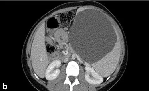
- Dalakta iyi sınırlı, oval kistik lezyon
- Tek odacıklı ve homojen sıvı dansiteli içerik
- İnce, düzgün veya BT'de seçilemeyen duvar
- İç septa, mural nodül ya da solid yumuşak doku bileşeni bulunmaması
- Duvar kalsifikasyonu bazı epidermoid kistlerde görülebilir
- Kanama veya protein içeriği artışı varsa iç atenüasyon basit sıvıdan yüksek olabilir
- Büyüyen kistte komşu mide, böbrek veya diyaframda bası ve dalak konturunda bombeleşme
Normalde
Normal dalakta parankim kesintisiz ve homojendir; parankim içinde sıvı dansiteli yuvarlak boşluk ya da duvar kalsifikasyonu görülmez. Kisti komşu mide fundusu ve sol böbrekten ayıran dalak kapsülünü izle; içerik homojenliğini ve lezyonun dalak dış konturuna yaptığı basıyı kıyasla.
Normal BT ile kıyasla
Aynı bölgenin, kaynak veri setinde patoloji içermediği belirtilen bir BT incelemesini kesit kesit kaydırın; organ sınırlarını açarak anatomiyi hasta görüntüsüyle karşılaştırın. Küçük rastlantısal bulgular tümüyle dışlanmamıştır.
Kaynak: TotalSegmentator v2.01 veri seti (Wasserthal ve ark., Radiology: Artificial Intelligence 2023), CC BY 4.0, vaka s0541; organ sınırları veri setinin otomatik segmentasyonundan, birleştirilerek sadeleştirildi. Görüntüler 2,5 mm kesit aralığına yeniden örneklendi.
Ayırıcı tanı
- Dalak psödokisti
- travma veya hematom öyküsü ve epitelyal döşeme olmaması ile ilişkilidir.
- Kistik ekinokokkozis
- kız kistler, ayrılmış membran veya çok odacıklı ana kist görünümü ayırır.
- Kistik lenfatik malformasyon
- dalakta görülebilir; tek veya çok kistik olabilir ve ince septaları hafif ya da orta derecede kontrastlanabilir.
- Dalak apsesi, kistik dalak lezyonlarının ayırıcı tanısında yer alabilir.
- Kistik nekrotik tümör
- kontrastlanan mural nodül veya periferik solid doku araştırılır.
Tuzaklar
- Kontrastsız seride içeriğin homojen düşük atenüasyonda olması kontrastlanmama kanıtı değildir; solid bileşen kuşkusu varsa kontrastlı karakterizasyon gerekir.
- Epidermoid kist ile psödokistin duvar görünümü örtüşebilir; epitel varlığı görüntüyle kesinleştirilemez.
- Yüksek atenüasyonlu içerik otomatik olarak solid tümör sayılmaz; kanama veya yoğun proteinli sıvı içeriği olabilir.
- Kist çevresindeki ince kalsifikasyonu kız vezikül sanmamak için kalsifikasyonun duvar boyunca uzanıp uzanmadığını izle.
Kendini dene
Tesadüfen saptanan dalak lezyonu düzgün sınırlı, homojen sıvı dansiteli ve tek boşluklu; membran veya nodül yok. En olası tanı?
Cevabı göster
Basit nonparaziter dalak kisti. Görüntüleme tek başına epitel döşemesini kanıtlamaz; bu nedenle tanı basit nonparaziter dalak kisti olarak verilir. Hidatik kistte kız kist veya membran, apsede inflamatuvar içerik, kistik tümörde ise kontrastlanan solid bileşen aranır.
Dalak içinde basit sıvı atenüasyonlu kistin ayrımında en çok hangi özellik şüphe uyandırır?
Cevabı göster
Mural yumuşak doku nodülü. Mural nodül basit kist görünümünü bozar ve kontrastlı karakterizasyon gerektirir; diğer seçenekler tipik benign kist morfolojisine uyar.
İlgili konular
Kaynaklar
Bu sayfadaki 34 cümle ve 2 quiz sorusu aşağıdaki kaynaklardan alıntılarla otomatik olarak karşılaştırıldı; 9 cümle bu karşılaştırmada düzeltildi (2026-10-09).
- Obstructive Pyelonephritis Secondary to Ureteral Compression by an Intra‐Abdominal Desmoid Tumor in Familial Adenomatous Polyposis: A Case Reportpmc.ncbi.nlm.nih.gov
- Recurrent splenic epidermoid cyst in a young adult: A case report and literature reviewpmc.ncbi.nlm.nih.gov
- Peritoneal Inclusion Cyst in an Unusual Location in a “Virgin” Abdomen: A Diagnostic Dilemma Expanding the Clinical Spectrumpmc.ncbi.nlm.nih.gov
- Laparoscopic Partial Splenectomy for a Giant Congenital Splenic Cyst in a Child: Case Report and Focused Literature Reviewpmc.ncbi.nlm.nih.gov
- Case Report: Primary splenic hydatid cystpmc.ncbi.nlm.nih.gov
- A case of ruptured splenic cyst with elevated serum levels of CEA treated by laparoscopic unroofing.pubmed.ncbi.nlm.nih.gov
- Obstructive Pyelonephritis Secondary to Ureteral Compression by an Intra-Abdominal Desmoid Tumor in Familial Adenomatous Polyposis: A Case Report.pubmed.ncbi.nlm.nih.gov
- Fetal MRI: abdominal cystic lesions.pubmed.ncbi.nlm.nih.gov
- A Comprehensive Review of Splenic Cysts: Case Reports and Clinical Insight.pubmed.ncbi.nlm.nih.gov
- 불분명한 소아 복부 종괴: 영상 기반 진단 접근법.pubmed.ncbi.nlm.nih.gov
- Peritoneal Inclusion Cyst in an Unusual Location in a "Virgin" Abdomen: A Diagnostic Dilemma Expanding the Clinical Spectrum.pubmed.ncbi.nlm.nih.gov
- Clinical, Radiological, and Endoscopic Features of Pancreatic Pseudocyst and Walled-Off Necrosis: How to Diagnose and How to Drain Them.pubmed.ncbi.nlm.nih.gov
- Special Issue: EACR 2026 Congress: Innovative Cancer Science, 8-11 June 2026.pubmed.ncbi.nlm.nih.gov
- Pathology of Aging in NOD scid gamma Female Mice.pubmed.ncbi.nlm.nih.gov
- ACG clinical guideline: Genetic testing and management of hereditary gastrointestinal cancer syndromes.pubmed.ncbi.nlm.nih.gov
- Severe Splenic Rupture After Minor Blunt Trauma in a Patient With an Epidermoid Splenic Cyst: A Case Report.pubmed.ncbi.nlm.nih.gov
- Upper gastrointestinal hemorrhage due to splenic artery pseudoaneurysm combined with pancreatic pseudocyst recurrence: A case report.pubmed.ncbi.nlm.nih.gov
- Our Experience in the Management of Splenic Arterial Thrombosis.pubmed.ncbi.nlm.nih.gov
- The Spectrum of Solitary Benign Splenic Lesions-Imaging Clues for a Noninvasive Diagnosis.pubmed.ncbi.nlm.nih.gov
- Tips and Tricks in Thoracic Radiology for Beginners: A Findings-Based Approach.pubmed.ncbi.nlm.nih.gov
- Imaging insights into pediatric liver masses: A comprehensive minireview for hepatology practice.pubmed.ncbi.nlm.nih.gov
- Imaging of renal pseudotumors in children: a comprehensive review.pubmed.ncbi.nlm.nih.gov
- Comparison of Virtual Non-Contrast Images Generated by Spectral Detector Computed Tomography and Conventional Computed Tomography Images of Histologically Confirmed Hepatic Pathologies in 28 Dogs.pubmed.ncbi.nlm.nih.gov
- Common and Uncommon Splenic Lesions: A Review.pubmed.ncbi.nlm.nih.gov
- A case series of radiologically misdiagnosed visceral lymphatic malformations.pubmed.ncbi.nlm.nih.gov
- Splenic pathologies: A pictorial review of common, wandering, twisting and rare presentations.pubmed.ncbi.nlm.nih.gov
- Left subphrenic lymphocele after total gastrectomy.pubmed.ncbi.nlm.nih.gov
- Incidental Focal Spleen Lesions: Integrated Imaging and Pattern Recognition Approach to the Differential Diagnosis.pubmed.ncbi.nlm.nih.gov
- Navigating Ovarian-Adnexal Reporting and Data System Magnetic Resonance Imaging (O-RADS MRI): A Review of Its Evolution, Current Advances, and Persistent Challenges in Ovarian Imaging.pubmed.ncbi.nlm.nih.gov
- Pancreatic Cystic Lesions: A Focused Review on Cyst Clinicopathological Features and Advanced Diagnostics.pubmed.ncbi.nlm.nih.gov
- Case Report: Isolated splenic hydatid cyst with negative serology managed by laparoscopic splenectomy.pubmed.ncbi.nlm.nih.gov
- Albendazole Monotherapy and Spleen Preservation in an Adolescent with Isolated Primary Splenic Cystic Echinococcosis: A Case Report.pubmed.ncbi.nlm.nih.gov
- Infected splenic artery pseudoaneurysm within a complex splenic cystic lesion: CT angiography pitfalls and surgical correlation.pubmed.ncbi.nlm.nih.gov
- Case Report: Two rare cases of aggressive fibromatosis originating in the intestinal tract.pubmed.ncbi.nlm.nih.gov
- Giant Congenital Splenic Cyst in a Pediatric Patient: A Case Report and Review of Surgical Management.pubmed.ncbi.nlm.nih.gov
- Splenoses and Other Ectopic and Heterotopic Splenic Tissue: The Use of Long-Lasting Enhancement in Contrast-Enhanced Ultrasound to Avoid Unnecessary Intervention.pubmed.ncbi.nlm.nih.gov
- Impact of structured reporting on interpretability of PSMA PET/CT in prostate cancer staging-towards standardised clinical implementation.pubmed.ncbi.nlm.nih.gov
- Differentiating walled-off pancreatic necrosis with no documented pancreatitis from mucinous cystic neoplasms: a multicenter LASSO-based nomogram.pubmed.ncbi.nlm.nih.gov
- MDCT Findings of Splenic Pathology.pubmed.ncbi.nlm.nih.gov
- Lymph node metastasis in cancer progression: molecular mechanisms, clinical significance and therapeutic interventions.pubmed.ncbi.nlm.nih.gov
- International Harmonization of Nomenclature and Diagnostic Criteria (INHAND): Nonproliferative and Proliferative Lesions of the Rabbit.pubmed.ncbi.nlm.nih.gov
- Giant adrenal pseudocyst with unclear preoperative origin: a case report and literature review.pubmed.ncbi.nlm.nih.gov
- Laparoscopic Partial Splenectomy for a Giant Congenital Splenic Cyst in a Child: Case Report and Focused Literature Review.pubmed.ncbi.nlm.nih.gov
- Incidental cystic pancreatic neuroendocrine neoplasm and differential diagnosis.pubmed.ncbi.nlm.nih.gov
- Approach to Cystic Lesions in the Abdomen and Pelvis, with Radiologic-Pathologic Correlation.pubmed.ncbi.nlm.nih.gov
- Primitive isolated hydatid cyst of the spleen: total splenectomy versus spleen saving surgical modalities.pubmed.ncbi.nlm.nih.gov
- Benign focal liver lesions: The role of magnetic resonance imaging.pubmed.ncbi.nlm.nih.gov
- Peritoneal disease: key imaging findings that help in the differential diagnosis.pubmed.ncbi.nlm.nih.gov
- Incidental findings in female pelvis MRI performed for gynaecological malignancies.pubmed.ncbi.nlm.nih.gov
- The Benign Side of the Abdominal Wall: A Pictorial Review of Non-Neoplastic Diseases.pubmed.ncbi.nlm.nih.gov
- A resected case of pancreatic neuroendocrine neoplasm with completely cystic degeneration mimicking a mucinous cystic neoplasm: a case report and literature review.pubmed.ncbi.nlm.nih.gov
- Endoscopic ultrasound-guided radiofrequency ablation for pancreatic cystic neoplasms: a comprehensive review.pubmed.ncbi.nlm.nih.gov
- Imaging and Management of Incidental Renal Lesions.pubmed.ncbi.nlm.nih.gov
- Case Report: Synchronous lingual osseous choristoma and suprahyoid thyroglossal duct cyst: insights into the embryological thyroglossal-hyoid axis.pubmed.ncbi.nlm.nih.gov
- Rapidly Progressive Merkel Cell Carcinoma with Atypical Metastatic Pattern-A Case Report and Brief Literature Review.pubmed.ncbi.nlm.nih.gov
- Rare pancreatic cystic neoplasms: A pictorial review.pubmed.ncbi.nlm.nih.gov
- Rare anatomical localizations of hydatid cysts (2020-2025): Narrative review of presentation, imaging, and management.pubmed.ncbi.nlm.nih.gov
- Primary Splenic Pregnancy: A Systematic Review of Published Case Reports.pubmed.ncbi.nlm.nih.gov
- Large Symptomatic Splenic Pseudocyst: Case Report of a Rare Entity.pubmed.ncbi.nlm.nih.gov
- A retrospective comparative study of CT and MRI findings of different types of pancreatic serous cystic neoplasms: Correlation with histopathology.pubmed.ncbi.nlm.nih.gov
- Imaging findings of fatal embolic hemorrhagic stroke secondary to right ventricular hydatid cyst: A case report.pubmed.ncbi.nlm.nih.gov
- Locally Aggressive Hepatic Echinococcosis With Colonic Involvement Requiring a Right Hemicolectomy: A Case Report and Focused Literature Review.pubmed.ncbi.nlm.nih.gov
- Endoscopic ultrasound-guided radiofrequency ablation for large branch-duct intraductal papillary mucinous neoplasms: Safety and efficacy trial.pubmed.ncbi.nlm.nih.gov
- An Overview for Clinicians on Intraductal Papillary Mucinous Neoplasms (IPMNs) of the Pancreas.pubmed.ncbi.nlm.nih.gov
- Recurrent splenic epidermoid cyst in a young adult: A case report and literature review.pubmed.ncbi.nlm.nih.gov
- Epidermoid Cysts in an Intrapancreatic Accessory Spleen Mimicking Pancreatic Neoplasms: Two Case Reports and a Literature Review of Japanese Cases.pubmed.ncbi.nlm.nih.gov
- Traumatic rupture of an isolated splenic hydatid cyst: a rare and challenging surgical emergency.pubmed.ncbi.nlm.nih.gov
- Cystic Neoplasms of the Pancreas: Differential Diagnosis and Radiology Correlation.pubmed.ncbi.nlm.nih.gov
- Cystic pancreatic lesions: MR imaging findings and management.pubmed.ncbi.nlm.nih.gov
- Minimally invasive surgery for giant splenic cysts.pubmed.ncbi.nlm.nih.gov
- Radiological Approach to Splenomegaly: Etiologies, Pathophysiologies, and Diagnostic Strategies.pubmed.ncbi.nlm.nih.gov
- Giant Intraabdominal Lymphangioma in a Pediatric Patient-A Challenging Diagnosis.pubmed.ncbi.nlm.nih.gov
- Culture-negative cryptogenic splenic abscess in an immunocompetent host with recurrent misdiagnosis as pleural effusion: a case report with a review of the literature.pubmed.ncbi.nlm.nih.gov
- Radiological Assessment After Pancreaticoduodenectomy for a Precision Approach to Managing Complications: A Narrative Review.pubmed.ncbi.nlm.nih.gov
- Evaluating Microsoft Bing with ChatGPT-4 for the assessment of abdominal computed tomography and magnetic resonance images.pubmed.ncbi.nlm.nih.gov
- Abdominal bronchogenic cyst: A rare case report.pubmed.ncbi.nlm.nih.gov
- A Case of Pancreatic Pseudocyst Complicated by Pseudoaneurysm.pubmed.ncbi.nlm.nih.gov
- International Harmonization of Nomenclature and Diagnostic Criteria (INHAND): Non-proliferative and Proliferative Lesions of the Non-human Primate ( M. fascicularis ).pubmed.ncbi.nlm.nih.gov
- Case Report: Delayed complications of endovascular treatment of recurrent erosive hemorrhage in necrotizing pancreatitis.pubmed.ncbi.nlm.nih.gov
- Venous congestive ischemic colitis following left hemicolectomy: A rare case of superior rectal vein occlusion.pubmed.ncbi.nlm.nih.gov
- Complications and expected imaging findings after endoscopic retrograde cholangiopancreatography.pubmed.ncbi.nlm.nih.gov
Eğitim amaçlıdır; hasta başında tanı ve tedavi kararının yerine geçmez, tıbbi tavsiye değildir. Metinler yapay zekâ desteğiyle yazıldı ve otomatik bir adımda kaynak alıntılarıyla karşılaştırıldı; bu bir tıbbi doğrulama değildir ve içerik uzman radyolog incelemesinden geçmedi. Klinik kararlarda güncel kılavuzları, kurum protokollerini ve radyoloji raporunu esas alın.