Özet
Peptik ülser değerlendirmesinde BT'nin mukozal ayrıntı düzeyi sınırlıdır. Kontrastlı BT'de mukozal hiper-kontrastlanmada fokal kesinti ya da lümene açılan niş, komşu duvar ödemi ve lokal yağ reaksiyonu görülebilir. Peptik ülser perforasyonla veya gastrik çıkış obstrüksiyonuyla komplike olabilir. Bu nedenle fokal bulguyu saptadıktan sonra incelemenin devamında perforasyon, kanama ve çıkış tıkanıklığına ait işaretleri aramak gerekir.
Faz ve pencere
- Portal tanısal
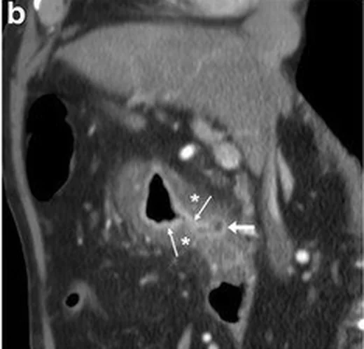
- Portal venöz fazda antrum, pilor ve duodenal bulbus duvarında ödemli kalınlaşma, kontrastlanan mukozada fokal kesinti ve lümene uzanan ülser nişi görülebilir; komşu yağlı dokuda stranding eşlik edebilir. Bu faz ülser çevresi ve perforasyon bulgularını gösterir, ancak ülserin doğrudan BT tanısı duyarlılığı sınırlıdır.
- Arteriyel
- Aktif gastrointestinal kanama şüphesinde arteriyel fazda damar dışına çıkan kontrastın lümen içindeki odağı izlenebilir; bu görünüm komplike kanamayı araştırmak içindir ve komplikasyonsuz ülserin rutin karakterizasyon fazı değildir.
Önerilen pencereler: Batın (G 400 / M 50), Yumuşak doku (G 400 / M 40).
BT bulguları
- Ülser nişi, mide veya duodenum lümeninden duvar içine ya da dışına uzanan küçük kontrastlı cepleşme şeklinde görülebilir.
- Mukoza kontrast çizgisindeki fokal kesinti ülser için doğrudan işaretlerden biridir.
- Antrum, pilor veya duodenal bulbus çevresindeki düzgün duvar kalınlaşması ve ödem ikincil bulgudur.
- Perigastrik veya periduodenal yağda lokal bulanıklık/stranding aktif inflamasyonu destekler.
- Pilorik ödem ya da sikatrisyel daralma ile birlikte proksimal mide genişlemesi çıkış obstrüksiyonu düşündürür.
- Perforasyonda ülser bölgesine komşu ekstraluminal hava, serbest intraperitoneal hava veya lokal sıvı görülebilir.
- Arteriyel fazda damar dışına yeni kontrast çıkışı aktif kanama odağına işaret eder; lümen içi yoğun içerik tek başına kanama kanıtı değildir.
Normalde
Normalde mide ve duodenal bulbus mukozası kesintisiz kontrastlanır; antrum/pilor geçişi açık, komşu yağ planları temiz ve proksimal mide aşırı genişlemiş değildir. Kıyasta aynı segmentte fokal niş veya mukozal kesinti ararken, düzgün yaygın duvar kalınlığını kollapsa bağlı görünümden ve tümör lehine asimetri/omuzlanmadan ayır.
Normal BT ile kıyasla
Aynı bölgenin, kaynak veri setinde patoloji içermediği belirtilen bir BT incelemesini kesit kesit kaydırın; organ sınırlarını açarak anatomiyi hasta görüntüsüyle karşılaştırın. Küçük rastlantısal bulgular tümüyle dışlanmamıştır.
Kaynak: TotalSegmentator v2.01 veri seti (Wasserthal ve ark., Radiology: Artificial Intelligence 2023), CC BY 4.0, vaka s0541; organ sınırları veri setinin otomatik segmentasyonundan, birleştirilerek sadeleştirildi. Görüntüler 2,5 mm kesit aralığına yeniden örneklendi.
Ayırıcı tanı
- Gastrik adenokarsinom
- fokal eksantrik/nodüler kalınlaşma, omuzlanma, sabit kitle veya perigastrik infiltrasyon ülser çevresindeki düzgün ödemden ayrılır.
- Duodenal divertikül
- duodenumdan dışa uzanan, sıklıkla hava veya kontrast içeren kese olabilir; bulbus ülseriyle karışırsa lümen bağlantısı ve çevre inflamasyon incelenir.
- Mide çıkış obstrüksiyonunda mide distansiyonu ve pilor düzeyinde daralma görülebilir; kronik peptik ülser olası nedenlerden biridir.
- Mide duvarı hematomu
- duvar içinde yüksek atenüasyonlu kalınlaşma, özellikle kontrastsız görüntüde, mukoza kaynaklı nişten farklıdır.
Tuzaklar
- Yüzeyel ülserde BT normal olabilir; doğrudan niş görülmemesi peptik ülseri dışlamaz.
- Kontrakte antrumda duvar kalınlaşması ülser ya da tümör görünümü taklit edebilir; mukozal hiper-kontrastlanma, kesinti ve komşu yağ reaksiyonunu birlikte ara.
- Uygunsa oral kontrast mide distansiyonunu sağlayarak perforasyon değerlendirmesine yardımcı olabilir; su negatif oral kontrast olarak mukozal kontrastlanmanın değerlendirilmesini bozmaz.
- Perforasyon değerlendirmesinde küçük ekstraluminal hava yalnız ülser düzeyinde kalmayabilir; üst abdomen ve perihepatik alanları da incele.
Kendini dene
Epigastrik ağrıda BT, duodenal bulbus mukozasında fokal kesinti ve dışa uzanan küçük niş gösteriyor. En olası tanı hangisidir?
Cevabı göster
Duodenal peptik ülser. Fokal mukozal kesinti ve lümene açılan niş peptik ülserin doğrudan BT işaretleridir. Lenfoma daha yaygın duvar/nodal hastalık yapar; hiatal herni hiatus anatomisini değiştirir; bezoar lümen içi materyaldir.
Pilorik duvar ödemiyle birlikte mide belirgin geniş ve distalde geçiş daralmış. Hangi komplikasyon gelişmiştir?
Cevabı göster
Mide çıkış obstrüksiyonu. Proksimal mide distansiyonu ile pilor düzeyindeki geçiş daralması çıkış obstrüksiyonunu gösterir. Divertikül keseleşmedir; mural stratifikasyon duvar görünümüdür; paraözofageal hernide fundus hiatus yanından toraksa yer değiştirir.
İlgili konular
Kaynaklar
Bu sayfadaki 37 cümle ve 2 quiz sorusu aşağıdaki kaynaklardan alıntılarla otomatik olarak karşılaştırıldı; 7 cümle bu karşılaştırmada düzeltildi (2026-10-09).
- Multimodality imaging of adult gastric emergencies: A pictorial reviewpmc.ncbi.nlm.nih.gov
- Dyspepsia: organic versus functional.pubmed.ncbi.nlm.nih.gov
- [Management of gastrinoma].pubmed.ncbi.nlm.nih.gov
- An Overview of Gastroduodenal Perforation.pubmed.ncbi.nlm.nih.gov
- ACR Appropriateness Criteria® Epigastric Pain.pubmed.ncbi.nlm.nih.gov
- Gastric ischaemia: a rare cause of gastric ulcers.pubmed.ncbi.nlm.nih.gov
- Imaging Assessment of Gastroduodenal Perforations.pubmed.ncbi.nlm.nih.gov
- CT of gastro-duodenal obstruction.pubmed.ncbi.nlm.nih.gov
- Gastroduodenal ulceration in dogs with liver disease.pubmed.ncbi.nlm.nih.gov
- Duodenal tuberculosis presenting as gastric outlet obstruction: A case report.pubmed.ncbi.nlm.nih.gov
- Gastric injury from (90)Y to left hepatic lobe tumors adjacent to the stomach: fact or fiction?pubmed.ncbi.nlm.nih.gov
- Perforated Duodenal Ulcer: Has Anything Changed?pubmed.ncbi.nlm.nih.gov
- Duodenal perforation due to compression necrosis by the tip of percutaneous endoscopic gastrostomy tube.pubmed.ncbi.nlm.nih.gov
- Modeling of influential predictors of gastric cancer incidence rates in Golestan province, North Iran.pubmed.ncbi.nlm.nih.gov
- Trauma-Associated Perforation of a Duodenal Ulcer With Concomitant Acute Appendicitis Presenting As Generalized Peritonitis: A Case Report.pubmed.ncbi.nlm.nih.gov
- Review of Peptic Ulcer Disease: Insights from Pathophysiology, Mechanisms, Pharmacotherapy, Phytotherapy, and Case Reports.pubmed.ncbi.nlm.nih.gov
- Pressure-induced posterior gastric perforation in a patient with severe gastroparesis and prior Nissen fundoplication.pubmed.ncbi.nlm.nih.gov
- Long-term safety and effectiveness of the "OptEase" vena cava filter.pubmed.ncbi.nlm.nih.gov
- Dynamic contrast-enhanced computed tomography for small bowel bleeding: Two cases of arteriovenous malformation and gastrointestinal stromal tumor.pubmed.ncbi.nlm.nih.gov
- Ruptured Peritoneal Dissemination of Hepatocellular Carcinoma Successfully Treated with Transcatheter Arterial Embolization: A Case Report and Literature Review.pubmed.ncbi.nlm.nih.gov
- Endovascular Treatment of an Iatrogenic Inferior Mesenteric Arteriovenous Fistula Presenting as Massive Lower Gastrointestinal Hemorrhage: A Case Report.pubmed.ncbi.nlm.nih.gov
- Sigmoid pneumatosis cystoides intestinalis secondary to chronic digestive hyperpressure on post-ulcer duodenal bulb stenosis, decompensated following recent endoscopic insufflation.pubmed.ncbi.nlm.nih.gov
- Complicated Jejunal Diverticulosis as a Rare Cause of Small Bowel Perforation in an Immunocompromised Patient: A Case Report.pubmed.ncbi.nlm.nih.gov
- Duodenal diverticulum perforation with pneumo-retroperitoneum: a case report of rare presentation.pubmed.ncbi.nlm.nih.gov
- A slow and silent return: a case of renal cell carcinoma recurring as gastrointestinal bleeding and small bowel obstruction.pubmed.ncbi.nlm.nih.gov
- Entero-Appendiceal Fistula Presenting with Overt Gastrointestinal Bleeding: A Case Report.pubmed.ncbi.nlm.nih.gov
- Perforated Gastric Cancer: Epidemiology, Diagnosis, and Surgical Management Strategies.pubmed.ncbi.nlm.nih.gov
- Stricturing Diseases of the Gastrointestinal Tract-Current Clinical Practice.pubmed.ncbi.nlm.nih.gov
- Brunner's gland hamartoma in the setting of a duodenal diverticulum: A case report.pubmed.ncbi.nlm.nih.gov
- Imaging Approach to Diverticular Disease of the Small Bowel.pubmed.ncbi.nlm.nih.gov
- Double gallbladder malformation with duodenal ulcer stricture leading to common bile duct dilation: a case report.pubmed.ncbi.nlm.nih.gov
- Evidence-based, cost-effective management of gastric outlet obstruction: An algorithm of the Journal of Trauma and Acute Care Surgery , Emergency General Surgery Algorithms Work Group.pubmed.ncbi.nlm.nih.gov
- Incidentally Identified Pneumatosis Cystoides Intestinalis in Patients With Gastric Outlet Obstruction From Addis Ababa, Ethiopia: A Case Report.pubmed.ncbi.nlm.nih.gov
- The Diagnostic Challenge of Crohn's Disease With Initial Duodenal Involvement: Two Cases and Literature Review.pubmed.ncbi.nlm.nih.gov
- Beyond Conventional CT: Role of Dual-Energy CT in Monitoring Response to Therapy in Abdominal Malignancies.pubmed.ncbi.nlm.nih.gov
- Spectrum of Multidetector Computed Tomography Imaging Findings in Iatrogenic Abdominopelvic Injuries: A Comprehensive Pictorial Review.pubmed.ncbi.nlm.nih.gov
- Complications after Nephron-sparing Interventions for Renal Tumors: Imaging Findings and Management.pubmed.ncbi.nlm.nih.gov
- <i>Mycobacterium abscessus</i> infection in a young man with cystic fibrosis: a case report and literature review.pubmed.ncbi.nlm.nih.gov
- Intractable hiccup leading to life-threatening malnutrition: case report.pubmed.ncbi.nlm.nih.gov
- Gut Microbiota in NSAID Enteropathy: Current Evidence and Future Perspectives for Therapeutic Strategies.pubmed.ncbi.nlm.nih.gov
- Gastric Antral Web as an Uncommon Cause of Gastric Outlet Obstruction in an Adult Female Patient: A Case Report of a Rare Medical Condition and a Review of the Literature.pubmed.ncbi.nlm.nih.gov
- Giant gastric trichobezoar (Rapunzel syndrome) presenting as gastric outlet obstruction with imaging features suggestive of secondary superior mesenteric artery syndrome in a 27-year-old female: surgical management and multidisciplinary approach.pubmed.ncbi.nlm.nih.gov
- Pseudomyxoma peritonei presenting with pyloric obstruction: a case report.pubmed.ncbi.nlm.nih.gov
- Research progress on the application of dual energy CT in gastric diseases.pubmed.ncbi.nlm.nih.gov
- Indian guidelines on imaging of the small intestine in Crohn's disease: A joint Indian Society of Gastroenterology and Indian Radiology and Imaging Association consensus statement.pubmed.ncbi.nlm.nih.gov
- International Society of Paediatric Surgical Oncology (IPSO) Surgical Practice Guidelines.pubmed.ncbi.nlm.nih.gov
- Smear-positive Pulmonary Tuberculosis with False-negative Loop-mediated Isothermal Amplification and T-SPOT.TB Assays.pubmed.ncbi.nlm.nih.gov
- A Focus on Inflammatory and Bacterial Biomarkers in Secondary Peritonitis.pubmed.ncbi.nlm.nih.gov
- Duodenal Diverticulosis as an Unusual Cause of Severe Abdominal Pain.pubmed.ncbi.nlm.nih.gov
- A Case Report Evaluating Gastric Emphysema versus Emphysematous Gastritis.pubmed.ncbi.nlm.nih.gov
- Practical concepts and strategies for early diagnosis and management of eosinophilic gastrointestinal disorders in East-Asian children.pubmed.ncbi.nlm.nih.gov
- Cholecystogastric fistula masquerading as gastrointestinal stromal tumour: Unusual presentation of gallbladder adenomyomatosis - a case report.pubmed.ncbi.nlm.nih.gov
- Gastric Adenocarcinoma Associated With CTLA-4 Haploinsufficiency in the Background of Common Variable Immunodeficiency: A Rare Case.pubmed.ncbi.nlm.nih.gov
- Geriatric Patient Successfully Managed With Conservative Therapy Despite Portal Venous Gas.pubmed.ncbi.nlm.nih.gov
- Complication imaging after laparoscopic Roux-en-Y gastric bypass: clues to the diagnosis and pitfalls.pubmed.ncbi.nlm.nih.gov
- <i>Sarcina ventriculi</i>: An incidental finding or a byproduct of a more concerning underlying disease?pubmed.ncbi.nlm.nih.gov
- Ga-68 FAPI-04 PET/CT detection of repeatedly biopsy-negative poorly differentiated gastric adenocarcinoma: a case report.pubmed.ncbi.nlm.nih.gov
- CT oral contrast and contemporary imaging pathways for suspected oesophageal perforation in the emergency department: a retrospective observational study.pubmed.ncbi.nlm.nih.gov
- Small bowel lymphatic malformation: Clinical presentation and a comprehensive literature review.pubmed.ncbi.nlm.nih.gov
- Gastric Outlet Obstruction from Brunner Gland Hyperplasia Mimicking Malignancy: A Case Report.pubmed.ncbi.nlm.nih.gov
- Guidelines on the Use of Therapeutic Apheresis in Clinical Practice-Evidence-Based Approach From the Writing Committee of the American Society for Apheresis: The Tenth Special Issue.pubmed.ncbi.nlm.nih.gov
- Best Practice Recommendations for Infertility Management.pubmed.ncbi.nlm.nih.gov
- Case report - giant duodenal ulcer with Candida infection in a relatively healthy child.pubmed.ncbi.nlm.nih.gov
- Gastric dysbiosis and more severe histological gastritis in symptomatic <i>Helicobacter pylori</i>-infected children and adolescents without ulcers: time to rethink early eradication?pubmed.ncbi.nlm.nih.gov
- Pneumoperitoneum revealing pneumatosis cystoides intestinalis and stenosing pyloro-duodenal ulcer following trauma: A case report.pubmed.ncbi.nlm.nih.gov
- Nephrocalcinosis Following Roux-en-Y Gastric Bypass Secondary to Suspected Enteric Hyperoxaluria: A Case Report.pubmed.ncbi.nlm.nih.gov
- Acute abdomen secondary to perforated jejunal diverticulitis: an unusual clinical presentation - case report.pubmed.ncbi.nlm.nih.gov
- Ileal perforation as the initial presentation of Crohn's disease in a 20-year-old female: a case report.pubmed.ncbi.nlm.nih.gov
Eğitim amaçlıdır; hasta başında tanı ve tedavi kararının yerine geçmez, tıbbi tavsiye değildir. Metinler yapay zekâ desteğiyle yazıldı ve otomatik bir adımda kaynak alıntılarıyla karşılaştırıldı; bu bir tıbbi doğrulama değildir ve içerik uzman radyolog incelemesinden geçmedi. Klinik kararlarda güncel kılavuzları, kurum protokollerini ve radyoloji raporunu esas alın.