Özet
Küçük PDAC, pankreas parankimiyle benzer attenüasyonda olduğundan rutin kontrastlı BT'de doğrudan seçilemeyebilir. Böyle olgularda kanalın ani sonlanması, fokal yukarı akım genişlemesi, segmental atrofi ve kontur değişikliği aramayı şüpheli düzeye taşır. İnce kesitli pankreas protokolü ve çok düzlemli değerlendirme görünür kitleyi bulmaya, damar ve kanal ilişkisini çözmeye yardımcı olur. İkincil işaretleri olmayan normal varyant gibi değerlendirmek erken tümörün tanısını geciktirebilir.
Faz ve pencere
- Pankreatik parankimal (geç arteriyel) tanısal
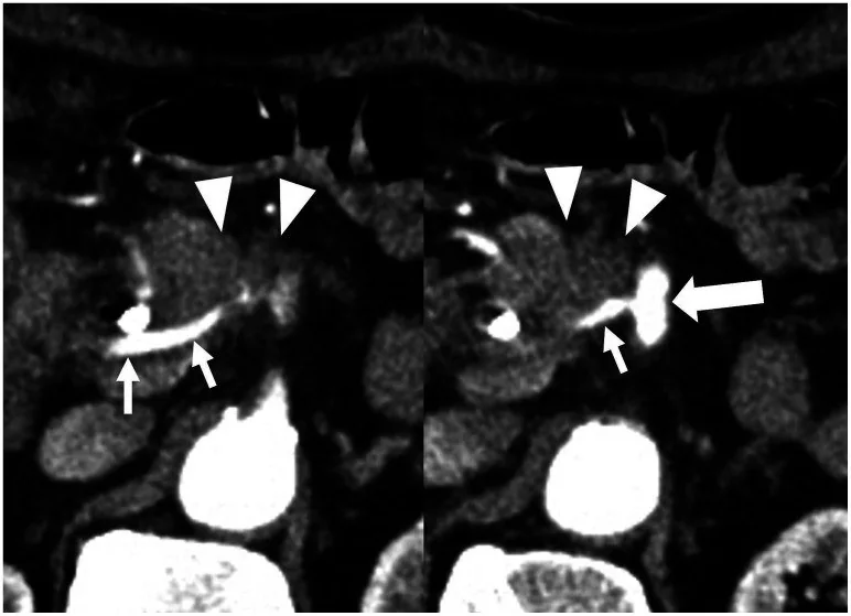
- Pankreatik parankim fazında küçük PDAC normal pankreasla benzer atenüasyonda kalabilir; fokal ana kanal stenozu veya obstrüksiyonu ve distal kanal genişlemesi doğrudan kitle seçilmese de tanıda ipucu olabilir.
- Portal tanısal
- Portal venöz fazı da içeren ince kesitli, çok fazlı BT'de damar tutulumu ve metastatik hastalık değerlendirilir; küçük PDAC normal pankreasla benzer atenüasyonda olduğunda kitle doğrudan seçilemeyebilir.
Önerilen pencereler: Batın (G 400 / M 50), Yumuşak doku (G 400 / M 40).
BT bulguları
- Ana pankreatik kanalda kısa segmentte ani daralma veya kesilme, kitle görünmeden önceki ipucu olabilir.
- Darlığın kuyruk tarafındaki kanalda fokal genişleme ve kesitler boyunca belirgin kalibre farkı görülür.
- Kanal tıkanıklığının distalindeki pankreas dokusunda segmental hacim kaybı veya lobülasyon azalması gelişebilir.
- Şüpheli düzeyde pankreasın ön konturunda hafif çökme, asimetri veya normal lobüler yapının silinmesi izlenebilir.
- Kitle, pankreasla aynı attenüasyonda olabilir; çevre yağ planı ve kanal yönündeki değişiklikler sınırını dolaylı biçimde belli eder.
- Tümör-damar teması, reseyabiliteyi belirlemede çokfazlı ince kesitli BT ile değerlendirilir.
Normalde
Normal bezde ana pankreatik kanal gövde ve kuyruk boyunca kesintisiz seyreder, parankim lobülasyonu düzenlidir ve segmental incelme beklenmez. İncelemeyi normal komşu segmentle kıyasla: kanal kalibre geçişinin fokal olup olmadığını, aynı düzeyde kontur çökmesi bulunup bulunmadığını ve distal parankimin hacmini değerlendir.
Normal BT ile kıyasla
Aynı bölgenin, kaynak veri setinde patoloji içermediği belirtilen bir BT incelemesini kesit kesit kaydırın; organ sınırlarını açarak anatomiyi hasta görüntüsüyle karşılaştırın. Küçük rastlantısal bulgular tümüyle dışlanmamıştır.
Kaynak: TotalSegmentator v2.01 veri seti (Wasserthal ve ark., Radiology: Artificial Intelligence 2023), CC BY 4.0, vaka s0541; organ sınırları veri setinin otomatik segmentasyonundan, birleştirilerek sadeleştirildi. Görüntüler 2,5 mm kesit aralığına yeniden örneklendi.
Ayırıcı tanı
- Kronik pankreatit darlığı
- düzensiz ve çok odaklı kanal değişikliği, parankimal kalsifikasyon veya yaygın atrofi eşlik edebilir.
- Ana kanal ve yan dal IPMN anatomik alt tipler olarak sınıflandırılır; kistik lezyonun pankreatik kanal sistemiyle bağlantısı değerlendirilir.
- Fokal otoimmün pankreatit
- bezde daha uzun segmentli tutulum ve gradual daralma ile kanal daralması görülebilir.
- Pankreas divisum, dorsal ve ventral pankreatik kanal sistemlerinin birleşme kusuruna bağlı doğuştan anatomik bir varyanttır.
- Küçük nöroendokrin tümör
- pankreatik fazda çoğunlukla parlak kontrastlanır; duktal tıkanma klasik küçük lezyon görünümü değildir.
Tuzaklar
- İzodens tümör, bezin doğal lobülasyonu içinde kaybolabilir; yalnız kitle aramak yerine tüm ana kanalı baştan kuyruğa takip et.
- Kronik pankreatit kanal darlığı tümörü taklit edebilir; yeni tek odaklı kesilme ve bu düzeyde kontur deformitesini kronik değişikliklerden ayrı kaydet.
- Küçük PDAC boyutu nedeniyle BT’de gözden kaçabilir; pankreası ince kesitli, çok fazlı BT’de dikkatle değerlendir.
Kendini dene
Küçük kitle seçilmiyor; ana kanal kısa segmentte aniden kesiliyor ve kuyrukta fokal genişleme var. En olası tanı nedir?
Cevabı göster
İzodens PDAC. Fokal kanal kesilmesiyle aynı seviyedeki yukarı akım genişlemesi küçük veya izodens PDAC için dolaylı bulgudur. Divisum doğuştan drenaj varyantı, kistadenom makrokistik kitle, anevrizma ise damar kökenli kontrastlanan yapıdır.
Kanal darlığına çok sayıda bez kalsifikasyonu ve düzensiz çok odaklı kanal yapısı eşlik ediyor. Daha olası alternatif nedir?
Cevabı göster
Kronik pankreatit. Yaygın kalsifikasyon ve çok odaklı duktal düzensizlik kronik pankreatit örüntüsünü destekler. Lipom yağ attenüasyonunda sınırlı odaktır; insulinoma tipik olarak arteriyel hipervaskülerdir, koledok kisti safra kanalında yerleşir.
İlgili konular
Kaynaklar
Bu sayfadaki 35 cümle ve 2 quiz sorusu aşağıdaki kaynaklardan alıntılarla tek tek karşılaştırıldı; 7 cümle bu karşılaştırmada düzeltildi (2026-10-09).
- Diagnostic Method Advancement to Improve the Prognosis of Pancreatic Ductal Adenocarcinomapmc.ncbi.nlm.nih.gov
- Artificial intelligence-assisted endoscopic ultrasonography for the detection of a small T1 pancreatic ductal adenocarcinoma in a difficult-to-puncture locationpmc.ncbi.nlm.nih.gov
- Multicenter Validation of Foundation Model Adaptation for Automated Pancreatic Tumor Delineation on CT Scanspmc.ncbi.nlm.nih.gov
- Pretherapeutic radiological sarcopenia in pancreatic ductal adenocarcinoma: a narrative reviewpmc.ncbi.nlm.nih.gov
- Concurrent EGFR exon 19 deletion-insertion and germline BRCA1 alteration in KRAS-wild-type pancreatic ductal adenocarcinoma: A case report and review of therapeutic implicationspmc.ncbi.nlm.nih.gov
- The isoattenuating pancreatic adenocarcinoma: Review of the literature and critical analysis.pubmed.ncbi.nlm.nih.gov
- Pitfalls in the Imaging Interpretation of Intracranial Hemorrhage.pubmed.ncbi.nlm.nih.gov
- Oncologic applications of dual-energy CT in the abdomen.pubmed.ncbi.nlm.nih.gov
- A Technique to Identify Isoattenuating Gallstones with Dual-Layer Spectral CT: An ex Vivo Phantom Study.pubmed.ncbi.nlm.nih.gov
- [Visually Isoattenuating Pancreatic Acinar Cell Carcinoma:Report of One Case].pubmed.ncbi.nlm.nih.gov
- Endoscopic Evaluation in the Workup of Pancreatic Cancer.pubmed.ncbi.nlm.nih.gov
- Primary solid pancreatic tumors: recent imaging findings updates with pathology correlation.pubmed.ncbi.nlm.nih.gov
- Diagnostic performance of endoscopic ultrasound for detection of pancreatic malignancy following an indeterminate multidetector CT scan: a systemic review and meta-analysis.pubmed.ncbi.nlm.nih.gov
- Plasmonic-doped melanin-mimic for CXCR4-targeted NIR-II photoacoustic computed tomography-guided photothermal ablation of orthotopic hepatocellular carcinoma.pubmed.ncbi.nlm.nih.gov
- Hypervascular subepithelial gastrointestinal masses: CT-pathologic correlation.pubmed.ncbi.nlm.nih.gov
- The appearance of canine insulinoma on dual phase computed tomographic angiography.pubmed.ncbi.nlm.nih.gov
- Computed tomography-guided percutaneous biopsy of isoattenuating focal liver lesions.pubmed.ncbi.nlm.nih.gov
- CT characteristics of primary hepatic mass lesions in dogs.pubmed.ncbi.nlm.nih.gov
- Pancreatic ductal adenocarcinoma concomitant with intraductal papillary mucinous neoplasm: a report of 8 cases.pubmed.ncbi.nlm.nih.gov
- Diagnostic Method Advancement to Improve the Prognosis of Pancreatic Ductal Adenocarcinoma.pubmed.ncbi.nlm.nih.gov
- New-Onset Diabetes and Pancreatic Ductal Adenocarcinoma: Implications for Early Recognition and Clinical Management.pubmed.ncbi.nlm.nih.gov
- Characteristics of ill-demarcated small pancreatic ductal adenocarcinoma on MRI: a multi-institutional retrospective cohort study.pubmed.ncbi.nlm.nih.gov
- CT and MR evaluation of main pancreatic duct dilation: A radiologist's guide to differentiating benign from malignant causes.pubmed.ncbi.nlm.nih.gov
- Case Report: A four-stage diagnostic cascade: from coronary stents to pancreatic cancer in a patient with recurrent chest pain.pubmed.ncbi.nlm.nih.gov
- Steroid-resistant Relapse of Autoimmune Pancreatitis with Marked Main Pancreatic Duct Dilatation Mimicking Multifocal Pancreatic Cancer.pubmed.ncbi.nlm.nih.gov
- Congenital pulmonary airway malformation mimicking lung cancer in adults: a pitfall in oncologic diagnosis.pubmed.ncbi.nlm.nih.gov
- Updates on Imaging Assessment of Pancreatic Cancer for Determining Anatomic and Biologic Resectability.pubmed.ncbi.nlm.nih.gov
- CT-based vascular invasion in pancreatic ductal adenocarcinoma compared with intraoperative and histological findings.pubmed.ncbi.nlm.nih.gov
- Fibrolamellar hepatocellular carcinoma: Advances, challenges and opportunities in a rare malignancy.pubmed.ncbi.nlm.nih.gov
- Imaging findings of a case of peliosis hepatis mimicking malignancies.pubmed.ncbi.nlm.nih.gov
- Pancreatic cancer, autoimmune or chronic pancreatitis, beyond tissue diagnosis: Collateral imaging and clinical characteristics may differentiate them.pubmed.ncbi.nlm.nih.gov
- Acinar cell carcinoma of the pancreas: can CT and MR features predict survival?pubmed.ncbi.nlm.nih.gov
- Morphological characterization of atypical pancreatic ductal adenocarcinoma with cystic lesion on DCE-CT: a comprehensive retrospective study.pubmed.ncbi.nlm.nih.gov
- Photon-counting CT for pancreatic cancer: advancing precision imaging and virtual surgical navigation.pubmed.ncbi.nlm.nih.gov
- Diagnosis of Perihilar Osteoclast Giant Cell Carcinoma in an Elderly Patient: A Report of Diagnostic Challenges Over Several Attempts.pubmed.ncbi.nlm.nih.gov
- Metachronous Primary Pancreatic Neuroendocrine Tumor and Adenocarcinoma: A Case Report.pubmed.ncbi.nlm.nih.gov
- Recent Advances in Non-Invasive Blood Markers for Intraductal Papillary Mucinous Neoplasm Grading.pubmed.ncbi.nlm.nih.gov
- Pancreatic Calculi: Two Case Reports and Literature Review.pubmed.ncbi.nlm.nih.gov
- Rapid Development of Diffuse-Type Remnant Pancreatic Cancer Following Obstructive Pancreatitis after Pancreaticoduodenectomy for Distal Bile Duct Carcinoma: A Case Report.pubmed.ncbi.nlm.nih.gov
- Successful endoscopic management of a pediatric intrapancreatic common bile duct stricture following blunt pancreatic trauma: A case report.pubmed.ncbi.nlm.nih.gov
- Pancreatic Atrophy: A Narrative Review and Surgical Interpretation.pubmed.ncbi.nlm.nih.gov
- The pseudocirrhotic liver: Hepar lobatum carcinomatosum in a patient with triple-negative breast cancer.pubmed.ncbi.nlm.nih.gov
- Intraductal Papillary Mucinous Neoplasm (IPMN) of the Pancreas: History, Myths, and Realities Between Past and Future.pubmed.ncbi.nlm.nih.gov
- Adaptive Radiotherapy in the Era of Image Guidance and Artificial Intelligence: A Critical Review.pubmed.ncbi.nlm.nih.gov
- Development of CBCT-Guided Online Adaptive Radiotherapy.pubmed.ncbi.nlm.nih.gov
- Assessment of dose distribution in pancreatic SABR in the presence of a metallic biliary stent using Monte Carlo modeling.pubmed.ncbi.nlm.nih.gov
- Pancreatic Adenocarcinoma: Imaging Modalities and the Role of Artificial Intelligence in Analyzing CT and MRI Images.pubmed.ncbi.nlm.nih.gov
- Dual-Energy CT in Oncologic Imaging.pubmed.ncbi.nlm.nih.gov
- Multidetector computer tomography in the pancreatic adenocarcinoma assessment: an update.pubmed.ncbi.nlm.nih.gov
- Distal cholangiocarcinoma mimicking pancreatic ductal adenocarcinoma successfully converted to resection following PD-1/CTLA-4 bispecific immunotherapy (cadonilimab) combined with nab-paclitaxel-based chemotherapy: a case report providing preliminary real-world insight.pubmed.ncbi.nlm.nih.gov
- A Case of Intraductal Tubulopapillary Neoplasm of the Pancreas Requiring Superior Mesenteric Vein-Portal Vein Axis Resection after Systemic Chemotherapy.pubmed.ncbi.nlm.nih.gov
- A super-elderly patient with distal biliary tract cancer and tumor-associated granuloma: a case report.pubmed.ncbi.nlm.nih.gov
- Dorsal Pancreatic Agenesis Associated With New-Onset Diabetes Mellitus and Acute Pancreatitis in an 18-Year-Old Man: A Case Report.pubmed.ncbi.nlm.nih.gov
- Pancreatic Cancer after Living-Donor Liver Transplantation: A Case Report of Long-Term Recurrence-Free Survival after Pancreaticoduodenectomy Followed by Adjuvant Chemotherapy.pubmed.ncbi.nlm.nih.gov
- Spontaneous Bacterial Peritonitis as a Rare Complication of a Pancreatic Duct Leak in a Patient Without Cirrhosis: A Case Report.pubmed.ncbi.nlm.nih.gov
- Common cystic pancreatic neoplasms: A comprehensive magnetic resonance imaging-based review of typical and atypical imaging features.pubmed.ncbi.nlm.nih.gov
- Endoscopic Ultrasound-Guided Radiofrequency Ablation for Pancreatic Metastases: Safety and Clinical Outcomes in a Single-Center Case Series.pubmed.ncbi.nlm.nih.gov
- High-Frequency Miniprobe Endoscopic Ultrasonography Across the Gastrointestinal Tract.pubmed.ncbi.nlm.nih.gov
- Artificial Intelligence-Based Prediction of Pancreatic Stone Clearance in Pancreatolithiasis Using Pretreatment CT Images and Clinical Features.pubmed.ncbi.nlm.nih.gov
- IgG4-related autoimmune pancreatitis mimicking pancreatic carcinoma: a case report.pubmed.ncbi.nlm.nih.gov
- Artificial intelligence in chronic and autoimmune pancreatitis: diagnosis, prognosis, and personalized management.pubmed.ncbi.nlm.nih.gov
- Diagnostic value of EUS-guided tissue acquisition in suspected autoimmune pancreatitis: A 15-year single-center retrospective study.pubmed.ncbi.nlm.nih.gov
- Role of endoscopic ultrasound in diagnosis and management of autoimmune pancreatitis.pubmed.ncbi.nlm.nih.gov
- Intraductal papillary mucinous neoplasm-derived pancreatic cancer of the ventral duct in patient with pancreas divisum: a case report.pubmed.ncbi.nlm.nih.gov
- A Rare Case of Pancreatic Neuroendocrine Tumor with Intraductal Extension in the Dorsal Duct of a Pancreas Divisum.pubmed.ncbi.nlm.nih.gov
- A rare case of pancreatitis caused by pancreas divisum in a middle-aged patient and treated with stent implantation: Case report.pubmed.ncbi.nlm.nih.gov
- Incidental cystic pancreatic neuroendocrine neoplasm and differential diagnosis.pubmed.ncbi.nlm.nih.gov
- Pancreatic Steatosis and Survival Outcomes in Pancreatic Ductal Adenocarcinoma: A Systematic Review of the Current Evidence.pubmed.ncbi.nlm.nih.gov
- Clinical Utility of Circulating Tumor DNA Analysis in Pancreatic Ductal Adenocarcinoma.pubmed.ncbi.nlm.nih.gov
- A case report of a composite pheochromocytoma that occurred 13 years after adrenalectomy for pheochromocytoma.pubmed.ncbi.nlm.nih.gov
- Resectable and Borderline Resectable Pancreatic Ductal Adenocarcinoma: Role of the Radiologist and Oncologist in the Era of Precision Medicine.pubmed.ncbi.nlm.nih.gov
- Imaging diagnosis and staging of pancreatic ductal adenocarcinoma: a comprehensive review.pubmed.ncbi.nlm.nih.gov
- Not All Pancreatic Masses are Cancer: Pancreatic Tuberculosis Masquerading as Malignancy.pubmed.ncbi.nlm.nih.gov
- A Tangled Tale: Type 2 Autoimmune Pancreatitis With a Sclerosing Twist.pubmed.ncbi.nlm.nih.gov
- Incidental Lung Lesions on the CT Component of Oncologic FDG PET/CT: A Pictorial Review of Interpretive Pitfalls and Diagnostic Clues.pubmed.ncbi.nlm.nih.gov
- Understanding Photon-Counting CT: Physics, Detector Technology, and Image Reconstructions.pubmed.ncbi.nlm.nih.gov
- Endoscopic ultrasound-guided radiofrequency ablation for pancreatic cystic neoplasms: a comprehensive review.pubmed.ncbi.nlm.nih.gov
- Centerline-guided reinforcement learning model for pancreatic duct identifications.pubmed.ncbi.nlm.nih.gov
- Using multidetector-row CT for the diagnosis of afferent loop syndrome following gastroenterostomy reconstruction.pubmed.ncbi.nlm.nih.gov
- Imaging of early pancreatic cancer on multidetector row helical computed tomography.pubmed.ncbi.nlm.nih.gov
- Synchronous Multifocal Intraductal Tubulopapillary Neoplasm of the Pancreas Treated with Total Pancreatectomy: A Case Report.pubmed.ncbi.nlm.nih.gov
- Granular cell tumor of the pancreas mimicking pancreatic ductal adenocarcinoma: a case report.pubmed.ncbi.nlm.nih.gov
- Pancreatic Collision Tumor Composed of Solid Pseudopapillary Neoplasm and Well-Differentiated Neuroendocrine Tumor: Diagnostic Challenges and Classification Considerations.pubmed.ncbi.nlm.nih.gov
- Small pancreatic ductal adenocarcinoma (≤ 2 cm): different imaging and clinicopathologic features according to extrapancreatic extension.pubmed.ncbi.nlm.nih.gov
- An update on pancreatic cancer imaging, staging, and use of the PACT-UK radiology template pre- and post-neoadjuvant treatment.pubmed.ncbi.nlm.nih.gov
- Pancreatic ductal adenocarcinoma staging: a narrative review of radiologic techniques and advances.pubmed.ncbi.nlm.nih.gov
- Changes Connected to Early Chronic Pancreatitis and Early Pancreatic Cancer in Endoscopic Ultrasonography (EUS): Clinical Implications.pubmed.ncbi.nlm.nih.gov
- Quantitative classification of pancreatic atrophy: the İstanbul morphometric pancreatic atrophy classification based on CT and MRI measurements.pubmed.ncbi.nlm.nih.gov
- Boerhaave Syndrome Caused by Pancreatitis-Induced Gastric Outlet Obstruction: A Case Report.pubmed.ncbi.nlm.nih.gov
- Coexisting autoimmune pancreatitis and pancreatic adenocarcinoma: Diagnostic challenges and case insights.pubmed.ncbi.nlm.nih.gov
- From Routine Imaging to Risk Stratification: Multimodal Vision-Language Survival Modeling for Pancreatic Cancer.pubmed.ncbi.nlm.nih.gov
- Development and validation of a clinical-radiomics nomogram for the differential diagnosis of focal pancreatic solid lesions: a retrospective cohort study.pubmed.ncbi.nlm.nih.gov
- Diagnosis and Surgical Management for Advanced Pancreatic Cancer Requiring Vascular Resection.pubmed.ncbi.nlm.nih.gov
- Case Report: Late repeat pancreatic irreversible electroporation in the long-term multidisciplinary management of unresectable pancreatic ductal adenocarcinoma.pubmed.ncbi.nlm.nih.gov
- Histotripsy Outcomes for Primary and Metastatic Hepatic Disease.pubmed.ncbi.nlm.nih.gov
- Portuguese Pancreatic Club Perspective on Epidemiology, Classification, and Natural History of Pancreatic Intraductal Papillary Mucinous Neoplasms.pubmed.ncbi.nlm.nih.gov
- Contrast enhanced CT and MRI interchangeably reflect tumor characteristics in murine pancreatic cancer.pubmed.ncbi.nlm.nih.gov
- A narrative review of imaging for pancreas adenocarcinoma: staging, surgical considerations, and surveillance.pubmed.ncbi.nlm.nih.gov
- Revolution APEX CT: preliminary assessment of its application value in pancreatic cancer T staging.pubmed.ncbi.nlm.nih.gov
- The blind spots on chest computed tomography: what do we miss.pubmed.ncbi.nlm.nih.gov
- Imaging of pancreatic ductal adenocarcinoma - An update for all stages of patient management.pubmed.ncbi.nlm.nih.gov
- Clinical significance of CD161<sup>+</sup>CD8<sup>+</sup> T cells in the tumor microenvironment of esophageal squamous cell carcinoma and their association with the 4-1BB signaling pathway.pubmed.ncbi.nlm.nih.gov
- Comprehensive Computed Tomography Evaluation of Gastric Cancer: 2026 Consensus Recommendation From the Korean Society of Abdominal Radiology.pubmed.ncbi.nlm.nih.gov
- Pulmonary nodules with explosive calcification were finally diagnosed as lung adenocarcinom: A case report.pubmed.ncbi.nlm.nih.gov
Eğitim amaçlıdır; hasta başında tanı ve tedavi kararının yerine geçmez. İçerik yapay zekâ destekli bir süreçle güncel literatüre göre cümle cümle kaynaklarla karşılaştırıldı, ancak uzman radyolog incelemesinden geçmedi. Klinik kararlarda güncel kılavuzları, kurum protokollerini ve radyoloji raporunu esas alın.