Özet
Kalsifiye psoas tüberküloz apsesi, çoğunlukla omurga tüberkülozundan psoas kompartımanına uzanan kronik enfeksiyonun kitle benzeri koleksiyonudur. Kontrastsız BT, psoas apsesiyle birlikte vertebra korpuslarında destrüksiyon ve kalsifikasyonu gösterir. Kalsifiye paravertebral veya psoas apsesi ve eşlik eden vertebra destrüksiyonu tüberkülozu güçlü biçimde düşündürür; kesin etken tanısı mikrobiyolojik veya histopatolojik incelemeyle doğrulanır. Spinal tüberkülozla ilişkili psoas apsesinde vertebral destrüksiyona paravertebral ve epidural uzanım eşlik edebilir; bu alanların değerlendirilmesi hastalığın yayılımını ortaya koymaya yardımcı olur.
Faz ve pencere
- Kontrastsız tanısal
- BT'de psoas apsesi içindeki kalsifikasyonlar ve eşlik eden vertebra destrüksiyonu görülebilir. Kalsifikasyonun yerini ve kemik korteksiyle ilişkisini bu fazda ayırt edin.
- Portal tanısal
- BT'de psoas koleksiyonu ve eşlik eden vertebra destrüksiyonu değerlendirilebilir.
Önerilen pencereler G genişlik, M merkez; değerler Hounsfield birimi (HU)
- KemikG 1800 / M +400
- BatınG 400 / M +50
- Yumuşak dokuG 400 / M +40
BT bulguları
- Psoas kasında büyük bir koleksiyon görülebilir.
- Duvar veya içerik kalsifikasyonu — koleksiyon çevresinde çizgisel/duvar biçimli ya da içerikte odaksal mineral yoğunluklar izlenir; kronik tüberküloz odağını destekler.
- Psoas apsesi, psoas kası içinde irin birikmesiyle oluşan bir koleksiyondur.
- Psoas koleksiyonu vertebra ve paravertebral dokularla birlikte tutulabilir; spinal tüberküloz olgusunda epidural uzanım da eşlik etmiştir.
- Psoas apsesine vertebra destrüksiyonu eşlik edebilir.
- Spinal tüberkülozda spondilodiskit görülebilir; bazı olgularda buna prevertebral veya psoas koleksiyonu eşlik eder.
- MRG, epidural ve paravertebral yumuşak doku komplikasyonlarını daha iyi gösterirken BT, sekestr ve paravertebral kalsifikasyon gibi kemik ve kalsifikasyon ayrıntılarını ortaya koyabilir.
Normalde
Psoas apsesi, psoas kası içinde irin birikmesiyle oluşan bir koleksiyondur.
Normal BT ile kıyasla
Aynı bölgenin, kaynak veri setinde patoloji içermediği belirtilen bir BT incelemesini kesit kesit kaydırın; organ sınırlarını açarak anatomiyi hasta görüntüsüyle karşılaştırın. Küçük rastlantısal bulgular tümüyle dışlanmamıştır.
Kaynak: TotalSegmentator v2.01 veri seti (Wasserthal ve ark., Radiology: Artificial Intelligence 2023), CC BY 4.0, vaka s0119; organ sınırları veri setinin otomatik segmentasyonundan, birleştirilerek sadeleştirildi. Görüntüler 2,5 mm kesit aralığına yeniden örneklendi.
Ayırıcı tanı
- Tüberküloz spondilodiskitinde torakal veya çok seviyeli tutulum ve görece korunmuş disk aralığı görülebilir; piyojenik spondilodiskitte erken disk-son plak yıkımı daha çok destekleyici özelliklerdendir.
- Psoas hematomu, spinal tüberkülozu taklit edebilen ayırıcı tanılar arasında bildirilmiştir.
- Paraspinal kitleler paraspinal yumuşak dokulardan veya vertebral kolondan kaynaklanabilir; bu bölgede primer ve sekonder tümörler de görülür.
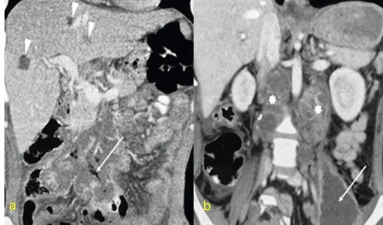
- Abdominal tüberkülozda pankreas tutulumu ile birlikte kalsifiye lenfadenopati görülebilir.
- Kalsifikasyon tek başına tüberküloz etkenini kanıtlamaz; spinal tüberküloz ile piyojenik spondilodiskit ayrımında tek bir görüntüleme bulgusu tanı koydurucu değildir.
Tuzaklar
- Kalsifikasyon tek başına tüberkülozu mikrobiyolojik olarak kanıtlamaz; raporda görüntüleme örüntüsünü güçlü şüphe olarak tanımlayın ve etken doğrulamasını görüntüden ayırın.
- Kitlelerin kökeni (kas içi, nodal veya vertebral) paraspinal bölgenin anatomik düzenine bağlıdır; doğru konumlandırma lenf nodu, tümör veya apse gibi ayırıcı tanıları birbirinden ayırmada kritik rol oynar.
- Vertebral kemik erozyonu kontrastsız yumuşak doku penceresinde silik kalabilir; aynı alanı kemik algoritmasında ve çok düzlemli kesitlerde inceleyin.
Kendini dene
Kalsifiye psoas apsesi ve spondilit saptanan olgu raporunda tüberküloz etkeni nasıl tanımlandı?
Cevabı göster
Psoas apsesine BT eşliğinde perkütan drenaj uygulandıktan sonra Mycobacterium tuberculosis saptandı. BT'de kalsifikasyonlu psoas apsesi görüldü; MTB, BT eşliğinde perkütan drenaj sonrası tanımlandı.
Olgu raporunda tedavi edilmemiş Pott hastalığına hangi görüntüleme bulguları eşlik ediyordu?
Cevabı göster
Bilateral kalsifiye psoas apseleri ve kifoz. Olgu raporunda tedavi edilmemiş Pott omurgasıyla birlikte bilateral kalsifiye psoas apseleri ve kifoz bildirilmiştir.
İlgili konular
Kaynaklar
Bu sayfadaki 33 cümle ve 2 quiz sorusu aşağıdaki kaynaklardan alıntılarla otomatik olarak karşılaştırıldı; 17 cümle bu karşılaştırmada düzeltildi (2026-10-09).
- Complete Bilateral Calcified Psoas Abscess- Rare Sequelae of Untreated Pott's Spine.pubmed.ncbi.nlm.nih.gov
- Iliopsoas Pyomyositis With Bacteremia at an Early Stage of Presentation in a Temperate Region.pubmed.ncbi.nlm.nih.gov
- Spinal lesions: a comprehensive radiologic overview.pubmed.ncbi.nlm.nih.gov
- Understanding a mass in the paraspinal region: an anatomical approach.pubmed.ncbi.nlm.nih.gov
- Extrapulmonary tuberculosıs: an old but resurgent problem.pubmed.ncbi.nlm.nih.gov
- Laparoscopic Living Donor Nephrectomy: Learning Curve Analysis Through 1446 Cases and Outcomes from 200 Consecutive Mastery-Phase Procedures-How I Do It.pubmed.ncbi.nlm.nih.gov
- Solid Primary Retroperitoneal Masses in Adults: An Imaging Approach.pubmed.ncbi.nlm.nih.gov
- Benign Renal Tumors in Pediatric Age Group: Retrospective Analysis.pubmed.ncbi.nlm.nih.gov
- Differential Diagnosis of Abdominal Tuberculosis in the Adult-Literature Review.pubmed.ncbi.nlm.nih.gov
- Imaging of Musculoskeletal Soft-Tissue Infections in Clinical Practice: A Comprehensive Updated Review.pubmed.ncbi.nlm.nih.gov
- Long-term safety and effectiveness of the "OptEase" vena cava filter.pubmed.ncbi.nlm.nih.gov
- Vertebral osteomyelitis caused by nontuberculous mycobacteria: a case series of 23 patients and a review of 108 cases in the literature.pubmed.ncbi.nlm.nih.gov
- Unmasking the mimic: vertebral alveolar echinococcosis diagnosed by metagenomic next-generation sequencing.pubmed.ncbi.nlm.nih.gov
- Infectious Spondylodiscitis of Bacterial Causes in Adults: Epidemiology, Pathophysiology, Diagnostic and Treatment Challenges.pubmed.ncbi.nlm.nih.gov
- Tuberculosis Beyond the Lungs: A Pictorial Review of Key Diagnostic Imaging Insights.pubmed.ncbi.nlm.nih.gov
- Bacterial Granulomatous Lung Diseases: Radiological Findings and Differential Diagnosis.pubmed.ncbi.nlm.nih.gov
- Comparative Review of Lateral and Oblique Lumbar Interbody Fusion: Technique, Outcomes, and Complications.pubmed.ncbi.nlm.nih.gov
- Emphysematous conditions of the abdomen and pelvis: pearls and pitfalls.pubmed.ncbi.nlm.nih.gov
- Minimally invasive prone lateral retropleural or retroperitoneal antepsoas approach spinal surgery using the rotatable radiolucent Jackson table.pubmed.ncbi.nlm.nih.gov
- Imaging clues for the diagnosis of various pathogenic causes of infectious spondylitis.pubmed.ncbi.nlm.nih.gov
- Non-Infectious Spinal Diseases Mimicking Infection: A Pictorial Essay.pubmed.ncbi.nlm.nih.gov
- Diagnostic value of percutaneous paramedian small-angle lateral intervertebral foramen Kambin's triangle approach for lumbar puncture biopsy combined with tNGS-based multimodal etiological diagnosis in early spinal infection: a multicenter retrospective diagnostic yield study.pubmed.ncbi.nlm.nih.gov
- Tuberculosis revisted: classic imaging findings in childhood.pubmed.ncbi.nlm.nih.gov
- Radiologic evaluation of the kidney transplant donor and recipient.pubmed.ncbi.nlm.nih.gov
- Lumbosacral Spinal Tuberculosis: Clinical Spectrum, Diagnostic Challenges, and Short-Term Management Outcomes in an Elderly Cohort.pubmed.ncbi.nlm.nih.gov
- Recommendations of the International Consensus Meeting (ICM) 2025 on Spinal Infections: Part B Tubercular Spondylodiscitis.pubmed.ncbi.nlm.nih.gov
- The clinical characteristics and outcomes of anaerobic spondylodiscitis: a retrospective observational study.pubmed.ncbi.nlm.nih.gov
- Acute sacral osteomyelitis mimicking hip pathology in a child: A diagnostic challenge.pubmed.ncbi.nlm.nih.gov
- Multifocal Skeletal Tuberculosis - A Case Report.pubmed.ncbi.nlm.nih.gov
- Feasibility and clinical efficacy of minimally invasive techniques in the treatment of type Ⅱ/Ⅲ brucellar spondylitis.pubmed.ncbi.nlm.nih.gov
- Imaging of lower extremity infections: predisposing conditions, atypical infections, mimics, and differentiating features.pubmed.ncbi.nlm.nih.gov
- Extravertebral low back pain: a scoping review.pubmed.ncbi.nlm.nih.gov
- The value of magnetic resonance imaging and computed tomography in the study of spinal disorders.pubmed.ncbi.nlm.nih.gov
- Diffuse cystic lung diseases-a primer for radiologists.pubmed.ncbi.nlm.nih.gov
- CT and MR in peritoneal malignancies: pearls and pitfalls at preoperative examination.pubmed.ncbi.nlm.nih.gov
- The Many Faces of the Angry Peritoneum.pubmed.ncbi.nlm.nih.gov
- Among the fibers: A multimodality imaging review of intramuscular mass lesions.pubmed.ncbi.nlm.nih.gov
- Imaging in spinal posterior epidural space lesions: A pictorial essay.pubmed.ncbi.nlm.nih.gov
- Differentiating Tuberculous and Pyogenic Spondylodiscitis: Part II: MRI/CT Features, Prediction Models, and the Role of PET/CT-A Narrative Review.pubmed.ncbi.nlm.nih.gov
- Imaging insights into systemic tuberculosis with cardiac involvement: A rare challenging case of tuberculous chronic constrictive pericarditis and myocarditis.pubmed.ncbi.nlm.nih.gov
- A Case of Pott's Disease Complicated by Tuberculous Meningitis and Psoas Abscess Diagnosed by Computed Tomography-Guided Percutaneous Drainage.pubmed.ncbi.nlm.nih.gov
- Spontaneous bacteremia and spontaneous bacterial peritonitis share similar prognosis in patients with cirrhosis: a cohort study.pubmed.ncbi.nlm.nih.gov
- A transformer-based deep learning algorithm for diagnosing spinal infections on axial non-contrast computed tomography images: a dual-center retrospective study.pubmed.ncbi.nlm.nih.gov
- Diagnostic values of peripheral blood T-cell spot of tuberculosis assay (T-SPOT.TB) and magnetic resonance imaging for osteoarticular tuberculosis: a case-control study.pubmed.ncbi.nlm.nih.gov
- State of the art of 18 F-FDG PET/CT application in inflammation and infection: a guide for image acquisition and interpretation.pubmed.ncbi.nlm.nih.gov
- Tuberculosis of the genitourinary system-Urinary tract tuberculosis: Renal tuberculosis-Part II.pubmed.ncbi.nlm.nih.gov
- Approach to Cystic Lesions in the Abdomen and Pelvis, with Radiologic-Pathologic Correlation.pubmed.ncbi.nlm.nih.gov
- Imaging of Sacroiliac Pain: The Current State-of-the-Art.pubmed.ncbi.nlm.nih.gov
- Imaging of presacral masses--a multidisciplinary approach.pubmed.ncbi.nlm.nih.gov
- A review of imaging modalities for the hip.pubmed.ncbi.nlm.nih.gov
- Pathogenic mechanisms of adult degenerative scoliosis.pubmed.ncbi.nlm.nih.gov
- Transverse Process Fractures in Athletes: Mechanisms, Management and Return-to-Play Considerations.pubmed.ncbi.nlm.nih.gov
- Morphometric analysis of safe working zones for minimally invasive lateral extrapleural/retroperitoneal approach via transdiaphragmatic and transpsoas to the thoracolumbar and lumbar spine.pubmed.ncbi.nlm.nih.gov
- Psoas Abscess From Spinal Tuberculosis Mimicking Appendicitis: A Case Report.pubmed.ncbi.nlm.nih.gov
- Brucella Spondylitis: Current Knowledge and Recent Advances.pubmed.ncbi.nlm.nih.gov
- A Severe Case of Disseminated Multifocal Methicillin-Susceptible Staphylococcal Infection in a Diabetic Patient.pubmed.ncbi.nlm.nih.gov
- Primary and secondary tumors of the peritoneum: key imaging features and differential diagnosis with surgical and pathological correlation.pubmed.ncbi.nlm.nih.gov
- Case Report: Primary gastric extraskeletal osteosarcoma and literature review.pubmed.ncbi.nlm.nih.gov
- CT and MR Imaging of Retroperitoneal Sarcomas: A Practical Guide for the Radiologist.pubmed.ncbi.nlm.nih.gov
- Incremental value of quantitative SPECT/CT integrated parameters in a multimodal machine-learning model for differentiating spinal tuberculosis from pyogenic spondylitis.pubmed.ncbi.nlm.nih.gov
- Computed tomography in the diagnosis of bilateral renal tuberculosis: diagnostic value, limitations, and future directions.pubmed.ncbi.nlm.nih.gov
- CT of acute abdomen in the elderly.pubmed.ncbi.nlm.nih.gov
- Purulent infectious myositis of deep core muscles, a MRSA infection in India.pubmed.ncbi.nlm.nih.gov
- Concomitant osteoporotic vertebral compression fracture and infectious spondylitis in a patient with herpes zoster: A case report.pubmed.ncbi.nlm.nih.gov
- Secondary Complications in COVID-19 Patients: A Case Series.pubmed.ncbi.nlm.nih.gov
- Unraveling a Complex Case: Brucellosis Manifesting as Fever of Unknown Origin, Septic Ankle Arthritis, and Iliacus Abscess.pubmed.ncbi.nlm.nih.gov
- Imaging of Spondylodiscitis: A Comprehensive Updated Review-Multimodality Imaging Findings, Differential Diagnosis, and Specific Microorganisms Detection.pubmed.ncbi.nlm.nih.gov
- Invited review: Recommendations for reporting intervention studies on reproductive performance in dairy cattle: Improving design, analysis, and interpretation of research on reproduction.pubmed.ncbi.nlm.nih.gov
- Case 295.pubmed.ncbi.nlm.nih.gov
- Vertical Medial Thigh Contouring.pubmed.ncbi.nlm.nih.gov
- The vertical medial thigh lift.pubmed.ncbi.nlm.nih.gov
- Acute pyelonephritis complicated by subcapsular and perinephric hematoma: Diagnostic role of CT scan and 6-month follow-up.pubmed.ncbi.nlm.nih.gov
- Appendiceal Mucinous Adenocarcinoma Presenting as Flank and Thigh Abscess: An Unusual Extra-abdominal Manifestation in a Rural Setting.pubmed.ncbi.nlm.nih.gov
- Clinical features of Q fever vertebral osteomyelitis: case reports and literature review.pubmed.ncbi.nlm.nih.gov
- Imaging features of mycotic aortic aneurysms.pubmed.ncbi.nlm.nih.gov
- Unmasking the great imitators-noninfectious conditions masquerading as spinal tuberculosis in a developing country: A single-center case series analysis.pubmed.ncbi.nlm.nih.gov
- MRI of Tumors and Tumor Mimics in the Female Pelvis: Anatomic Pelvic Space-based Approach.pubmed.ncbi.nlm.nih.gov
- Pitfalls in paediatric oncology imaging.pubmed.ncbi.nlm.nih.gov
- Kyphoplasty in the setting of corynebacterium striatum septicemia with postoperative osteomyelitis requiring salvage vertebrectomy: a case report.pubmed.ncbi.nlm.nih.gov
- Unilateral biportal endoscopic debridement with antibiotic-loaded cement beads implantation for lumbar spinal infection: a preliminary report on feasibility and early clinical outcomes.pubmed.ncbi.nlm.nih.gov
- Evaluation of Apparent Diffusion Coefficient Values in Spinal Tuberculosis by MRI.pubmed.ncbi.nlm.nih.gov
- Imaging in Hip Arthroplasty Management Part 2: Postoperative Diagnostic Imaging Strategy.pubmed.ncbi.nlm.nih.gov
- Soft-Tissue Sarcomas of the Abdomen and Pelvis: Radiologic-Pathologic Features, Part 1-Common Sarcomas: From the Radiologic Pathology Archives.pubmed.ncbi.nlm.nih.gov
- Postpartum intramuscular abscess of the biceps femoris in a patient with a history of intravenous drug use.pubmed.ncbi.nlm.nih.gov
- The Method of Portal Making in Lumbar Unilateral Biportal Endoscopic Surgery with Different Operative Approaches According to the Constant Anatomical Landmarks of the Lumbar Spine: A Review of the Literature.pubmed.ncbi.nlm.nih.gov
- Comprehensive treatment strategy for improving surgical resection rate of retroperitoneal sarcomas: a histology-specific approach narrative review.pubmed.ncbi.nlm.nih.gov
- Multidetector CT Imaging Biomarkers as Predictors of Prognosis in Shock: Updates and Future Directions.pubmed.ncbi.nlm.nih.gov
- Spontaneous bleeding in COVID-19: A retrospective experience of an Italian COVID-19 hospital.pubmed.ncbi.nlm.nih.gov
- Hybrid eclipse: a case of isolated renal tuberculosis with an unusual infectious-inflammatory-neoplastic triad.pubmed.ncbi.nlm.nih.gov
- Delayed Ileal Perforation With Psoas Abscess and Fistulization Into the Inferior Vena Cava Post-Dilation and Curettage-A Case Report.pubmed.ncbi.nlm.nih.gov
- Acute left ovarian vein thrombosis following percutaneous renal biopsy: A case report.pubmed.ncbi.nlm.nih.gov
- Spondylodiscitis and Its Mimickers: A Pictorial Review.pubmed.ncbi.nlm.nih.gov
- The Diagnostic Deceiver: Radiological Pictorial Review of Tuberculosis.pubmed.ncbi.nlm.nih.gov
- Chronic active non-lethal human-type tuberculosis in a high royal Bavarian officer of Napoleonic times-a mummy study.pubmed.ncbi.nlm.nih.gov
- Multidetector computed tomography has replaced conventional intravenous excretory urography in imaging of the kidneys: A scoping review of multidetector computed tomography findings in renal tuberculosis.pubmed.ncbi.nlm.nih.gov
- The silent pandemic in South Africa: Extra-pulmonary tuberculosis from head to heel.pubmed.ncbi.nlm.nih.gov
- Paraspinal synovial sarcoma mimicking tuberculosis: A case report and literature review.pubmed.ncbi.nlm.nih.gov
- Imaging Spectrum of Granulomatous Diseases of the Abdomen and Pelvis.pubmed.ncbi.nlm.nih.gov
- Sarcopenia in Inflammatory Bowel Disease: Prevalence, Mechanisms, Detection, Adverse Clinical Impact and Targetable Care Gaps-A Narrative Review Supported by a Structured Literature Search.pubmed.ncbi.nlm.nih.gov
- Peritoneal Tuberculosis Complicated by Abdominal Abscesses: Diagnostic Challenges and Clinical Insights From Two Cases.pubmed.ncbi.nlm.nih.gov
- Efficacy analysis of anterior debridement and bone graft fusion in the treatment of sacroiliac joint tuberculous arthritis: a retrospective analysis of 17 patients.pubmed.ncbi.nlm.nih.gov
- Pott's disease with extensive bilateral psoas abscesses in a Nigerian woman: an unusual case.pubmed.ncbi.nlm.nih.gov
- Bacterial, fungal, parasitic, and viral myositis.pubmed.ncbi.nlm.nih.gov
- Incidental findings in female pelvis MRI performed for gynaecological malignancies.pubmed.ncbi.nlm.nih.gov
- Imaging of Strictures in Crohn's Disease.pubmed.ncbi.nlm.nih.gov
- Per-Vertebra Prediction of Future Osteoporotic Fractures from Routine Computed Tomography Using a Two-Stage Machine Learning Framework.pubmed.ncbi.nlm.nih.gov
- Exercise-responsive crosstalk within local muscle-bone units in osteosarcopenia: from mechanotransduction to secretome signalling.pubmed.ncbi.nlm.nih.gov
- The Anterolateral Transpedicular Transverse Process Screw Technique: Surgical Techniques, Biomechanical study and Preliminary Outcomes.pubmed.ncbi.nlm.nih.gov
- Romosozumab-Induced Epidural Calcification in a Hemodialysis Patient.pubmed.ncbi.nlm.nih.gov
- Tuberculous pyomyositis presenting as septic arthritis and multiple site pyomyositis.pubmed.ncbi.nlm.nih.gov
Eğitim amaçlıdır; hasta başında tanı ve tedavi kararının yerine geçmez, tıbbi tavsiye değildir. Metinler yapay zekâ desteğiyle yazıldı ve otomatik bir adımda kaynak alıntılarıyla karşılaştırıldı; bu bir tıbbi doğrulama değildir ve içerik uzman radyolog incelemesinden geçmedi. Klinik kararlarda güncel kılavuzları, kurum protokollerini ve radyoloji raporunu esas alın.