Özet
Diyabetik ayak osteomiyeliti, sıklıkla cilt ülseriyle başlayan enfeksiyonun alttaki kemiğe ilerlemesiyle gelişir; periferik nöropati eşlik edebilir. Düz radyografide kortikal bozulma, periosteal yükselme, sekestr ve kortikal kemikte belirgin yıkım osteomiyelitin tipik bulguları arasındadır; kuşku sürerse ileri görüntüleme kemik tutulumunun değerlendirilmesine yardımcı olur. Tanı kuşkusu sürüyorsa, MR yüksek duyarlılığıyla erken osteomiyelitte kemik iliği tutulumunun değerlendirilmesinde en iyi görüntüleme yöntemidir. Görüntülemede enfeksiyonun eklem, tendon kılıfı ve derin yumuşak doku kompartımanlarına uzanımı değerlendirilmelidir.
Okuma sırası
- Plantar cilt ve ülser tabanı. Ülserin alttaki kemiğe veya ekleme uzanıp uzanmadığını değerlendirin.
- Kalkaneus plantar korteksi. MRG’de kortikal bozulma ve sinüs traktı değerlendirilebilir; ilk basamak incelemelerden sonra kuşku sürerse MRG kullanılabilir, kesin tanı kemik örneğinin histoloji ve kültür incelemesine dayanır.
- Kalkaneus medullası. İlk basamak değerlendirmeden sonra kuşku sürerse MRG kullanılabilir; MRG kontrendikeyse lökosit sintigrafisi veya FDG-PET/BT düşünülebilir.
- Kloaka ve involukrum. Görüntülemede kortikal bozulma ve sekestr gibi kemik bulgularını değerlendirin.
- Subtalar eklem ve tendon kılıfları. Görüntülemede enfeksiyonun anatomik yayılımı ve olası komplikasyonlar; septik artrit ve enfeksiyöz tenosinovit dahil değerlendirilir.
Faz ve pencere
- Radyografi
- Düz radyografide kortikal düzensizlik, periosteal reaksiyon ve sekestr görülebilir; bu değişiklikler başlangıçtan haftalar sonra belirginleşebileceğinden erken dönemde normal radyografi osteomiyeliti dışlamaz.
- MRG (gerektiğinde IV kontrast)
- MRG, kemik ve yumuşak doku tutulumunun yaygınlığını değerlendirmeye yardımcı olur; sinüs traktı oluşumu DFO’nun tipik MRG bulgularındandır. Bulgular klinik bulgularla birlikte yorumlanır.
Önerilen pencereler G genişlik, M merkez; değerler Hounsfield birimi (HU)
- KemikG 1800 / M +400
- Yumuşak dokuG 400 / M +40
BT bulguları
- Sinüs traktı, diyabetik ayak osteomiyelitinde enfeksiyonun ülserden alttaki kemiğe uzanımıyla uyumlu bir MRG bulgusudur; diyabetik ayakta osteomiyelit sıklıkla ülser veya derin yumuşak doku enfeksiyonundan komşuluk yoluyla kemiğe yayılır.
- Fokal kortikal destrüksiyon, diyabetik ayak osteomiyelitinde ülserle ilişkili kemikte kortikal bozulma şeklinde izlenebilir.
- Kemik yıkımı, osteomiyelit ilerlediğinde düz radyografide görülebilir; bulgular özgül değildir ve Charcot nöro-osteoartropatisiyle karışabilir. Normal erken radyografi osteomiyeliti dışlamaz.
- Korteks bozulmasının yanı sıra sekestr, yani nekrotik kemik fragmanı, enfeksiyonun tipik radyografik bulguları arasındadır.
- Periosteal yükselme düz radyografide, sinüs traktı oluşumu ise MRG’de diyabetik ayak osteomiyelitinde görülebilir. Bulgular klinik ve diğer görüntüleme özellikleriyle birlikte yorumlanmalıdır.
- Periosteal yükselme veya reaksiyon, enfekte kemiğin korteks kenarında izlenen tipik bulgulardandır.
- Komşu eklem tutulumu olasılığı görüntülemede değerlendirilmelidir.
- Derin yumuşak doku uzanımı — sinüs, sıvı koleksiyonu, flegmon veya gaz, kemik odağına eşlik eden yayılımı gösterir.
- Başlangıç değerlendirmesinde klinik muayene, düz radyografi, probla kemiğe ulaşma testi ve ESR/CRP birlikte ele alınır; tanı kuşkusu sürerse MRG önerilir.
- MRG’de ülserle ilişkili sinüs traktı ve komşu kemikte T1 sinyal azalması osteomiyelit lehine olabilir; Charcot nöro-osteoartropatisinde orta ayak, periartiküler veya subkondral tutulum öne çıkar ve kemik iliği ödemi travma gibi başka durumlarda da görülebilir.
Normalde
Karşı taraf veya komşu sağlıklı kemiklerle kıyaslanmalıdır; korteks bütünlüğü ve trabeküler yapıda ani değişimler veya sekestr benzeri yoğunluklar normalde görülmez. Görüntülemede kemik ve eklem enfeksiyonunun anatomik yayılımı değerlendirilmelidir.
Normal BT ile kıyasla
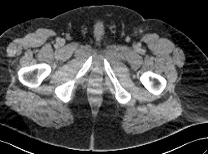
Aynı bölgenin, kaynak veri setinde patoloji içermediği belirtilen bir BT incelemesini kesit kesit kaydırın; organ sınırlarını açarak anatomiyi hasta görüntüsüyle karşılaştırın. Küçük rastlantısal bulgular tümüyle dışlanmamıştır.
Kaynak: TotalSegmentator v2.01 veri seti (Wasserthal ve ark., Radiology: Artificial Intelligence 2023), CC BY 4.0, vaka s0685; organ sınırları veri setinin otomatik segmentasyonundan, birleştirilerek sadeleştirildi. Görüntüler 2,5 mm kesit aralığına yeniden örneklendi.
Ayırıcı tanı
- Charcot nöro-osteoartropatisi
- orta ayakta birden çok eklem merkezli subkondral parçalanma ve hizalanma kaybı öne çıkar; ülserle doğrudan kemik bağlantısı beklenmez.
- Akut kırık
- keskin fraktür çizgisi ve mekanik yüklenmeye uygun fragman dizilimi vardır; ülser tabanından kemiğe uzanan trakt tipik değildir.
- Charcot nöro-osteoartropatisinde orta ayak tutulumu, periartiküler veya subkondral parçalanma, eklem deformitesi ya da subluksasyon ve üzerindeki derinin görece korunması öne çıkar.
- Osteomiyelit ve septik artrit değerlendirmesinde dejeneratif değişiklikler, Charcot nöro-osteoartropatisi, inflamatuar hastalıklar ve neoplazi ayırıcı tanıda göz önünde bulundurulmalıdır.
Tuzaklar
- Charcot nöro-osteoartropatisinde orta ayak ağırlıklı, periartiküler veya subkondral parçalanma ve eklem deformitesi görülebilir; diyabetik ayak osteomiyelitinde ise sinüs traktı ve kortikal bozulma tipik MRG bulgularındandır.
- Nöropatik artropati, kırık ve önceki ortopedik cerrahi enfeksiyon tanısını güçleştirebilir; bu durumlarda klinik bulgular ve ileri görüntüleme birlikte değerlendirilmelidir.
- Ortopedik implantlar ve önceki cerrahi müdahaleler, enfeksiyonun radyografik bulgularını gölgelendirebilir veya taklit edebilir; tanı için histopatoloji ve kültür desteği gerekebilir.
- BT'de belirgin kemik yıkımının olmaması erken enfeksiyonu dışlamaz; medüller sinyal ve yayılım sorusu sürüyorsa MR'ın rolünü koruyun.
Kendini dene
Diyabetik ayak ülserinden kemiğe uzanan sinüs traktı ve komşu kemikte kortikal bozulma görülüyor. En olası tanı hangisidir?
Cevabı göster
Diyabetik ayak osteomiyeliti. Ülserle ilişkili sinüs traktı ve kortikal bozulma DFO'yu düşündürür; MRG bulguları klinik değerlendirmeyle birlikte yorumlanmalıdır.
Nöropatik ayakta orta ayakta periartiküler veya subkondral parçalanma, eklem deformitesi ve görece korunmuş üzerindeki deri görülüyor. En olası tanı hangisidir?
Cevabı göster
Charcot nöro-osteoartropatisi. Bu dağılım Charcot nöro-osteoartropatisini düşündürür; DFO tanısında ülserle ilişkili kemik ve yumuşak doku bulguları önem taşır.
İlgili konular
Kaynaklar
Bu sayfadaki 41 cümle ve 2 quiz sorusu aşağıdaki kaynaklardan alıntılarla otomatik olarak karşılaştırıldı; 15 cümle bu karşılaştırmada düzeltildi (2026-10-09).
- Concordance of Microbiological and Histopathological Examination of Bone Biopsies in Diagnosing Diabetic Foot Osteomyelitis, a Systematic Reviewpmc.ncbi.nlm.nih.gov
- Plain Radiography for Diagnosing and Monitoring Foot Osteomyelitis in Persons With Diabetes: Accuracy, Limitations and Clinical Utilitypmc.ncbi.nlm.nih.gov
- From serum inflammatory markers to fluid, tissue, and molecular assays: current advances in the laboratory diagnosis of bone and joint infectionspmc.ncbi.nlm.nih.gov
- State-of-the-art update for diagnosing diabetic foot osteomyelitis: a narrative reviewpmc.ncbi.nlm.nih.gov
- Clinical Use of S53P4 Bioactive Glass in the Treatment of Bone Defects and Infected Bone: A Systematic Review of the Quality of Clinical Outcomes and A Grade Assessmentpmc.ncbi.nlm.nih.gov
- The role of nuclear medicine in the diagnosis of common and specific diabetic infections.pubmed.ncbi.nlm.nih.gov
- Diagnosis and treatment of necrotising otitis externa and diabetic foot osteomyelitis - similarities and differences.pubmed.ncbi.nlm.nih.gov
- Clinical Inquiry: What's the best test for underlying osteomyelitis in patients with diabetic foot ulcers?pubmed.ncbi.nlm.nih.gov
- [18F]FDG PET/CT for the diagnosis of diabetic foot osteomyelitis.pubmed.ncbi.nlm.nih.gov
- [ 18 F]Fluorodeoxyglucose ([ 18 F]FDG) positron emission tomography and conventional imaging modalities in the diagnosis of diabetic foot osteomyelitis: a meta-analysis.pubmed.ncbi.nlm.nih.gov
- Diagnosing osteomyelitis in diabetic foot by diffusion-weighted imaging and dynamic contrast material-enhanced magnetic resonance imaging: a systematic review and meta-analysis.pubmed.ncbi.nlm.nih.gov
- Diagnosing diabetic foot osteomyelitis.pubmed.ncbi.nlm.nih.gov
- Bone and joint infection.pubmed.ncbi.nlm.nih.gov
- Imaging of diabetic foot infections.pubmed.ncbi.nlm.nih.gov
- Differential Diagnosis of Diabetic Foot Osteomyelitis and Charcot Neuropathic Osteoarthropathy with Deep Learning Methods.pubmed.ncbi.nlm.nih.gov
- Diagnostic and therapeutic update on diabetic foot osteomyelitis.pubmed.ncbi.nlm.nih.gov
- Diagnosis of infection in the foot in diabetes: a systematic review.pubmed.ncbi.nlm.nih.gov
- Expert opinion on the management of infections in the diabetic foot.pubmed.ncbi.nlm.nih.gov
- Ultrasonography Cannot Exclude Osteomyelitis in Diabetic Foot Ulcers: A Cross-Sectional Diagnostic Accuracy Study Against Magnetic Resonance Imaging.pubmed.ncbi.nlm.nih.gov
- Bone‑origin repair in diabetic foot ulcers: Mechanisms of callus formation and endocrine effects in healing (Review).pubmed.ncbi.nlm.nih.gov
- Diabetic Foot Ulcer-Associated Versus Other Lower-Extremity Necrotising Soft-Tissue Infections: A Retrospective Cohort Study.pubmed.ncbi.nlm.nih.gov
- Current updates in MSK infection imaging: A narrative review.pubmed.ncbi.nlm.nih.gov
- Osteomyelitis Masquerading as Cellulitis: A Case Report.pubmed.ncbi.nlm.nih.gov
- Musculoskeletal infections through direct inoculation.pubmed.ncbi.nlm.nih.gov
- Diffusion-Weighted Imaging in the Musculoskeletal System: Evolving Role in Modern Imaging Practice.pubmed.ncbi.nlm.nih.gov
- Advances in the application of artificial intelligence-driven multi-modal imaging technologies in the comprehensive diagnosis and treatment of diabetic foot ulcers.pubmed.ncbi.nlm.nih.gov
- Staged antibiotic-loaded cement-based reconstruction versus flap-based reconstruction for complex diabetic foot defects in patients with osteoporosis: a retrospective cohort study.pubmed.ncbi.nlm.nih.gov
- Lower extremity infections: Essential anatomy and multimodality imaging findings.pubmed.ncbi.nlm.nih.gov
- Guidelines on multidisciplinary approaches for the prevention and management of diabetic foot disease (2020 edition).pubmed.ncbi.nlm.nih.gov
- Long-term safety and effectiveness of the "OptEase" vena cava filter.pubmed.ncbi.nlm.nih.gov
- Investigating and Practicing Orthopedics at the Intersection of Sex and Gender: Understanding the Physiological Basis, Pathology, and Treatment Response of Orthopedic Conditions by Adopting a Gender Lens: A Narrative Overview.pubmed.ncbi.nlm.nih.gov
- Radiologic Mimics of Osteomyelitis and Septic Arthritis: A Pictorial Essay.pubmed.ncbi.nlm.nih.gov
- A Guide to Improving the Care of Patients with Fragility Fractures, Edition 2.pubmed.ncbi.nlm.nih.gov
- In vitro expanded human CD4+CD25+ regulatory T cells suppress effector T cell proliferation.pubmed.ncbi.nlm.nih.gov
- Concordance of Microbiological and Histopathological Examination of Bone Biopsies in Diagnosing Diabetic Foot Osteomyelitis, a Systematic Review.pubmed.ncbi.nlm.nih.gov
- Hand Osteomyelitis: Exploratory Associations of Admission C-Reactive Protein and MRI with Surgical Burden and Hospital Length of Stay.pubmed.ncbi.nlm.nih.gov
- Evaluating chronic bone and soft tissue infections with [<sup>68</sup>Ga]Ga-Pentixafor PET/CT: a head-to-head comparison with scintigraphy.pubmed.ncbi.nlm.nih.gov
- From serum inflammatory markers to fluid, tissue, and molecular assays: current advances in the laboratory diagnosis of bone and joint infections.pubmed.ncbi.nlm.nih.gov
- Sculpting the Future of Bone: The Evolution of Absorbable Materials in Orthopedics.pubmed.ncbi.nlm.nih.gov
- Conference Proceedings: Select Abstracts Presented at the 2025 Advocate Aurora Scientific Day.pubmed.ncbi.nlm.nih.gov
- Imaging Atlas of Osseous Pathology in the Ankle and Foot.pubmed.ncbi.nlm.nih.gov
- The added value of ZTE MRI in the assessment of diabetic foot disease.pubmed.ncbi.nlm.nih.gov
- Musculoskeletal ultrasound in the emergency department: a narrative review for general radiologists.pubmed.ncbi.nlm.nih.gov
- Osteomyelitis of the Jaw Bones and Its Mimics: Resolving the Diagnostic Enigma.pubmed.ncbi.nlm.nih.gov
- A Journey into Animal Models of Human Osteomyelitis: A Review.pubmed.ncbi.nlm.nih.gov
- Pathophysiology and Molecular Imaging of Diabetic Foot Infections.pubmed.ncbi.nlm.nih.gov
- MRI nomenclature for musculoskeletal infection.pubmed.ncbi.nlm.nih.gov
- Enigmatic Cases of Finger Proximal Interphalangeal Joint Swelling: Case Series and Focused Review of the Literature.pubmed.ncbi.nlm.nih.gov
- Combining anthropology and imaging to reconstruct antemortem trauma for identification purposes.pubmed.ncbi.nlm.nih.gov
- Radiological approach to metatarsalgia in current practice: an educational review.pubmed.ncbi.nlm.nih.gov
- Musculoskeletal Infection: The Great Mimickers on Imaging.pubmed.ncbi.nlm.nih.gov
- Imaging of acute musculoskeletal infections in children and their differential diagnoses.pubmed.ncbi.nlm.nih.gov
- Global Burden of Deep Neck Space Abscesses: Epidemiology, Challenges, and Outcomes.pubmed.ncbi.nlm.nih.gov
- An implantable system to restore hemodynamic stability after spinal cord injury.pubmed.ncbi.nlm.nih.gov
- Patterns of injury and violence in Yaoundé Cameroon: an analysis of hospital data.pubmed.ncbi.nlm.nih.gov
- Nonproliferative and Proliferative Lesions of the Gastrointestinal Tract, Pancreas and Salivary Glands of the Rat and Mouse.pubmed.ncbi.nlm.nih.gov
- Emerging and re-emerging infectious disease in otorhinolaryngology.pubmed.ncbi.nlm.nih.gov
- Review of imaging modalities and radiological findings of calvarial lesions.pubmed.ncbi.nlm.nih.gov
- Glossary of terms for musculoskeletal radiology.pubmed.ncbi.nlm.nih.gov
- The SLC-ome of membrane transport: From molecular discovery to physiology and clinical applications.pubmed.ncbi.nlm.nih.gov
- Purely osteolytic bone lesions in the early osteoarticular phase of synovitis, acne, pustulosis, hyperostosis and osteitis syndrome.pubmed.ncbi.nlm.nih.gov
- The quiet flame: whole-body MRI and the underestimated burden of pediatric chronic non-bacterial osteomyelitis (CNO).pubmed.ncbi.nlm.nih.gov
- The imaging of osteomyelitis.pubmed.ncbi.nlm.nih.gov
- Outcome of Surgical Fixation for Midfoot Charcot Neuroarthropathy - A Systematic Review.pubmed.ncbi.nlm.nih.gov
- Magnetic resonance imaging of the ankle and foot.pubmed.ncbi.nlm.nih.gov
- Prevalence and Associated Factors of Sarcopenia in Patients With Diabetic Foot Ulcers: A Systematic Review and Meta-Analysis.pubmed.ncbi.nlm.nih.gov
- Imaging of lower extremity infections: predisposing conditions, atypical infections, mimics, and differentiating features.pubmed.ncbi.nlm.nih.gov
- Effectiveness and Safety of Antibiotic Therapy Combined with NSAIDs or SAIDs in Osteomyelitis of the Oral and Maxillofacial Region: A Systematic Review.pubmed.ncbi.nlm.nih.gov
- Japanese clinical practice guidelines for vascular anomalies 2017.pubmed.ncbi.nlm.nih.gov
- Japanese clinical practice guidelines for vascular anomalies 2017.pubmed.ncbi.nlm.nih.gov
- High-affinity alphavbeta3 integrin targeted optical probe as a new imaging biomarker for early atherosclerosis: initial studies in Watanabe rabbits.pubmed.ncbi.nlm.nih.gov
- Comparing the Effectiveness of Different Types of Orthoses in Preventing Diabetic Foot Ulcer Recurrence: A Systematic Review of Randomized Clinical Trials.pubmed.ncbi.nlm.nih.gov
- Ultrasound-guided continuous parasacral ischial plane block for diabetic foot ulcer healing: study protocol for a single-centre, prospective, randomised, open-label, blinded-endpoint controlled trial.pubmed.ncbi.nlm.nih.gov
- Offloading orthotic insoles on healing of diabetic foot ulcers: a protocol for systematic review and meta-analysis.pubmed.ncbi.nlm.nih.gov
- Four-Week vs Six-Week Antibiotic Therapy in the Management of Nonsurgically Treated Diabetic Foot Osteomyelitis: Protocol for a Multicentric, Single-Blind Randomized Clinical Trial.pubmed.ncbi.nlm.nih.gov
- Role of Biomarkers in Diabetic Foot Osteomyelitis.pubmed.ncbi.nlm.nih.gov
- S1 Guideline on Infected Interdigital Intertrigo (also called Gram-Negative Toe Web Infection).pubmed.ncbi.nlm.nih.gov
- Minimally Invasive Drainage for Diabetic Foot Phlegmon.pubmed.ncbi.nlm.nih.gov
- A Brush With the Lake: Recurrent Lower Extremity Cellulitis in a 50-Year-Old Male.pubmed.ncbi.nlm.nih.gov
- Successful Limb Salvage in a Diabetic Septic Foot Using Anterolateral Thigh Free Flap Reconstruction: A Case Report.pubmed.ncbi.nlm.nih.gov
- Calciphylaxis Mimicry in End-Stage Renal Disease: Acute Limb Ischemia From Calcific Peripheral Arterial Disease.pubmed.ncbi.nlm.nih.gov
- Surgical resection of a ruptured left frontal mycotic aneurysm localized with ICG angiography: a case report.pubmed.ncbi.nlm.nih.gov
- Musculoskeletal Infection Reporting and Data System (MSKI-RADS): reviewed and explained.pubmed.ncbi.nlm.nih.gov
- Madura foot: Atypical MRI presentation beyond the classic dot-in-circle sign.pubmed.ncbi.nlm.nih.gov
- Risk factors for prolonged hospitalization and disease course in osteomyelitis: a single-centre cohort study.pubmed.ncbi.nlm.nih.gov
- Clinical, etiological, and microbiological differences between foot and non-foot osteomyelitis.pubmed.ncbi.nlm.nih.gov
- Early postoperative C-reactive protein-to-albumin ratio for risk stratification of osteomyelitis recurrence after surgical treatment.pubmed.ncbi.nlm.nih.gov
- Serratia Marcescens Septic Arthritis of the Ankle in a Compromised Host: Ring Fixator Arthrodesis as Salvage, a Case Report.pubmed.ncbi.nlm.nih.gov
- Hand Infections Across Tissue Planes: A Pictorial Review.pubmed.ncbi.nlm.nih.gov
- Quantitative Bone SPECT/CT in Diabetic Foot Osteomyelitis: Diagnostic Performance Within-Patient Lesion-Contralateral Separation and Associations with Inflammatory Burden.pubmed.ncbi.nlm.nih.gov
- Decoding Chronic Charcot Arthropathy: Molecular Mechanisms, Predictive Biomarkers, and Emerging Therapies.pubmed.ncbi.nlm.nih.gov
- Asymptomatic Charcot foot in longstanding diabetes.pubmed.ncbi.nlm.nih.gov
- Guideline for the Management of Diabetes Mellitus in the Elderly in China (2024 Edition).pubmed.ncbi.nlm.nih.gov
- Dufourmentel Rhomboid Flap for Plantar Charcot Midfoot Ulcer: A Novel Reconstructive Approach.pubmed.ncbi.nlm.nih.gov
- Charcot Neuroarthropathy Masquerading as Severe Acute Orthopedic Trauma: Severe Ramifications of Untreated Diabetes Mellitus.pubmed.ncbi.nlm.nih.gov
- Collagen-Based Regenerative Therapy in a Critical Ischemic Diabetic Foot Ulcer: A Limb-Salvaging Case Report.pubmed.ncbi.nlm.nih.gov
- PMMA-induced membranous angiogenesis accelerates wound healing in refractory diabetic foot ulcers: A comparative cohort study.pubmed.ncbi.nlm.nih.gov
- An Unexpected Cause of Diabetic Foot Ulcer Infection: Pasteurella multocida.pubmed.ncbi.nlm.nih.gov
- The Great Mimicker in Bone: A Rare Case of Melioidosis With Multifocal Skeletal Manifestations and Diagnostic Challenges.pubmed.ncbi.nlm.nih.gov
- The Role of Weight-Bearing Computed Tomography in the Assessment and Management of Charcot Foot Deformity: A Narrative Review.pubmed.ncbi.nlm.nih.gov
- Diagnostic Performance of Integrated <sup>18</sup> F FDG PETMR in the Diagnosis of Recurrent Foot Infection-Comparison with <sup>18</sup> F FDG PETCT and Conventional <sup>99m</sup> Tc MDP Bone Scan.pubmed.ncbi.nlm.nih.gov
- When Glycaemic Control Comes Too Fast: Treatment-Induced Neuropathy of Diabetes and Charcot Neuro-Osteoarthropathy as Emerging Iatrogenic Complications of Rapid Metabolic Correction.pubmed.ncbi.nlm.nih.gov
- Hexapod-assisted limb salvage after talectomy: outcomes of tibiocalcaneal arthrodesis using the TL-hex system.pubmed.ncbi.nlm.nih.gov
- Arthroscopic Management for Hip Joint Septic Arthritis: A Retrospective Study of 28 Adult Patients With a Minimum 3-Year Follow-Up.pubmed.ncbi.nlm.nih.gov
- A Multi-Centre, Randomised, Controlled Clinical Trial Assessing Cryopreserved Ultra-Thick Human Amniotic Membrane in the Treatment of Complex Diabetic Foot Ulcers.pubmed.ncbi.nlm.nih.gov
- A three-stage sequential surgical approach to a more efficient management of clinical stage 4 diabetic foot ulcers.pubmed.ncbi.nlm.nih.gov
- Prevalence and Microbial Association of Onychomycosis in Patients with Diabetic Foot Ulcers: A Retrospective Observational Study.pubmed.ncbi.nlm.nih.gov
- GLP-1 receptor agonists vs DPP-4 inhibitors and neuropathic complications in type 2 diabetes.pubmed.ncbi.nlm.nih.gov
- Risk Factors Associated With Multidrug Resistant Organism (MDRO) Isolates in Infected Diabetic Foot Ulcers (DFU): An Analytical Cross-sectional Study.pubmed.ncbi.nlm.nih.gov
- Severe Chronic Suppurative Osteomyelitis Following Dental Implant Placement.pubmed.ncbi.nlm.nih.gov
- Antibiotic and Surgical Treatment of Diabetic Foot Osteomyelitis: The Histopathological Evidence.pubmed.ncbi.nlm.nih.gov
- Limb Salvage Using Local Antibiotic Beads for Calcaneal Osteomyelitis With Multidrug-resistant Infection and Chronic Limb-threatening Ischemia.pubmed.ncbi.nlm.nih.gov
- Squamous Cell Carcinoma Arising from Chronic Osteomyelitis: A Single-Center Case Series of Six Patients.pubmed.ncbi.nlm.nih.gov
- A Study of 29 Pediatric Patients With Osteomyelitis Resulting From Neuropathy and Pressure Effects.pubmed.ncbi.nlm.nih.gov
- Efficacy and Safety of Empirically Used Levonadifloxacin (Intravenous and Oral) in the Treatment of Patients with Bone and Joint Infection: A Multicentric, Investigator-Initiated Study.pubmed.ncbi.nlm.nih.gov
Eğitim amaçlıdır; hasta başında tanı ve tedavi kararının yerine geçmez, tıbbi tavsiye değildir. Metinler yapay zekâ desteğiyle yazıldı ve otomatik bir adımda kaynak alıntılarıyla karşılaştırıldı; bu bir tıbbi doğrulama değildir ve içerik uzman radyolog incelemesinden geçmedi. Klinik kararlarda güncel kılavuzları, kurum protokollerini ve radyoloji raporunu esas alın.