Özet
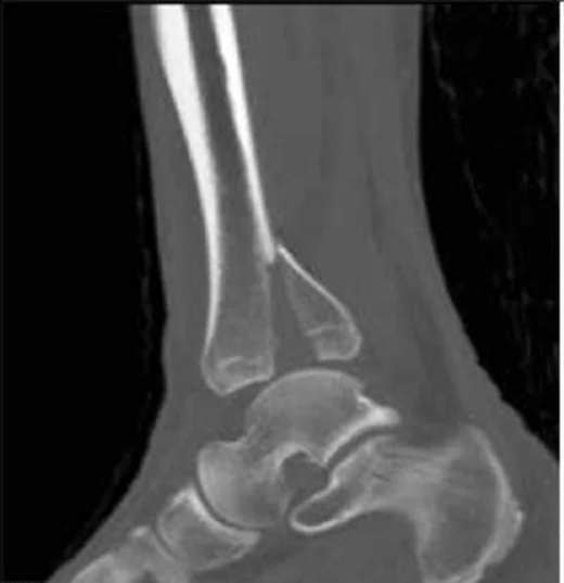
Posterior malleol kırığı distal tibianın arka eklem kenarını tutar ve tek başına ya da başka malleol kırıklarıyla birlikte görülebilir. Kontrastsız BT fragmanın yönünü, incisura fibularis ile eklem yüzüne uzanımını, deplasmanı ve eşlik eden impaksiyonu tanımlar; yalnız grafideki görünür boyut tüm morfolojiyi yansıtmayabilir. Bu fragman posterior sindezmoz yapışmasını da taşıyabileceğinden yalnız bir kemik parçası olarak ele alınmamalıdır. Incisura veya eklem yüzü tutulumu atlanırsa yaralanmanın distal tibiofibular ilişkisi eksik tarif edilir.
Faz ve pencere
- Kontrastsız tanısal
- Posterior distal tibia korteksinden ayrılan fragmanın eklem yüzüne, medial tarafa ve fibular incisuraya uzanımı; fragmanın rotasyonu, çökmesi, eklem basamağı ve varsa aradaki küçük interkaler parça izlenir. Kemik kırığı için kontrast gerekmez.
- Kemik algoritması tanısal
- Aksiyel rekonstrüksiyon fragmanın incisura ve posterior kenarla ilişkisini, koronal kesitler eklem yüzü basamağını, sagittal kesitler ön-arka deplasman ve plafond çökmesini gösterir; sınıflama için bu düzlemler birlikte değerlendirilir.
Önerilen pencereler G genişlik, M merkez; değerler Hounsfield birimi (HU)
- KemikG 1800 / M +400
BT bulguları
- Posterior malleol fragmanı — posterior tibial plafond korteksinden ayrılıp eklem kenarını içeren kemik parçası.
- Posterior malleol fragmanının deplasmanı, BT’de değerlendirilen morfolojik özellikler arasındadır.
- Eklem basamağı — fragman ile ana plafond arasındaki subkondral yüzey aynı hizada değildir.
- Fibular incisura tutulumu — posterior fragman distal fibulanın oturduğu tibial çentiğe uzanır.
- Volkmann fragmanı — PITFL yapışma bölgesini taşıyan posterolateral tibial kenar avulsiyonu.
- Chaput fragmanı — anterolateral tibia kenarında AITFL yapışma yerinin karşı taraftaki avulsiyon bileşeni; posterior fragmanla karıştırılmamalıdır.
- İnterkaler fragman — ana posterior parçayla tibia arasında eklem içi küçük kemik parçası.
- Talar posterior subluksasyon — talus eklem içinde arkaya yer değiştirir ve posterior eklem aralığı daralır ya da kaybolur.
- Posterior plafond impaksiyonu — BT'de eklem yüzü impaksiyonunun varlığını ve eklem uyumsuzluğunu değerlendirin.
Ölçütler ve sınıflamalar
- Bartoníček-Rammelt BT sınıflaması
- Tip I ekstra-insisural fragman ve korunmuş incisura; tip II posterolateral fragmanın incisuraya uzanması; tip III medial malleole uzanan iki parçalı posteromedial kırık; tip IV incisuranın geniş bölümünü tutan büyük posterolateral üçgensi fragman; tip V diğer tiplere girmeyen düzensiz osteoporotik kırık olarak tanımlanır.
- Haraguchi BT sınıflaması
- Tip I posterolateral oblik kama fragmanı; tip II fibular çentikten medial malleole uzanan transvers-medial çizgi; tip III posterior plafond dudağında küçük kabuk biçimli fragmanları tanımlar.
Normalde
Normal posterior plafondda tibianın arka eklem korteksi kesintisizdir ve distal fibula incisura içinde düzgün biçimde oturur. Aynı aksiyel, koronal ve sagittal düzeylerde posterior kenar konturunu, fragman kaynaklı yüzey basamağını ve talusun merkezlenmesini karşılaştırın.
Normal BT ile kıyasla
Aynı bölgenin, kaynak veri setinde patoloji içermediği belirtilen bir BT incelemesini kesit kesit kaydırın; organ sınırlarını açarak anatomiyi hasta görüntüsüyle karşılaştırın. Küçük rastlantısal bulgular tümüyle dışlanmamıştır.
Kaynak: TotalSegmentator v2.01 veri seti (Wasserthal ve ark., Radiology: Artificial Intelligence 2023), CC BY 4.0, vaka s0685; organ sınırları veri setinin otomatik segmentasyonundan, birleştirilerek sadeleştirildi. Görüntüler 2,5 mm kesit aralığına yeniden örneklendi.
Ayırıcı tanı
- Pilon kırığı
- posterior malleol parçası pilon yaralanmasının yalnız bir bileşeni olabilir; plafondun diğer kısımlarında ve metafizde de parçalanma/impaksiyon aranır.
- Eski avulsiyon fragmanı
- tamamen kortike kenar ve komşu skleroz kronikliği destekler; akut kırıkta keskin kortikal uçlar görülebilir.
- Talar posterior proses kırığı
- BT'de fragmanın tibia yerine talusun posterior prosesinden köken aldığını ve talus gövdesine uzanımını değerlendirin.
- Arka ayak osteokondral lezyonu
- defekt talus kubbesinde bulunur, posterior tibia kenarında ayrılmış fragman oluşturmaz.
Tuzaklar
- Fragman boyutunu tek bir lateral grafi kesitinden tahmin etme; BT'de incisura uzanımı, eklem basamağı ve parçalanma farklı düzlemlerde belirginleşebilir.
- Posterior tibial fragmanı otomatik olarak tek parça Volkmann avulsiyonu sayma; medial uzanım ve ikinci fragman paterni sınıflamayı değiştirir.
- İnterkaler küçük parçayı ana fragmanın kenar düzensizliği sanma; seri aksiyel ve sagittal kesitlerde ayrı kemik yoğunluğu olarak izini sür.
Kendini dene
Posterior plafond fragmanı fibular incisuraya uzanıyor ve talar kubbe hafif arkaya kaymış. Hangi özelliği özellikle raporlamak gerekir?
Cevabı göster
İncisura katılımı ve talar uyum. Posterior fragmanın incisuraya uzanımı distal tibiofibular eklem biçimini, talar kayma da mortis uyumunu etkiler. Diğer seçenekler bu posterior malleol yaralanmasının anatomik hedefleri değildir.
Posterior tibial fragman için BT'de incisura intakt ve parça ekstra-insisural olarak görülüyor. Bartoníček-Rammelt tipi hangisidir?
Cevabı göster
Tip I. Tip I, incisura korunmuşken ekstra-insisural posterior fragmanı tanımlar. Tip II incisuraya uzanan posterolateral parçadır; tip III medial malleole uzanır; tip IV geniş incisura katılımlı büyük posterolateral parçadır.
İlgili konular
Kaynaklar
Bu sayfadaki 42 cümle ve 2 quiz sorusu aşağıdaki kaynaklardan alıntılarla otomatik olarak karşılaştırıldı; 2 cümle bu karşılaştırmada düzeltildi (2026-10-09).
- Posterior Malleolar Fracture Assessment: An Independent Interobserver and Intraobserver Validation of Three Computed Tomography-Based Classificationspmc.ncbi.nlm.nih.gov
- Intercalary fragments in posterior malleolar fractures: incidence, treatment implications, and distribution within CT-based classification systemspmc.ncbi.nlm.nih.gov
- A Comprehensive Review of Recent Trends in Posterior Malleolus Fracture Managementpmc.ncbi.nlm.nih.gov
- Bosworth ankle fracture-dislocation: current concept review.pubmed.ncbi.nlm.nih.gov
- Posterior Malleolar Fractures: A Critical Analysis Review.pubmed.ncbi.nlm.nih.gov
- Traumatic injuries of the distal tibiofibular syndesmosis.pubmed.ncbi.nlm.nih.gov
- A systematic review of posterior pilon variant fractures.pubmed.ncbi.nlm.nih.gov
- Fracture obliquity angle and distance from plafond: novel radiographic predictors of posterior malleolar involvement in tibial shaft fractures.pubmed.ncbi.nlm.nih.gov
- Posterior interosseous ligament of the ankle: current evidence and knowledge gaps.pubmed.ncbi.nlm.nih.gov
- Posterior Malleolar Ankle Fractures: An Effort at Improving Outcomes.pubmed.ncbi.nlm.nih.gov
- Posterior Malleolar Fracture Patterns.pubmed.ncbi.nlm.nih.gov
- Posterior Malleolar Fracture Assessment: An Independent Interobserver and Intraobserver Validation of Three Computed Tomography-Based Classifications.pubmed.ncbi.nlm.nih.gov
- [The isolated posterior malleolus fracture].pubmed.ncbi.nlm.nih.gov
- Clinical outcomes and trans-syndesmotic screw frequency after posterior malleolar fracture osteosynthesis.pubmed.ncbi.nlm.nih.gov
- Guesstimation of posterior malleolar fractures on lateral plain radiographs.pubmed.ncbi.nlm.nih.gov
- Characteristics of intercalary fragment in posterior malleolus fractures.pubmed.ncbi.nlm.nih.gov
- Pathoanatomy and Injury Mechanism of Typical Maisonneuve Fracture.pubmed.ncbi.nlm.nih.gov
- Pilon Fracture with Associated Achilles Tendon Rupture: Case Report and Review of the Literature.pubmed.ncbi.nlm.nih.gov
- Isolated Displaced Chaput Fracture (Rammelt Type III) With Significant Intra-articular Displacement: A Case Report.pubmed.ncbi.nlm.nih.gov
- Injury Characteristics and Prognostic Outcomes of Ankle Fracture-Dislocation: The Importance of Posteromedial Extension of Posterior Malleolar Fractures.pubmed.ncbi.nlm.nih.gov
- Guide to lower extremity radiologic measurements: part 3 ankle and foot.pubmed.ncbi.nlm.nih.gov
- Treatment Strategy for Posterior Malleolar Fractures: Different Operative Strategies Are Needed for Each Morphological Type.pubmed.ncbi.nlm.nih.gov
- Biomechanical Efficacy of Three Methods for the Fixation of Posterior Malleolar Fractures: A Three-Dimensional Finite Element Study.pubmed.ncbi.nlm.nih.gov
- Buttress plating versus trans-syndesmotic stabilisation for posterolateral malleolus fractures: outcomes with early weight-bearing.pubmed.ncbi.nlm.nih.gov
- The Diagnosis, Treatment, and Aftercare of Ankle Fractures.pubmed.ncbi.nlm.nih.gov
- Clinical and CT-Based Outcomes of Anterior and Posterior Syndesmotic Augmentation Using Nonabsorbable Suture Tape for Acute Syndesmotic Instability: A Preliminary Case Series of Seven Patients.pubmed.ncbi.nlm.nih.gov
- MRI analysis of distal tibiofibular joint and ankle anatomy to assess lateral ankle sprain risk.pubmed.ncbi.nlm.nih.gov
- Anterior Incisura Fibularis Corner Landmarks Can Safely Validate the Optimal Distal Tibiofibular Reduction in Malleolar Fractures-Prospective CT Study.pubmed.ncbi.nlm.nih.gov
- Modified 3D-Controlled Inside-Out Compression Screw Fixation Technique in Posterior Malleolar Fractures: A Narrative Review and Case Report.pubmed.ncbi.nlm.nih.gov
- A Systematic Approach to the Evaluation of Acute Musculoskeletal Injuries in the Emergency Department.pubmed.ncbi.nlm.nih.gov
- Emergency radiology of severe acute injury in extreme alpine summer sports: a pictorial essay.pubmed.ncbi.nlm.nih.gov
- Imaging Anatomy of the Ankle in Normal and Pathological States: A Clinically Focused Pictorial Review.pubmed.ncbi.nlm.nih.gov
- What is the desired screw trajectory for fixation of noncontiguous posterior distal articular fractures associated with distal tibial shaft fractures?pubmed.ncbi.nlm.nih.gov
- Effect of fibular rotation and sagittal shift on postoperative function in ankle fractures with syndesmotic injury.pubmed.ncbi.nlm.nih.gov
- CT-Based Three-Dimensional Volumetric Analysis of Posterior and Lateral Malleolar Fragments in SER-Type Trimalleolar Ankle Fractures: Correlation and Reproducibility Study.pubmed.ncbi.nlm.nih.gov
- Clinical and radiological outcomes of surgical treatment using a posterolateral approach based on computed tomography-based fracture morphology in posterior malleolar fractures: A single-center retrospective analysis.pubmed.ncbi.nlm.nih.gov
- Associations of Bartoníček classification and fragment size with treatment selection and reduction quality in posterior malleolar fractures.pubmed.ncbi.nlm.nih.gov
- Concomitant Factors Associated With Tillaux-Chaput Fractures in Adults: A Case-Control Study.pubmed.ncbi.nlm.nih.gov
- Dance Around the Fibula with a Twisted Plate: A Technical Note.pubmed.ncbi.nlm.nih.gov
- Ultrasound of the distal tibiofibular syndesmosis.pubmed.ncbi.nlm.nih.gov
- Chronic Syndesmotic Instability Associated with a Complex Lesion of the Posterior Inferior Tibiofibular Ligament: Review, Case Report, and Surgical Report.pubmed.ncbi.nlm.nih.gov
- Comparative finite element analysis of a novel distal tibiofibular locking plate versus conventional fixation methods for Tillaux-Chaput fractures.pubmed.ncbi.nlm.nih.gov
- Surgical approaches for the treatment of posterior malleolar fracture: which one to choose?pubmed.ncbi.nlm.nih.gov
- Reduction and outcome of posterior pilon fractures with intercalary fragments: a retrospective cohort study comparing the transfibular and posteromedial approaches.pubmed.ncbi.nlm.nih.gov
- Does lateral column lengthening reach the ankle? Lateral calcaneal lengthening for flatfoot and medial talar dome osteochondral lesions: A weight-bearing CT 3D modelling analysis.pubmed.ncbi.nlm.nih.gov
- Deltoid ligament injury: thinking beyond the ankle - an expert opinion.pubmed.ncbi.nlm.nih.gov
- Medial Subtalar Joint Dislocation in an 18-Year-Old Athlete with Pes Planus: Beyond A Simple Misstep.pubmed.ncbi.nlm.nih.gov
- Peroneal Artery Danger Zone With Syndesmotic Screw Fixation: A Computed Tomography Angiography Study.pubmed.ncbi.nlm.nih.gov
- A Novel Lateral Tibiofibular Index for Simulated Posterior Fibular Translation: A Three-Dimensional CT Study.pubmed.ncbi.nlm.nih.gov
- Posterior Malleolar Fragment Size as a Predictor of Ligamentous Injuries in Trimalleolar and Quadrimalleolar Ankle Fractures: A Novel Correlative Cohort Study in a South Indian Population.pubmed.ncbi.nlm.nih.gov
- Integrating Quadrimalleolar Fractures of the Ankle in a Johannesburg Academic Center: A New Classification.pubmed.ncbi.nlm.nih.gov
- A comparative analysis of the therapeutic outcomes between nickel-titanium shape memory staples and miniplate for Bartoníček-Rammelt type III and IV posterior malleolar fractures.pubmed.ncbi.nlm.nih.gov
- Prevalence and risk factors of concomitant malleolar and fibular fractures in patients with distal spiral tibial shaft fractures.pubmed.ncbi.nlm.nih.gov
- [Related factors and analysis of prevention strategies for functional ankle instability after ankle fracture surgery].pubmed.ncbi.nlm.nih.gov
- Inter-observer and Intra-observer Reliability of Posterior Malleolus Fracture Classification Systems.pubmed.ncbi.nlm.nih.gov
- Advancements in the diagnosis and management of complex trimalleolar ankle fractures: A comprehensive review.pubmed.ncbi.nlm.nih.gov
- Preoperative Risk Score for Predicting Delayed Wound Healing After Plate Fixation in Patients With Closed Distal Fibular Fractures.pubmed.ncbi.nlm.nih.gov
- Early weight-bearing following surgical fixation of ankle fractures without trans-syndesmotic fixation: safety and early functional outcomes - a systematic review and meta-analysis.pubmed.ncbi.nlm.nih.gov
- Posterolateral plating increases peroneal tendon thickness without affecting clinical outcomes: a ultrasonographic comparative study of 76 SER ankle fractures.pubmed.ncbi.nlm.nih.gov
- Flexible Flatfoot-Epidemiology, Pathophysiology and Clinical Aspects: A Review Focused on Less-Invasive Surgical Treatment in Children.pubmed.ncbi.nlm.nih.gov
- Imaging modalities for non-acute pathologies of the foot and ankle.pubmed.ncbi.nlm.nih.gov
- Enhanced subfibular ossicle diagnosis: CT-MRI integration of morphology and ligament attachments.pubmed.ncbi.nlm.nih.gov
- Achilles, peroneus longus and brevis ruptures with lateral malleolus fracture: A case report and review of literature.pubmed.ncbi.nlm.nih.gov
- Moth-Eaten Osteolysis of Multiple Foot Bones After a Conservatively Treated "Stable" Ankle Fracture: A Case Report and Literature Review.pubmed.ncbi.nlm.nih.gov
- An Acta Orthopaedica educational article: Weightbearing assessment to guide nonoperative treatment of lateral malleolar fractures: the paradigm change.pubmed.ncbi.nlm.nih.gov
- ChatGPT-o1 Preview Outperforms ChatGPT-4 as a Diagnostic Support Tool for Ankle Pain Triage in Emergency Settings.pubmed.ncbi.nlm.nih.gov
- Donor Site Morbidity After Talar Mosaicplasty: Arthroscopic Medial Femoral Condyle vs Mini-Open Lateral Femoral Condyle Harvesting.pubmed.ncbi.nlm.nih.gov
- The Evolution of Sinus Tarsi Syndrome-What Is the Underlying Pathology?-A Critical Review.pubmed.ncbi.nlm.nih.gov
- Artificial intelligence in orthopedic trauma surgery: a scoping review of current applications and research gaps.pubmed.ncbi.nlm.nih.gov
- Fracture of the lateral tubercle of the posterior talar process caused by a rock-climbing fall: a case report.pubmed.ncbi.nlm.nih.gov
- Computed Tomography-Based Comparison of Anterior and Posterior Cortical Thickness Around the Interfragmentary Screw Fixation Level in Weber B Lateral Malleolar Fractures.pubmed.ncbi.nlm.nih.gov
- Reduction Quality in Posterior Malleolar Fractures Using a Modified Posteromedial Ankle Approach.pubmed.ncbi.nlm.nih.gov
- Outcome analysis of modified lateral malleolar osteotomy for the management of talar body fractures associated with tibial pilon fractures.pubmed.ncbi.nlm.nih.gov
- Association Between Talar Morphology and Subchondral Bone Sclerosis in the Medial Gutter of Patients With Chronic Lateral Ankle Instability.pubmed.ncbi.nlm.nih.gov
- Operative Treatment of Posterior Malleolar Fractures.pubmed.ncbi.nlm.nih.gov
- Ankle Sprains in Athletes: Current Epidemiological, Clinical and Imaging Trends.pubmed.ncbi.nlm.nih.gov
- Direct visualization for syndesmotic stabilization of ankle fractures.pubmed.ncbi.nlm.nih.gov
- [Joint-preserving osteotomy of malunited ankle and pilon fractures].pubmed.ncbi.nlm.nih.gov
- Influence of syndesmotic injuries and posterior malleolar ankle fractures on fibula position in the ankle joint: a cadaveric study.pubmed.ncbi.nlm.nih.gov
- Computer-assisted surgery and navigation in foot and ankle: state of the art and fields of application.pubmed.ncbi.nlm.nih.gov
- A fibular notch approach for the treatment of ankle fractures involving the distal tibial plafond.pubmed.ncbi.nlm.nih.gov
- Glossary of terms for musculoskeletal radiology.pubmed.ncbi.nlm.nih.gov
- Imaging of pediatric foot disorders.pubmed.ncbi.nlm.nih.gov
- The state of trial registrations in the field of Orthopaedics in years 2015-2020. A meta-epidemiological study.pubmed.ncbi.nlm.nih.gov
- Avulsion fracture of the posterior calcaneal tuberosity: anatomy, injury patterns, and an approach to management.pubmed.ncbi.nlm.nih.gov
- The role of imaging in the diagnosis, staging, and management of the osteochondral lesions of the talus.pubmed.ncbi.nlm.nih.gov
- Osteochondral Lesions of the Subtalar Joint: Clinical Outcomes in 11 Patients.pubmed.ncbi.nlm.nih.gov
- Diagnosis and Treatment of Peritalar Injuries in the Acute Trauma Setting: A Review of the Literature.pubmed.ncbi.nlm.nih.gov
- Review of running injuries of the foot and ankle: clinical presentation and SPECT-CT imaging patterns.pubmed.ncbi.nlm.nih.gov
- Arthroscopic surgery for the treatment of a geriatric bipartite talus: A case report.pubmed.ncbi.nlm.nih.gov
- Foot pain in children and adolescents: a problem-based approach in musculoskeletal ultrasonography.pubmed.ncbi.nlm.nih.gov
- Treatment of Intra-Articular Lesions After Posterior Inferior Tibiofibular Ligament Injury: A Case Series of Elite Rugby Players.pubmed.ncbi.nlm.nih.gov
- Computed Tomography Scan Architectural Measurements in Adult Foot and Ankle Surgery: A Narrative Review for Orthopaedic Trainees.pubmed.ncbi.nlm.nih.gov
- Salvage treatment of traumatic total talar loss: a case report on the eventual application of 3D printing technology.pubmed.ncbi.nlm.nih.gov
- Open fracture dislocation of the talus with nearly total extrusion and with tri-malleolar fracture: A case report and literature review.pubmed.ncbi.nlm.nih.gov
- Clinical study on arthroscopic reduction and internal fixation for malunion and nonunion talus fractures classified as type II by Zwipp.pubmed.ncbi.nlm.nih.gov
Eğitim amaçlıdır; hasta başında tanı ve tedavi kararının yerine geçmez, tıbbi tavsiye değildir. Metinler yapay zekâ desteğiyle yazıldı ve otomatik bir adımda kaynak alıntılarıyla karşılaştırıldı; bu bir tıbbi doğrulama değildir ve içerik uzman radyolog incelemesinden geçmedi. Klinik kararlarda güncel kılavuzları, kurum protokollerini ve radyoloji raporunu esas alın.