Özet
Trimalleoler kırıkta medial ve lateral malleole posterior tibial kenar kırığı eklenir; yaralanmaya mortis içinde talar subluksasyon veya çıkık eşlik edebilir. BT, üç parçanın yönelimini, posterior fragmanın incisura ve eklem yüzü ilişkisini ve arada kalmış serbest parçaları gösterir. Düz grafiye göre BT, posterior malleolus kırıklarını daha doğru şekilde saptar ve karakterize eder. Talar kayma veya eklem içi parça gözden kaçarsa kırıklı çıkığın eklem uyumu ve kemik bileşenleri eksik belgelenir.
Okuma sırası
- Posterior malleol ve tibial plafond. Posterior malleolü aksiyel ve sagittal BT kesitlerinde izleyerek fragman boyutunu ve tibial eklem yüzü tutulumunu değerlendir.
- Posterior malleol ve incisura. Posterior tibial kenarı aksiyel kesitlerde tarayın; fragmanın medial uzanımını ve fibular incisuranın arka sınırını tutup tutmadığını belirleyin.
- Talar kubbe ve tibiotalar aralık. Ayak bileği kırıklı çıkığında BT ile eklem içi serbest parça bulunup bulunmadığını değerlendirin.
- Eklem içi yapılar ve sindezmoz. Son olarak tibiotalar eklemdeki osteokondral lezyonlar, intra-artiküler serbest parçalar ve sinezdzmotik uyumsuzluk değerlendirilir.
Faz ve pencere
- Kontrastsız tanısal
- Trimalleoler kırıkta BT ile posterior malleol morfolojisi, incisura tutulumu ve eklem içi serbest parçalar değerlendirilir. Aksiyel, koronal ve sagittal kemik algoritmalı rekonstrüksiyonlar fragman yönelimini ve eklem yüzü uzanımını gösterir; intravenöz kontrast gerekmez.
Önerilen pencereler: Kemik (G 1800 / M 400).
BT bulguları
- Medial malleol kırığı — ayak bileği mortisini oluşturan medial malleolde gelişen kırık.
- Lateral malleol kırığı — distal fibulada kırık; seviyesi sindezmoza göre tarif edilir.
- Posterior malleol kırığı — posterior tibial eklem dudağında ayrılmış, deplase veya parçalı fragman.
- Posterior incisura uzanımı — posterior fragman distal fibulanın oturduğu çentiğin arka sınırını içerir.
- Talar subluksasyon/çıkık — talar kubbe mortis merkezinden kayar; eklem aralığı asimetrikleşir veya temas kaybolur.
- Eklem içi serbest parça — kırık kaynaklı kemik veya osteokondral parça tibia-talus eklem aralığında kalır.
- Talar kubbe osteokondral lezyonu — ayak bileği kırıklı çıkıklarına talus ve distal tibial plafondda osteokondral lezyonlar eşlik edebilir.
- Distal fibulanın incisura içindeki konumu ve rotasyonu BT'de değerlendirilebilir; sindezmoz uyumunun değerlendirilmesinde karşı taraf referans olarak kullanılabilir.
Ölçütler ve sınıflamalar
- Lauge–Hansen
- Ayak pozisyonu ve kuvvet yönüne dayalı mekanizma sınıflamasıdır; bazı bimalleoler ve trimalleoler örüntüler bu sisteme uymayabilir, posterior fragmanın BT morfolojisini tek başına tarif etmez.
- Bartoníček–Rammelt posterior malleol sınıflaması
- BT tabanlı beş tipli sınıflama posterior fragmanın incisura ve medial uzanımını da morfolojik olarak tarif eder. Haraguchi, Bartoníček–Rammelt ve Mason sistemleri kullanılmaktadır; literatürde evrensel olarak yerleşmiş tek bir sınıflama yoktur.
Normalde
Normal anatomide ayak bileği mortisini medial ve lateral malleoller ile distal tibia plafondu oluşturur. Aynı düzeylerde karşı tarafı kullanarak posterior eklem dudağını, tibiotalar aralığı ve sindezmoz kemik ilişkisini kıyaslayın; kırık kenarları ile düzgün normal korteksi ayırın. Ayak bileği mortisini oluşturan malleoller ve distal tibial plafond ile talusu mortis içinde stabilize eden bağlar dikkate alınarak talar konumu değerlendirilir; sindezmoz uyumunu değerlendirirken karşı taraf referans alınabilir.
Normal BT ile kıyasla
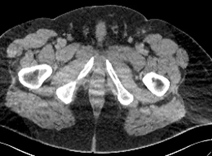
Aynı bölgenin, kaynak veri setinde patoloji içermediği belirtilen bir BT incelemesini kesit kesit kaydırın; organ sınırlarını açarak anatomiyi hasta görüntüsüyle karşılaştırın. Küçük rastlantısal bulgular tümüyle dışlanmamıştır.
Kaynak: TotalSegmentator v2.01 veri seti (Wasserthal ve ark., Radiology: Artificial Intelligence 2023), CC BY 4.0, vaka s0685; organ sınırları veri setinin otomatik segmentasyonundan, birleştirilerek sadeleştirildi. Görüntüler 2,5 mm kesit aralığına yeniden örneklendi.
Ayırıcı tanı
- Bimalleoler kırık
- medial ve lateral malleol kırıkları birlikte bulunur; posterior malleol kırığı da eşlik edebileceğinden ayrıca değerlendirilmelidir.
- Pilon kırığı
- plafondun daha geniş eklem yüzü ve metafiz bölgesi parçalanır; yalnız üç malleol fragmanıyla sınırlı değildir.
- Posterior malleol kırığı
- posterior tibial kenar fragmanı vardır, ancak diğer iki malleol kırık olmayabilir.
- Talar osteokondral kırık
- defekt talar kubbe yüzeyindedir ve tibial/medial/lateral malleol kırıklarının yerini tutmaz.
- Eski malleol avulsiyon fragmanları
- nonkaynamış lateral malleol avulsiyon fragmanları, os subfibulareye göre daha düzensiz şekilli ve pürüzlü kenarlı olabilir.
Tuzaklar
- Lateral ve posterior fragmanların üst üste geldiği grafi görünümünden posterior bileşeni atlama; BT'de posterior plafondu aksiyel ve sagittal düzlemde izle.
- Redüksiyon sonrası grafiler redüksiyonun yeterliliğini değerlendirmek için kullanılır; ayak bileği kırıklı çıkıklarında BT, grafilerde gözden kaçabilen eklem içi serbest parçaları gösterebilir.
- Posterior parçayı yalnız büyüklüğüne göre özetleme; incisura, medial uzanım, eklem basamağı ve interkaler fragman olup olmadığını ekle.
- Mortis içindeki her küçük kemik yoğunluğunu serbest parça sayma; kırık donör alanını ve parça çevresindeki kortikal kenarı takip et.
Kendini dene
Medial ve lateral malleol kırıklarına posterior plafond fragmanı da eşlik ediyor. Yaralanma örüntüsü nasıl adlandırılır?
Cevabı göster
Trimalleoler kırık. Üçüncü bileşen posterior malleol kırığıdır; medial, lateral ve posterior malleolün birlikte tutulması trimalleoler örüntüyü tanımlar. Bimalleoler iki malleolü içerir; pilon plafond-metafiz parçalanmasını, Maisonneuve ise proksimal fibula yaralanmasını ifade eder.
Üç malleol kırığına ek olarak talus posteriora kaymış ve eklem aralığında ayrı bir kemik parçası görülüyor. BT raporunda hangi iki bulgu özellikle belirtilmeli?
Cevabı göster
Talar translasyon ve eklem içi parça. Talar translasyon mortis uyumunu, eklem içi parça ise olası mekanik engel oluşturan kemik bileşenini tarif eder. Diğer seçenekler ayak bileğinin trimalleoler yaralanma örüntüsüne ait değildir.
İlgili konular
Kaynaklar
Bu sayfadaki 44 cümle ve 2 quiz sorusu aşağıdaki kaynaklardan alıntılarla otomatik olarak karşılaştırıldı; 10 cümle bu karşılaştırmada düzeltildi (2026-10-09).
- Republication of “Ankle Fracture-Dislocations: A Review”pmc.ncbi.nlm.nih.gov
- Posterior Malleolar Fracture Assessment: An Independent Interobserver and Intraobserver Validation of Three Computed Tomography-Based Classificationspmc.ncbi.nlm.nih.gov
- Etablierte Klassifikationssysteme der posterioren Malleolus-Frakturpmc.ncbi.nlm.nih.gov
- Advances and disputes of posterior malleolus fracture.pubmed.ncbi.nlm.nih.gov
- The Cunéo and Picot fracture-dislocation of the ankle: A case report and review of the literature.pubmed.ncbi.nlm.nih.gov
- Posterior Malleolar Fractures: From the "Forgotten Fragment" to Modern Concepts in Management.pubmed.ncbi.nlm.nih.gov
- Ankle fracture controversies: Do the foot and ankle specialists have a different vision?pubmed.ncbi.nlm.nih.gov
- The role of a small posterior malleolar fragment in trimalleolar fractures: a biomechanical study.pubmed.ncbi.nlm.nih.gov
- Use of a Locked Fibular Plate for Fixation of a Vertical Shear Medial Malleolus Fracture: A Case Report.pubmed.ncbi.nlm.nih.gov
- High incidence of osteochondral lesions after open reduction and internal fixation of displaced ankle fractures: Medium-term follow-up of 100 cases.pubmed.ncbi.nlm.nih.gov
- Interobserver variation in classification of malleolar fractures.pubmed.ncbi.nlm.nih.gov
- Outcomes of posterior malleolar fractures with intra-articular impacted fragment.pubmed.ncbi.nlm.nih.gov
- [APPLICATION OF POSTEROLATERAL APPROACH FOR TREATMENT OF BIMALLEOLAR AND TRIMALLEOLAR FRACTURES].pubmed.ncbi.nlm.nih.gov
- Leave it or fix it? How fixation of a small posterior malleolar fragment neutralizes rotational forces in trimalleolar fractures.pubmed.ncbi.nlm.nih.gov
- Open reduction and internal fixation of the posterior malleolus fragment frequently restores syndesmotic stability.pubmed.ncbi.nlm.nih.gov
- Outcomes of Posterior Arthroscopic Reduction and Internal Fixation (PARIF) for the Posterior Malleolar Fragment in Trimalleolar Ankle Fractures.pubmed.ncbi.nlm.nih.gov
- Fragment-Specific Fixation for Trimalleolar Fractures: Functional and Radiological Outcomes.pubmed.ncbi.nlm.nih.gov
- Comparison of screw and suture-button techniques in the treatment of ankle syndesmotic injuries.pubmed.ncbi.nlm.nih.gov
- Single-Stage Hybrid Fixation of a Grade III Open, Unstable Trimalleolar Ankle Fracture with Syndesmotic Disruption and Posterior Talar Subluxation.pubmed.ncbi.nlm.nih.gov
- Development and incremental value assessment of a risk prediction model for post-traumatic osteoarthritis following ankle fracture surgery: a prospective study.pubmed.ncbi.nlm.nih.gov
- The Diagnosis, Treatment, and Aftercare of Ankle Fractures.pubmed.ncbi.nlm.nih.gov
- Posterior Malleolar Fragment Size as a Predictor of Ligamentous Injuries in Trimalleolar and Quadrimalleolar Ankle Fractures: A Novel Correlative Cohort Study in a South Indian Population.pubmed.ncbi.nlm.nih.gov
- Inter-observer and Intra-observer Reliability of Posterior Malleolus Fracture Classification Systems.pubmed.ncbi.nlm.nih.gov
- Injury Characteristics and Prognostic Outcomes of Ankle Fracture-Dislocation: The Importance of Posteromedial Extension of Posterior Malleolar Fractures.pubmed.ncbi.nlm.nih.gov
- Diagnostic and Surgical Planning Value of Computed Tomography (CT) in Adult Ankle Fractures: A Narrative Review.pubmed.ncbi.nlm.nih.gov
- Additional table for easier access to ankle fracture: A retrospective study of traditional positioning versus modified positioning.pubmed.ncbi.nlm.nih.gov
- Application of Multislice Spiral CT and Three-Dimensional Image Reconstruction Technology in the Observation of Ankle Sports Injury under the Microscope.pubmed.ncbi.nlm.nih.gov
- Surgical versus conservative interventions for treating ankle fractures in adults.pubmed.ncbi.nlm.nih.gov
- Emergency radiology of severe acute injury in extreme alpine summer sports: a pictorial essay.pubmed.ncbi.nlm.nih.gov
- Imaging modalities for non-acute pathologies of the foot and ankle.pubmed.ncbi.nlm.nih.gov
- Prospective comparison of WBC SPECT/CT and conventional MRI for diagnosing osteomyelitis and monitoring treatment response in diabetic foot infections.pubmed.ncbi.nlm.nih.gov
- Can Anatomical Differences Contribute to the Etiology of Danis-Weber Type B Lateral Malleolus Fractures?pubmed.ncbi.nlm.nih.gov
- Rehabilitation for ankle fractures in adults.pubmed.ncbi.nlm.nih.gov
- Calcaneal fractures: radiological and CT evaluation and classification systems.pubmed.ncbi.nlm.nih.gov
- Evolution of a traditional technique: Comparison of a 4-mm lag screw and Kirschner wire technique versus a 4-mm lag screw and Kirschner technique with anti-gliding miniplate fixation for the treatment of medial malleolar fractures.pubmed.ncbi.nlm.nih.gov
- Gait Alterations in Adults after Ankle Fracture: A Systematic Review.pubmed.ncbi.nlm.nih.gov
- Medial Subtalar Joint Dislocation in an 18-Year-Old Athlete with Pes Planus: Beyond A Simple Misstep.pubmed.ncbi.nlm.nih.gov
- Bosworth fracture-dislocation of the ankle: a case report and literature review.pubmed.ncbi.nlm.nih.gov
- Arthroscopic Targeted Screw Placement: A Minimally Invasive Approach for Hindfoot Fractures.pubmed.ncbi.nlm.nih.gov
- All-Inside Arthroscopic Lateral Capsular Shift for Chronic Lateral Ankle Instability With Poor Ligament Remnants: A Retrospective Comparative Study With the Arthroscopic Modified Bröstrom Procedure.pubmed.ncbi.nlm.nih.gov
- Republication of "Osteochondral Lesions of the Talus: Current Concepts in Diagnosis and Treatment".pubmed.ncbi.nlm.nih.gov
- Management of Juvenile Osteochondral Fractures Utilising Absorbable PLGA Implants.pubmed.ncbi.nlm.nih.gov
- Magnetic resonance imaging of the ankle and foot.pubmed.ncbi.nlm.nih.gov
- Clinical and CT-Based Outcomes of Anterior and Posterior Syndesmotic Augmentation Using Nonabsorbable Suture Tape for Acute Syndesmotic Instability: A Preliminary Case Series of Seven Patients.pubmed.ncbi.nlm.nih.gov
- Incisura groove depth is associated with reduced rotational fibular displacement after syndesmotic injury: cadaveric validation of an automated three-dimensional incisura morphometric pipeline.pubmed.ncbi.nlm.nih.gov
- A Novel Lateral Tibiofibular Index for Simulated Posterior Fibular Translation: A Three-Dimensional CT Study.pubmed.ncbi.nlm.nih.gov
- Guide to lower extremity radiologic measurements: part 3 ankle and foot.pubmed.ncbi.nlm.nih.gov
- Surgical Management of Chronic Distal Tibiofibular Syndesmotic Instability: A Scoping Review of Techniques and Outcomes.pubmed.ncbi.nlm.nih.gov
- Clinical and radiological outcomes of surgical treatment using a posterolateral approach based on computed tomography-based fracture morphology in posterior malleolar fractures: A single-center retrospective analysis.pubmed.ncbi.nlm.nih.gov
- Association Between Stiffness of the Deep Fibres of the Tibialis Anterior Muscle and <i>Seiza</i> Posture Performance After Ankle Fracture Surgery.pubmed.ncbi.nlm.nih.gov
- Primary Ankle Fracture Dislocation Is Not a Negative Prognostic Factor for the Surgical Treatment of Syndesmotic Injury-A Retrospective Analysis of 246 Patients.pubmed.ncbi.nlm.nih.gov
- Weight-Bearing Versus Non-Weight-Bearing After Ankle Fracture: A Systematic Review and Meta-Analysis of Patient-Reported Outcome.pubmed.ncbi.nlm.nih.gov
- Comparative Analysis of Work Relative Value Units by Procedural Complexity in the Surgical Treatment of Ankle Fractures and Syndesmosis Injury: A Propensity Score-matched Study.pubmed.ncbi.nlm.nih.gov
- Healthcare Utilization and Recovery Duration After Ankle Fracture: A Claims-Based Study of Timeline, Costs and Complications.pubmed.ncbi.nlm.nih.gov
- Tibiotalocalcaneal arthrodesis versus open reduction and internal fixation in treatment of trimalleolar ankle fractures: A propensity-matched TriNetX database study.pubmed.ncbi.nlm.nih.gov
- Knee pain and mobility impairments: meniscal and articular cartilage lesions.pubmed.ncbi.nlm.nih.gov
- Buttress plating versus trans-syndesmotic stabilisation for posterolateral malleolus fractures: outcomes with early weight-bearing.pubmed.ncbi.nlm.nih.gov
- End-Stage Ankle Osteoarthritis: Exploring the Precedent Causes and Time Course of Joint Degeneration.pubmed.ncbi.nlm.nih.gov
- Deep learning for radiographic differentiation between lateral malleolar avulsion fractures and subfibular ossicles.pubmed.ncbi.nlm.nih.gov
- YOLOv12 Algorithm-Aided Detection and Classification of Lateral Malleolar Avulsion Fracture and Subfibular Ossicle Based on CT Images: Multicenter Study.pubmed.ncbi.nlm.nih.gov
- Enhanced subfibular ossicle diagnosis: CT-MRI integration of morphology and ligament attachments.pubmed.ncbi.nlm.nih.gov
- Relationship between talar dome osteochondral lesions and medial malleolus fractures, and their diagnostic significance for occult medial malleolus fractures.pubmed.ncbi.nlm.nih.gov
- Machine learning prediction of post-traumatic osteoarthritis based on three-dimensional printing-derived joint congruence biomechanics in ankle fractures.pubmed.ncbi.nlm.nih.gov
- Acute syndesmotic injuries in ankle fractures: From diagnosis to treatment and current concepts.pubmed.ncbi.nlm.nih.gov
- A Retrospective Study on the Morphology of Posterior Malleolar Fractures Based on a CT Scan: Whether We Ignore the Importance of Fracture Height.pubmed.ncbi.nlm.nih.gov
- Allograft Deltoid Ligament Reconstruction and Z-Lengthening Fibular Osteotomy for Residual Valgus Instability After Ankle Fracture Fixation: A Case Report.pubmed.ncbi.nlm.nih.gov
- Longitudinal 3D Shape and Alignment Analysis of Post-traumatic Osteoarthritis After Tibial Plafond Fractures.pubmed.ncbi.nlm.nih.gov
- Radiographic comparison of weight-bearing and non-weight-bearing positions in evaluating the distal tibiofibular syndesmosis: a single-center study of 72 ankles.pubmed.ncbi.nlm.nih.gov
- Computed Tomography Scan Architectural Measurements in Adult Foot and Ankle Surgery: A Narrative Review for Orthopaedic Trainees.pubmed.ncbi.nlm.nih.gov
- Early weight-bearing following surgical fixation of ankle fractures without trans-syndesmotic fixation: safety and early functional outcomes - a systematic review and meta-analysis.pubmed.ncbi.nlm.nih.gov
- Return to sport in athletes after surgical ankle fractures: A systematic review.pubmed.ncbi.nlm.nih.gov
- Weber Classification Does Not Correlate with PROMIS Outcome Measures.pubmed.ncbi.nlm.nih.gov
- Treatment of Intra-Articular Lesions After Posterior Inferior Tibiofibular Ligament Injury: A Case Series of Elite Rugby Players.pubmed.ncbi.nlm.nih.gov
- Classifications of posterior malleolar fractures: a systematic literature review.pubmed.ncbi.nlm.nih.gov
- Position-Dependent Stability of an Unconstrained Total Talar Replacement and Its Suture Reinforcement: A Human Cadaveric Weight-Bearing Computed Tomography Study.pubmed.ncbi.nlm.nih.gov
- CT-Based Three-Dimensional Volumetric Analysis of Posterior and Lateral Malleolar Fragments in SER-Type Trimalleolar Ankle Fractures: Correlation and Reproducibility Study.pubmed.ncbi.nlm.nih.gov
- Validity and prognostic value of a novel intraoperative assessment tool for reduction in trimalleolar fractures: the SGSC checklist approach.pubmed.ncbi.nlm.nih.gov
- Routine 6-Week Outpatient Visit in Patients After Osteosynthesis of Lower Extremity Fractures: Is It Truly Necessary?pubmed.ncbi.nlm.nih.gov
- Fluoroscopic Corner Congruency and Tibiotalar Symmetry Confirm Syndesmotic Reduction.pubmed.ncbi.nlm.nih.gov
- The Effect of Flexible Suture Button Fixation on the Reduction of the Syndesmosis: A Cadaver Study.pubmed.ncbi.nlm.nih.gov
- Decision-Making and Management of Acute Isolated Syndesmosis Injuries in Athletes.pubmed.ncbi.nlm.nih.gov
- Hindfoot Nail Positioning: WBCT-Based Simulation Indicates Valgus Angulation of Straight Nails Is Necessary to Obtain Appropriate Hindfoot Alignment.pubmed.ncbi.nlm.nih.gov
- Photon-counting CT: image quality evaluation in patients with tibial plateau fracture treated with metallic osteosynthesis material.pubmed.ncbi.nlm.nih.gov
- Medial Malleolar Fracture Fixation with Stainless Steel, Titanium, Magnesium, and PLGA Screws: A Finite Element Analysis.pubmed.ncbi.nlm.nih.gov
- Use of Fibula Nails With Proximal and Distal Fixation.pubmed.ncbi.nlm.nih.gov
- Zero Echo Time MRI for Osseous Assessment of Sports-Related Pathology in Athletes: A Pictorial Essay.pubmed.ncbi.nlm.nih.gov
- Open Lift, Drill, Fill, and Fix (LDFF) for Chronic Osteochondral Lesions of the Talus: Favorable 2-Year Clinical Outcomes.pubmed.ncbi.nlm.nih.gov
- The role of imaging in the diagnosis, staging, and management of the osteochondral lesions of the talus.pubmed.ncbi.nlm.nih.gov
- Glossary of terms for musculoskeletal radiology.pubmed.ncbi.nlm.nih.gov
- Posterior malleolar stabilization of syndesmotic injuries is equivalent to screw fixation.pubmed.ncbi.nlm.nih.gov
- Medial wall/arthroscopy/posterior facet (MAP) strategy for displaced intra-articular calcaneal fractures: evaluation of a novel minimally invasive protocol in a retrospective comparative cohort.pubmed.ncbi.nlm.nih.gov
- Surgical approaches for the treatment of posterior malleolar fracture: which one to choose?pubmed.ncbi.nlm.nih.gov
- Hypothesis-Generating Magnetic Resonance Imaging (MRI) Observation of Maisonneuve Fractures With Discontinuous Interosseous Membrane Injury: A Three-Case Series.pubmed.ncbi.nlm.nih.gov
- Bosworth ankle fracture-dislocation: current concept review.pubmed.ncbi.nlm.nih.gov
- Modified 3D-Controlled Inside-Out Compression Screw Fixation Technique in Posterior Malleolar Fractures: A Narrative Review and Case Report.pubmed.ncbi.nlm.nih.gov
- Nonoperative treatment of Maisonneuve fractures.pubmed.ncbi.nlm.nih.gov
- Integrating Quadrimalleolar Fractures of the Ankle in a Johannesburg Academic Center: A New Classification.pubmed.ncbi.nlm.nih.gov
- A Review of Syndesmosis Injuries and Preferred Treatment in Football Players.pubmed.ncbi.nlm.nih.gov
- Pilon Fracture with Associated Achilles Tendon Rupture: Case Report and Review of the Literature.pubmed.ncbi.nlm.nih.gov
- High incidence of (osteo)chondral lesions in ankle fractures.pubmed.ncbi.nlm.nih.gov
- Talar fractures: radiological and CT evaluation and classification systems.pubmed.ncbi.nlm.nih.gov
- The value of arthroscopy in the treatment of complex ankle fractures - a protocol of a randomised controlled trial.pubmed.ncbi.nlm.nih.gov
- Rotational Dynamics of the Distal Tibiofibular Joint After Operative Treatment of Ankle Fractures With Syndesmosis Injury.pubmed.ncbi.nlm.nih.gov
- Computed Tomography-Based Comparison of Anterior and Posterior Cortical Thickness Around the Interfragmentary Screw Fixation Level in Weber B Lateral Malleolar Fractures.pubmed.ncbi.nlm.nih.gov
- Association Between Hypothyroidism and Postoperative Complications Following Operative Ankle Fracture Fixation: A Retrospective Cohort Study.pubmed.ncbi.nlm.nih.gov
- Influence of posterior malleolar fragment size on clinical function and spatiotemporal gait parameters in trimalleolar ankle fractures.pubmed.ncbi.nlm.nih.gov
- Therapeutic progress in the management of triplane fractures in adolescents.pubmed.ncbi.nlm.nih.gov
- OUTCOMES AFTER UNSTABLE FRACTURES OF THE ANKLE: WHAT'S NEW? A SYSTEMATIC REVIEW.pubmed.ncbi.nlm.nih.gov
- Posterior Arthroscopic Talar Body Nonunion Repair With Subtalar Arthrodesis.pubmed.ncbi.nlm.nih.gov
- Fresh Talus Allograft Preparation for the Treatment of a Large Off-Track Hill-Sachs Lesion.pubmed.ncbi.nlm.nih.gov
- Os peroneum imaging: normal appearance and pathological findings.pubmed.ncbi.nlm.nih.gov
- Physeal Bar Formation After Salter-Harris III and IV Medial Malleolus Fractures in Children Aged ≤10 Years.pubmed.ncbi.nlm.nih.gov
- Computed tomography Osteoabsorptiometry: Review of bone density, mechanical strength of material and clinical application.pubmed.ncbi.nlm.nih.gov
Eğitim amaçlıdır; hasta başında tanı ve tedavi kararının yerine geçmez, tıbbi tavsiye değildir. Metinler yapay zekâ desteğiyle yazıldı ve otomatik bir adımda kaynak alıntılarıyla karşılaştırıldı; bu bir tıbbi doğrulama değildir ve içerik uzman radyolog incelemesinden geçmedi. Klinik kararlarda güncel kılavuzları, kurum protokollerini ve radyoloji raporunu esas alın.