Özet
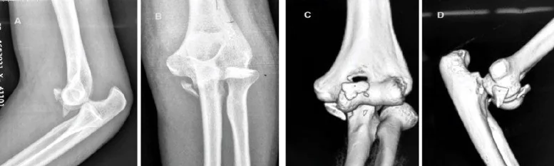
Koronoid ucundaki küçük fragman, dirseğin ön kemik desteğini zayıflatabilir ve basit çıkık ya da terrible triad örüntüsünün parçası olabilir. Kontrastsız BT kırık apeksinin boyutunu, yönünü ve ulnohumeral eklemle ilişkisini gösterirken radyografide üst üste gelen küçük parçaları da açığa çıkarır. Radial baş kırığı ve çıkık eşlik ediyorsa yaralanma tek bir kemik bulgusundan daha geniştir. Fragmanı veya eklemdeki eksantrik yerleşimi atlamak instabilite örüntüsünün eksik tanımlanmasına yol açabilir.
Faz ve pencere
- Kontrastsız tanısal
- Aksiyel ve sagittal kemik rekonstrüksiyonlarda koronoid apeksinden ayrılan kortike olmayan küçük avulsiyon fragmanı, kırık tabanının ön-arka uzanımı ve fragman deplasmanı görülür; sagittal planda ulnohumeral eklem merkezlenmesi, aksiyel planda radial baş ve dirsek çıkığı ilişkisi değerlendirilir. Kemik parçacığının tanısı ince kesitli BT kesitlerinde konulur; kırık örüntüsünü değerlendirmek ve ameliyat öncesi planlama için üç boyutlu BT rekonstrüksiyonu önerilir.
Önerilen pencereler: Kemik (G 1800 / M 400), Yumuşak doku (G 400 / M 40).
BT bulguları
- Koronoid apeksinde ince avulsiyon parçası, uç korteksinden ayrılmış küçük kemik fragmanı olarak izlenir.
- Sagittal rekonstrüksiyonda fragmanın yüksekliği ve kırık tabanının koronoid gövdesine ne kadar uzandığı görülür.
- Parçanın öne ya da distale yer değiştirmesi ve ana ulna ile arasında kalan açıklık multiplanar kesitlerde tanımlanır.
- Ulnohumeral eklemde trochleanın trochlear çentik içindeki merkezlenmesi, uç fragmanı varlığından bağımsız olarak kontrol edilir.
- Radial başta eşlik eden kırık ve radius başı-şaft ilişkisi, terrible triad bileşenini tamamlayabilecek yaralanmayı gösterir.
- Ön ya da arka ulnohumeral çıkık, eklem yüzlerinin karşılıklı temas kaybıyla belirlenir; indirgenmiş BT'de hafif eksantriklik de önemlidir.
- Küçük kortikal fragmanın kaynağını değerlendirirken eşlik eden bulgular tek başına yeterli değildir; üst üste binen patolojilerin ayrımında BT görüntülemesinin tanısal rolü öne çıkar.
Ölçütler ve sınıflamalar
- O’Driscoll tip I alt tip 1 (küçük uç fragmanı)
- ≤2 mm
- O’Driscoll tip I alt tip 2 (büyük uç fragmanı)
- >2 mm
- O’Driscoll koronoid sınıflaması, tip I (uç)
- Alt tip 1 küçük uç fragmanını (<2 mm), alt tip 2 daha büyük uç fragmanını (>2 mm) tanımlar. O’Driscoll tip I alt tip 1, 2 mm ve altında fragmanları; alt tip 2 ise 2 mm’den büyük fragmanları tanımlar. Uç kırığı posterolateral rotatuvar instabilite veya terrible triad örüntüsünde görülebilir.
- Regan–Morrey
- Regan–Morrey sınıflaması koronoid kırıklarını fragman yüksekliğine göre sınıflar; bu tipler fragman hacmi ve eklem yüzü tutulumu bakımından da farklılaşır. BT’de fragmanın yeri ve eklem uyumu ayrıca tarif edilir.
Normalde
Normal koronoid çıkıntı, ulnanın trochlear çentiğinin ön kenarında düzgün kortike bir tepe oluşturur; sagittal BT'de trochlea ile uyumlu ulnohumeral temas korunur. Normal koronoid çıkıntıda korteks bütünlüğü devam eder; avulsiyon parçasının tanısında eklem içindeki eksantrik yerleşim, kortikal bütünlüğün kesilmesi ve ince kesitli sagittal BT'de fragmanın ana kemikle ilişkisi esas alınır.
Ayırıcı tanı
- Koronoid anteromedial faset kırığı
- parça yalnız apeksi değil medial eklem kenarını da izler ve oblik eklem yüzü oluşturur.
- Koronoid baz kırığı
- kırık koronoid tabanını tutar; fragman hacmi ve eklem yüzü tutulumu uç kırığından büyüktür ve proksimal ulna kırığıyla birlikte trans-ulnar kırık-çıkık örüntüsü oluşturabilir.
- Osteokondral serbest cisim
- eklem içinde bulunur ve dirsek çıkıklarında sık gözden kaçan posterolateral kapitellum kaynaklı osteokondral lezyonlardan köken alabilir; koronoid tepesinde donör korteks defekti bulunmayabilir.
- Olekranon apofiz varyantı
- iskelet olgunlaşmamış hastada sekonder kemikleşme merkezleri kırık izlenimi verebilir; buna karşılık koronoid, dirseğin varus ve arkaya yönelik kuvvetlere karşı direnç gösteren önemli ön destek çıkıntısıdır.
- Ulnohumeral çıkık sonrası eklem uyumsuzluğu
- belirgin ayrılmış kemik parçası olmadan trochlea ile trochlear çentik merkezlenmesi bozulur.
Tuzaklar
- Küçük uç fragmanı tek planda okumada gözden kaçabilir; koronoid apeksinden geçen sagittal rekonstrüksiyonlar ve ardışık ince kesitler (0,5–1,0 mm) fragmanın gösterilmesi ve ölçülmesinde kullanılır.
- İndirgenmiş dirsekte normal görünen tek kesit hafif ulnohumeral eksantrikliği saklayabilir; trochlear çentik boyunca seri sagittal görüntüleri tarayarak eklem merkezini doğrula.
- Uç avulsiyonu ile faset yarılması birlikte olabilir; fragmanın yalnız tepeden mi yoksa medial eklem kenarından mı çıktığını aksiyel ve koronal planlarda ayır.
Kendini dene
Dirsek çıkığı redükte edildikten sonra BT’de koronoid tepesinden küçük bir fragman ve radial baş kırığı görülüyor. Hangi yaralanma örüntüsü özellikle düşünülür?
Cevabı göster
Terrible triad. Koronoid uç ve radial baş kırıkları dirsek çıkığı ile birlikte terrible triad bileşenlerini oluşturabilir. Diğer örüntüler sırasıyla proksimal ulna-radial baş ilişkisi, olekranon üzerinden eklem ayrılması veya anteromedial faset ağırlıklı yaralanma tarif eder.
Sagittal BT'de trochlea trochlear çentiğin merkezinde değil; ufak kemik parçası da var. İlk değerlendirme hangisi olmalı?
Cevabı göster
Ulnohumeral subluksasyon. Trochleanın çentik içinde eksantrik kalması ulnohumeral eklem uyum kaybını gösterir ve küçük koronoid fragmanıyla birlikte önem taşır. Diğer seçenekler bu eklem yüzlerinin konum bozukluğunu açıklamaz.
İlgili konular
Kaynaklar
Bu sayfadaki 46 cümle ve 2 quiz sorusu aşağıdaki kaynaklardan alıntılarla tek tek karşılaştırıldı; 10 cümle bu karşılaştırmada düzeltildi (2026-10-08).
- Medial elbow approach via a medial epicondyle osteotomypmc.ncbi.nlm.nih.gov
- Concurrent Acute Fracture and Dysplasia‐Related Fragmentation of the Medial Coronoid Process of the Ulna in a Dog—A Case Reportpmc.ncbi.nlm.nih.gov
- Articular varus angles of the elbow are not associated with coronoid fracture typepmc.ncbi.nlm.nih.gov
- Biomechanical stability of coronoid fractures with different fragment heights in the terrible triad of the elbow: CT measurement and finite element analysispmc.ncbi.nlm.nih.gov
- Three‐Dimensional Fracture Mapping of the Terrible Triad of the Elbow: Morphological Characteristics and Clinical Implicationspmc.ncbi.nlm.nih.gov
- An anatomic study of coronoid cartilage thickness with special reference to fractures.pubmed.ncbi.nlm.nih.gov
- Factors associated with the choice of treatment for coronoid fractures.pubmed.ncbi.nlm.nih.gov
- Fracture of the Anteromedial Facet of the Coronoid is More Common Than Previously Thought in Combined Fractures of the Coronoid and Radial Head.pubmed.ncbi.nlm.nih.gov
- Look for the POLESTAR (POsteroLateral Engagement of Soft Tissue And Radial head) while navigating around elbow dislocation.pubmed.ncbi.nlm.nih.gov
- Osteochondral reconstruction for post-traumatic coronoid deficiency.pubmed.ncbi.nlm.nih.gov
- Coronoid fractures and traumatic elbow instability.pubmed.ncbi.nlm.nih.gov
- Irreducible Elbow Dislocation Associated with Hill-Sachs-like Lesion over the Capitellum.pubmed.ncbi.nlm.nih.gov
- Fractures of the coronoid: morphology based upon computer tomography scanning.pubmed.ncbi.nlm.nih.gov
- Computed tomography analysis of the relationship between the coronoid and the radial head.pubmed.ncbi.nlm.nih.gov
- Quantitative 3-dimensional computed tomography measurements of coronoid fractures.pubmed.ncbi.nlm.nih.gov
- Morphometric feature description of the proximal ulna based on quantitative measurement: a key consideration for implant design.pubmed.ncbi.nlm.nih.gov
- Morphological characteristics of fractures of the anteromedial facet of the coronoid in posteromedial rotatory instability of the elbow: a three-dimensional CT remodeling study.pubmed.ncbi.nlm.nih.gov
- The shape match of the olecranon tip for reconstruction of the coronoid process: influence of side and osteotomy angle.pubmed.ncbi.nlm.nih.gov
- Micro-CT Structural Analysis of the Canine Medial Coronoid Disease.pubmed.ncbi.nlm.nih.gov
- Biomechanical stability of coronoid fractures with different fragment heights in the terrible triad of the elbow: CT measurement and finite element analysis.pubmed.ncbi.nlm.nih.gov
- Three-Dimensional Fracture Mapping of the Terrible Triad of the Elbow: Morphological Characteristics and Clinical Implications.pubmed.ncbi.nlm.nih.gov
- The effects of coronoid fracture fixation on functional outcomes and complication rates following terrible triad injuries of the elbow.pubmed.ncbi.nlm.nih.gov
- Current State-of-the-Art 3D MRI Sequences for Assessing Bone Morphology with Emphasis on Cranial and Spinal Imaging: A Narrative Review.pubmed.ncbi.nlm.nih.gov
- Mechanobiologically-optimized non-resorbable artificial bone for patient-matched scaffold-guided bone regeneration.pubmed.ncbi.nlm.nih.gov
- Imaging of Bone in the Head and Neck Region, is There More Than CT?pubmed.ncbi.nlm.nih.gov
- The "Terrible Tetrad" of the Elbow: A Case Series of Two Patients with a Rare Quadruple-Component Injury and a Proposed New Classification Term.pubmed.ncbi.nlm.nih.gov
- The anconeus approach is safe and effective for lateral elbow trauma: a consecutive case series of clinical and functional outcomes.pubmed.ncbi.nlm.nih.gov
- Clinical anatomy and biomechanics of the elbow.pubmed.ncbi.nlm.nih.gov
- Acute and overuse elbow trauma: radio-orthopaedics overview.pubmed.ncbi.nlm.nih.gov
- Comparison of all suture fixation with tension band wiring and plate fixation of the olecranon.pubmed.ncbi.nlm.nih.gov
- Optimal Radial Head Prosthesis Sizing in Elbow Trauma: A Review of Anatomical Principles and Clinical Techniques.pubmed.ncbi.nlm.nih.gov
- Fractures of the coronoid process: state of the art.pubmed.ncbi.nlm.nih.gov
- The Role of the Critical Coronoid Angle in Simple Elbow Dislocation: A Computed Tomography-Based Index to Stratify Elbow Dislocation Risk.pubmed.ncbi.nlm.nih.gov
- Photon-Counting CT: Technology, Current and Potential Future Clinical Applications, and Overview of Approved Systems and Those in Various Stages of Research and Development.pubmed.ncbi.nlm.nih.gov
- CT Angiography of the Upper Extremities: Review of Acute Arterial Entities.pubmed.ncbi.nlm.nih.gov
- Segmental Megaesophagus Secondary to Extraluminal Esophageal Stenosis Caused by Transitional Seventh Cervical Vertebra and Supernumerary Ribs in a Goat.pubmed.ncbi.nlm.nih.gov
- Integrated management of mandibular fractures and traumatic dental injuries: a literature review and clinical algorithm proposal.pubmed.ncbi.nlm.nih.gov
- The Application of Cone Beam Computed Tomography (CBCT) on the Diagnosis and Management of Maxillofacial Trauma.pubmed.ncbi.nlm.nih.gov
- Pre- and postmortem tyrannosaurid bite marks on the remains of Daspletosaurus (Tyrannosaurinae: Theropoda) from Dinosaur Provincial Park, Alberta, Canada.pubmed.ncbi.nlm.nih.gov
- Autologous Tissue Grafts for Chin Augmentation With or Without Genioplasty: A Systematic Review.pubmed.ncbi.nlm.nih.gov
- Temporomandibular Joint Defects: Etiology and Reconstruction Strategy: A Narrative Review.pubmed.ncbi.nlm.nih.gov
- Elbow Varus posteromedial rotatory instability: Reflections from a multidimensional perspective.pubmed.ncbi.nlm.nih.gov
- Valgus mechanism inducing a unique "terrible triad injury" pattern in the elbow: a detailed clinical and surgical analysis.pubmed.ncbi.nlm.nih.gov
- A Rare Repair: Trochlear Fracture Fixed With Headless Screws and Medial Epicondyle Osteotomy - A Case Report.pubmed.ncbi.nlm.nih.gov
- 오버헤드 투구 선수에서의 팔꿈치 외반-신전 과부하 증후군.pubmed.ncbi.nlm.nih.gov
- A Systematic Approach to the Evaluation of Acute Musculoskeletal Injuries in the Emergency Department.pubmed.ncbi.nlm.nih.gov
- Musculoskeletal Ultrasound and Protein-Based Hydrogels: Novel Approaches for the Diagnosis and Treatment of Sports Injuries.pubmed.ncbi.nlm.nih.gov
- Musculoskeletal ultrasound in the emergency department: a narrative review for general radiologists.pubmed.ncbi.nlm.nih.gov
- Dislocations of the elbow - An instructional review.pubmed.ncbi.nlm.nih.gov
- Precision of virtual surgical planning employing screw-holes locator and patient-specific implants versus conventional plate osteosynthesis for parasymphyseal mandibular fracture: a double-blinded randomized control trial.pubmed.ncbi.nlm.nih.gov
- Radiographic Evaluation of Common Pediatric Elbow Injuries.pubmed.ncbi.nlm.nih.gov
- Articular varus angles of the elbow are not associated with coronoid fracture type.pubmed.ncbi.nlm.nih.gov
- Anatomic, Radiographic, and Computed Tomographic Study of the Skull in the Persian Squirrel (Sciurus anomalus).pubmed.ncbi.nlm.nih.gov
- Paleontological and paleoecological significance of the oldest highly productive Upper Cretaceous (lowermost Maastrichtian) bonebed of Haţeg Basin (western Romania; Densuş-Ciula Formation).pubmed.ncbi.nlm.nih.gov
- Evidence for deliberate burial of the dead by <i>Homo naledi</i>.pubmed.ncbi.nlm.nih.gov
- Implant Survivorship, Radiographic Evaluation, and Clinical Outcomes of Radial Head Arthroplasty Using The Depuy Synthes Radial Head Replacement System.pubmed.ncbi.nlm.nih.gov
- Nonoperative management of terrible triad injuries: clinical outcomes of 39 cases.pubmed.ncbi.nlm.nih.gov
- Clinical and radiological outcome of Mason-Johnston types III and IV radial head fractures treated by an on-table reconstruction.pubmed.ncbi.nlm.nih.gov
- Surgical Treatment of Coronoid Fracture With Elbow Varus Posteromedial Rotatory Instability: An Instructional Review.pubmed.ncbi.nlm.nih.gov
- Repair Versus Non-Repair of Lateral Ulnar Collateral Ligament in Elbow Varus Posteromedial Rotatory Instability Treatment: A Comparative Study.pubmed.ncbi.nlm.nih.gov
- Arthroscopic Reduction and Fixation of Coronoid Fracture with Screws and Modified Pull Out Configuration.pubmed.ncbi.nlm.nih.gov
- Biomechanical investigation of elbow dislocation: comparative analysis using <i>Papio anubis</i> baboon and human cadaver models.pubmed.ncbi.nlm.nih.gov
- The value of elbow arthroscopy in diagnosing and treatment of radial head fractures.pubmed.ncbi.nlm.nih.gov
- Elbow Instability: Anatomy, Biomechanics, Diagnostic Maneuvers, and Testing.pubmed.ncbi.nlm.nih.gov
- Magnetic resonance imaging of the elbow: a structured approach.pubmed.ncbi.nlm.nih.gov
- Geology and paleontology of the Upper Cretaceous Kem Kem Group of eastern Morocco.pubmed.ncbi.nlm.nih.gov
- Cranial anatomy, palaeoneurology, palaeobiology and stratigraphic age of the large-bodied ornithopod, <i>Muttaburrasaurus langdoni</i> Bartholomai and Molnar, 1981, from the mid-Cretaceous of Australia.pubmed.ncbi.nlm.nih.gov
- Anatomy and relationships of the early diverging Crocodylomorphs Junggarsuchus sloani and Dibothrosuchus elaphros.pubmed.ncbi.nlm.nih.gov
- Arthroscopically-assisted fixation of anteromedial coronoid facet fracture and lateral ulnar collateral ligament repair for acute posteromedial rotatory fracture dislocation of the elbow.pubmed.ncbi.nlm.nih.gov
- Impact of immobilization period and anterior capsular injury on flexion contracture in distal humerus coronal shear fractures.pubmed.ncbi.nlm.nih.gov
- Postoperative Outcomes of Anteromedial Approach in Patients with Elbow Varus Posteromedial Rotatory Instability: A Retrospective Study.pubmed.ncbi.nlm.nih.gov
- Trauma of the midface.pubmed.ncbi.nlm.nih.gov
- Management of a Nonunion Type IIC Monteggia Fracture.pubmed.ncbi.nlm.nih.gov
- Concurrent Acute Fracture and Dysplasia-Related Fragmentation of the Medial Coronoid Process of the Ulna in a Dog-A Case Report.pubmed.ncbi.nlm.nih.gov
- Fracture mapping of coronal shear fractures of the distal humerus: morphological analysis and fixation efficacy.pubmed.ncbi.nlm.nih.gov
- Evaluation of treatment outcomes of zygomaticomaxillary complex (ZMC) fractures using Foley catheter ballooning technique.pubmed.ncbi.nlm.nih.gov
- Prevalence and Morphology of the Palatal Bone Reservoir in the Posterior Maxilla as an Alternative to Maxillary Sinus Lift: A Cross-Sectional Retrospective Analysis Determined by Helical CT Scan.pubmed.ncbi.nlm.nih.gov
- The role of forensic imaging in the allegations of torture in asylum seekers.pubmed.ncbi.nlm.nih.gov
- Rugby Injury Imaging.pubmed.ncbi.nlm.nih.gov
- Novel Radiographic Indexes for Elbow Stability Assessment: Part B-Preliminary Clinical Study.pubmed.ncbi.nlm.nih.gov
- Surgical Outcomes and Complications in Treatment of Terrible Triad of the Elbow: Comparisons of 3 Surgical Approaches.pubmed.ncbi.nlm.nih.gov
- Salmonella Typhi Osteomyelitis of the Ulnar Diaphysis in an Immunocompetent Adult: A Case Report.pubmed.ncbi.nlm.nih.gov
- Management of Pediatric Elbow Fractures and Dislocations.pubmed.ncbi.nlm.nih.gov
- Prediction and Potential Preventions for the Development of Posttraumatic Osteoarthritis after the Terrible Triad Injury: A Multicenter Risk Factors Study.pubmed.ncbi.nlm.nih.gov
- Postcranial anatomy of <i>Besanosaurus leptorhynchus</i> (Reptilia: Ichthyosauria) from the Middle Triassic Besano Formation of Monte San Giorgio (Italy/Switzerland), with implications for reconstructing the swimming styles of Triassic ichthyosaurs.pubmed.ncbi.nlm.nih.gov
- Osteochondritis Dissecans of the Elbow in Overhead Athletes: A Comprehensive Narrative Review.pubmed.ncbi.nlm.nih.gov
- Irreducible anteromedial radial head fracture-dislocation: value of pre-operative magnetic resonance imaging.pubmed.ncbi.nlm.nih.gov
- Temporary Cemented Spacer for Complex Radial Head Fractures: A Case Report.pubmed.ncbi.nlm.nih.gov
- Reliability of the CURL classification for proximal ulna fracture-dislocations of the elbow.pubmed.ncbi.nlm.nih.gov
- A novel internal fixation technique for chronic elbow dislocations: Technical note and case report.pubmed.ncbi.nlm.nih.gov
- The inter-individual anatomical variation of the trochlear notch as a predisposition for simple elbow dislocation.pubmed.ncbi.nlm.nih.gov
- Terrible Triad Injuries around the Elbow: It is Still a Puzzle? Prospective Study.pubmed.ncbi.nlm.nih.gov
- The coronoid as the key fragment of trans-ulnar fracture-dislocations of the elbow: Insights from a retrospective cohort comparison using the coronoid-centric Mayo classification system.pubmed.ncbi.nlm.nih.gov
- Treatment of Proximal Trans-ulnar Fracture-Dislocations Based on a Step-By-Step Coronoid-Centric Surgical Technique: Proposal of a Surgical Algorithm Based on Coronoid Management and Windows Approach.pubmed.ncbi.nlm.nih.gov
- Outcomes Associated With Osteochondral Allograft Transplantation in Dogs.pubmed.ncbi.nlm.nih.gov
- Surgical treatment of rheumatoid arthritis of the elbow.pubmed.ncbi.nlm.nih.gov
- A review on artificial intelligence for the diagnosis of fractures in facial trauma imaging.pubmed.ncbi.nlm.nih.gov
- A comparative study assessing the efficacy and safety of radiofrequency ablation versus surgical treatment for osteoid osteoma: retrospective analysis in a single institution.pubmed.ncbi.nlm.nih.gov
- Quantifying Vascular Changes Surrounding Bone Regeneration in a Porcine Mandibular Defect Using Computed Tomography.pubmed.ncbi.nlm.nih.gov
- Osteology of the Hamadryas Baboon ( Papio hamadryas ).pubmed.ncbi.nlm.nih.gov
- Description of the Pliocene marsupial Ambulator keanei gen. nov. (Marsupialia: Diprotodontidae) from inland Australia and its locomotory adaptations.pubmed.ncbi.nlm.nih.gov
- Clinical and Radiographic Outcomes of Total Elbow Arthroplasty Using a Semi-constrained Prosthesis with a Triceps-preserving Approach over a Minimum Follow-up Period of 4 Years.pubmed.ncbi.nlm.nih.gov
- Elbow terrible triad: Advancing current concepts in the treatment of 13 cases.pubmed.ncbi.nlm.nih.gov
Eğitim amaçlıdır; hasta başında tanı ve tedavi kararının yerine geçmez. İçerik yapay zekâ destekli bir süreçle güncel literatüre göre cümle cümle kaynaklarla karşılaştırıldı, ancak uzman radyolog incelemesinden geçmedi. Klinik kararlarda güncel kılavuzları, kurum protokollerini ve radyoloji raporunu esas alın.