Özet
Atravmatik diz ağrısı olan erişkinde subkondral yetersizlik kırığı (SIF) ve osteonekrozla komplike SIF (SIF-ON); sistemik riskleri olan hastada ise epifiz osteonekrozu veya distal femur/proksimal tibia medüller kemik enfarktı ayrıştırılmalıdır. Erken subkondral kemik iliği değişikliklerinin değerlendirilmesinde MR öne çıkar; osteonekrozla komplike SIF eklem yüzü çökmesine ilerleyebilir. Medüller kemik enfarktı çoğu kez serpijinöz sklerotik kenarlı, merkezinde yağlı medüller yoğunluğu koruyan bir odak olarak görünür.
Faz ve pencere
- Kontrastsız tanısal
- SIF’te subkondral kırık çizgisi görülebilir; SIF-ON eklem yüzünde geri dönüşümsüz çökme ile ilerleyebilir. Erken SIF ve SIF-ON’da kemik iliği ödemi-benzeri sinyal MR’da değerlendirilir; tek başına özgül değildir.
- Kemik algoritması tanısal
- Erken SIF ve SIF-ON’da kemik iliği ödemi-benzeri sinyal MR’da görülebilir; SIF’in saptanması ve izleminde MR en iyi yöntemdir.
Önerilen pencereler: Kemik (G 1800 / M 400), Yumuşak doku (G 400 / M 40).
BT bulguları
- Subkondral skleroz — femoral kondillerde belirgin skleroz görülebilir.
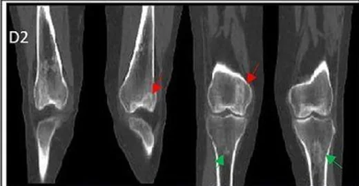
- Serpijinöz medüller sınır — metafiz/şaft medullasında kıvrımlı sklerotik kenarın çevrelediği, merkezinde medüller yağ yoğunluğunu koruyabilen enfarkt odağı.
- Subkondral yarımay kırığı — MR’da subkondral hipointens kırık çizgisi SIF ile uyumludur; SIF-ON eklem yüzünde geri dönüşümsüz çökme ile ilerleyebilir.
- Eklem yüzü çökmesi — SIF-ON, eklem yüzünde geri dönüşümsüz çökme ile ilerleyebilir.
- İkincil eklem değişikliği — subkondral çökme sonrası osteoartrit gelişebilir.
- Birden fazla kemikte benzer medüller serpijinöz odaklar çok odaklı kemik enfarktı örüntüsünü düşündürür; dağılım klinik bağlamla birlikte yorumlanır.
- Eşlik eden kırık veya serbest osteokondral parça — subkondral kemik plağı kesintisi ve eklem içi fragman ayrıca kaydedilir.
Normalde
Normal femoral kondil ve tibial platoda subkondral kemik plağı düzgün ve kesintisizdir; eklem yüzü olağan konturunu korur. SIF-ON'da subkondral alanda, değişmiş kemik iliğine bağlı sıvıya duyarlı MR sekanslarında düşük sinyal görülebilir.
Normal BT ile kıyasla
Aynı bölgenin, kaynak veri setinde patoloji içermediği belirtilen bir BT incelemesini kesit kesit kaydırın; organ sınırlarını açarak anatomiyi hasta görüntüsüyle karşılaştırın. Küçük rastlantısal bulgular tümüyle dışlanmamıştır.
Kaynak: TotalSegmentator v2.01 veri seti (Wasserthal ve ark., Radiology: Artificial Intelligence 2023), CC BY 4.0, vaka s0685; organ sınırları veri setinin otomatik segmentasyonundan, birleştirilerek sadeleştirildi. Görüntüler 2,5 mm kesit aralığına yeniden örneklendi.
Ayırıcı tanı
- Subkondral yetersizlik kırığı (SIF), subkondral kırık çizgisiyle ilişkilidir; MR'da kemik iliği ödemi-benzeri sinyal eşlik edebilir. SIF osteonekroz ile komplike olduğunda SIF-ON olarak adlandırılır. Medüller kemik enfarktı metafiz/şaft medullasında, sistemik kökenli osteonekroz ise epifizde yerleşebilir; klinik bağlamla birlikte değerlendirilir.
- Osteokondritis dissekans
- subkondral kemik ve eklem kıkırdağını içeren fokal osteokondral lezyondur; juvenil olgularda en sık femoral kondilin posterolateral bölümünde görülür.
- Kronik osteomiyelit
- sekestr, kortikal kloaka, periostal yeni kemik veya yumuşak doku sinüs yolu eşlik edebilir.
- Osteoartrit
- yaygın kompartman daralması, osteofit ve subkondral skleroz oluşturur; tek başına serpijinöz medüller sınır beklenmez.
Tuzaklar
- Erken SIF'te MR'da kemik iliği ödemi-benzeri sinyal ve subkondral kırık çizgisi görülebilir; erken tanı ve değerlendirmede MR en uygun yöntemdir. Tarihsel “SONK” terimi yerine bulgu uygunsa SIF veya SIF-ON kullanın.
- SIF'te subkondral kırık çizgisi MR'da görülebilir; değerlendirmede eşlik eden kemik iliği ödemi de dikkate alınır.
- Medüller enfarktın çevresel sklerozu agresif lezyon sanılabilir; merkezde korunmuş medüller yağ ve düzgün serpijinöz sınır, kortikal yıkım olmamasıyla birlikte benign enfarkt lehinedir.
- Subkondral yetersizlik kırığı ilerleyerek eklem yüzü çökmesine ve ikincil osteoartrite yol açabilir.
- SIF’te MR’da subkondral hipointens kırık çizgisi ve kemik iliği ödemi-benzeri sinyal görülebilir; menisküs arka kök yırtıkları ve ekstrüzyon birçok hastada bildirilmiştir.
Kendini dene
Diz MR'ında kondil altında hipointens kırık çizgisi ve kemik iliğinde ödem-benzeri sinyal görülüyor. Bu bulgular en çok hangi lezyonla uyumludur?
Cevabı göster
Subkondral stres kaynaklı kırık. Subkondral hipointens kırık çizgisi ve kemik iliği ödemi-benzeri sinyal SIF için tipik MR bulgularıdır.
Distal femur medullasında iyi sınırlı, serpentinöz kenarlı bir odak görülüyor. Tanı nedir?
Cevabı göster
Kemik enfarktı. Serpentin biçimli, iyi sınırlı medüller odak kemik enfarktını destekler; kemik iliği ödemi-benzeri sinyal MR’da değerlendirilir. Brodie apsesi kemikte litik, sklerotik kenarlı bir kavite olarak görülebilir.
İlgili konular
Kaynaklar
Bu sayfadaki 39 cümle ve 2 quiz sorusu aşağıdaki kaynaklardan alıntılarla otomatik olarak karşılaştırıldı; 18 cümle bu karşılaştırmada düzeltildi (2026-10-09).
- Subchondral insufficiency fractures, subchondral insufficiency fractures with osteonecrosis, and other apparently spontaneous subchondral bone lesions of the knee—pathogenesis and diagnosis at imaginglink.springer.com
- ACR Appropriateness Criteria® Osteonecrosis: 2022 Updateacsearch.acr.org
- Multiple bone infarcts with intra-articular extensionpubmed.ncbi.nlm.nih.gov
- Imaging of bone marrow pitfalls with emphasis on MRIpmc.ncbi.nlm.nih.gov
- An update on osteochondritis dissecans of the kneepmc.ncbi.nlm.nih.gov
- Common Skeletal Neoplasms and Nonneoplastic Lesions at 18F-FDG PET/CTpubs.rsna.org
- Imaging of knee osteoarthritis: a review of multimodal diagnostic approachqims.amegroups.org
- Bone and Joint Infections: The Role of Imaging in Tailoring Diagnosis to Improve Patients’ Carepmc.ncbi.nlm.nih.gov
- Diagnostic Perspectives in Bone and Soft Tissue Infectionsjournals.sagepub.com
- MRI features of pancreatitis, panniculitis, polyarthritis and the fourth p: The “polka dot sign.” A case series and imaging reviewpmc.ncbi.nlm.nih.gov
- Mesenchymal stem cell-derived extracellular vesicles: current advances in preparation and therapeutic applications for neurological disorderspmc.ncbi.nlm.nih.gov
- Imaging of lower extremity infections: predisposing conditions, atypical infections, mimics, and differentiating featurespmc.ncbi.nlm.nih.gov
- Proceedings of the 33rd European Paediatric Rheumatology Congresspmc.ncbi.nlm.nih.gov
- Bilateral Knee Osteonecrosis in an Adolescent With Leukemia on Prolonged Corticosteroids: Discordant Femoral-Tibial Findings on Bone Scan SPECT-CT.pubmed.ncbi.nlm.nih.gov
- Osteonecrosis in Youth with Sickle Cell Disease-A Narrative Review.pubmed.ncbi.nlm.nih.gov
- Association of anterior necrotic boundary location with collapse progression in symptomatic osteonecrosis of the femoral head: analysis of ARCO 2021 type 2 lesions.pubmed.ncbi.nlm.nih.gov
- Case Report: A case of surgical and enzyme replacement therapy for type I Gaucher disease complicating femoral shaft pathological fracture.pubmed.ncbi.nlm.nih.gov
- A Primer on Anatomy, Biophysics, Pathology, Imaging and Treatment of the Intervertebral Disc and the Anterior Spinal Column: The Discogenic, Intervertebral, Spinal Column (DISC) ASPN Workgroup.pubmed.ncbi.nlm.nih.gov
- The role of imaging in extremity sarcoma surgery.pubmed.ncbi.nlm.nih.gov
- Utility of SPECT/CT in Non-Traumatic Osteonecrosis: Revisited with New Insights from a Prospective Diagnostic Study.pubmed.ncbi.nlm.nih.gov
- A Review of Pediatric Orthopedic Disorders: Diagnosis and Treatment Updates.pubmed.ncbi.nlm.nih.gov
- Relationship between DXA measured systemic bone mineral density and subchondral bone cysts in postmenopausal female patients with knee osteoarthritis: a cross-sectional study : Osteoarthritis cysts and bone mineral density.pubmed.ncbi.nlm.nih.gov
- Medial femoral condyle bone mineral density is associated with subchondral insufficiency fractures in female patients with medial meniscus posterior root tears.pubmed.ncbi.nlm.nih.gov
- Association between radial meniscal tears with and without posterior root involvement and subchondral insufficiency fracture of the knee on MRI: The mediating role of meniscal extrusion.pubmed.ncbi.nlm.nih.gov
- Beyond Cartilage Regeneration: Targeting the Subchondral Bone with Extracorporeal Shock Wave Therapy and Regenerative Medicine in Knee Osteoarthritis.pubmed.ncbi.nlm.nih.gov
- Treatment of Steroid-Induced Osteonecrosis of the Distal Femoral Condyle Using Structural Bone Grafting.pubmed.ncbi.nlm.nih.gov
- Subchondral Insufficiency Fracture of the Knee Previously Diagnosed as Spontaneous Osteonecrosis: A Case Report With Histological Analysis.pubmed.ncbi.nlm.nih.gov
- Multimodality imaging considerations for genicular artery embolization in knee osteoarthritis.pubmed.ncbi.nlm.nih.gov
- Intraosseous bone marrow concentrate delays total hip arthroplasty in osteoarthritis: A fifteen year matched cohort study with dose-response analysis.pubmed.ncbi.nlm.nih.gov
- Multisite Osteonecrosis to Long-term Corticoid Treatment: A Case Report.pubmed.ncbi.nlm.nih.gov
- Avascular Necrosis Secondary to Traumatic Posterior Hip Dislocation.pubmed.ncbi.nlm.nih.gov
- Clinical and Radiological Outcomes of Oral Bisphosphonate Therapy Combined with Conservative Treatment in Subchondral Bone Marrow Edema of the Knee: A Retrospective Cohort Study.pubmed.ncbi.nlm.nih.gov
- Arthroscopic Autologous Minced Cartilage Implantation for Subchondral Insufficiency Fracture of the Knee.pubmed.ncbi.nlm.nih.gov
- Association between low bone mineral density and subchondral insufficiency fractures of the knee in women, and influence of radial meniscal root tears.pubmed.ncbi.nlm.nih.gov
- Pharmacotherapy of Kienböck Disease and the Carpal Avascular Necrosis Family: A Narrative Review with a Four-Axis Translation Framework.pubmed.ncbi.nlm.nih.gov
- Intraosseous tumours of the knee.pubmed.ncbi.nlm.nih.gov
- Bone lesions of the tibia: Multimodal iconographic review and diagnostic algorithms, Part 2: Metaphyseal and epiphyseal lesions.pubmed.ncbi.nlm.nih.gov
- Radiological manifestation of avascular necrosis (AVN) in sickle cell disease (SCD): a review of diagnostic imaging.pubmed.ncbi.nlm.nih.gov
- Pharmacological Effects and Mechanisms of Tanshinone IIA in Bone Injury Repair.pubmed.ncbi.nlm.nih.gov
- Dysbaric osteonecrosis of the humeral head in a patient with type 2 decompressive sickness.pubmed.ncbi.nlm.nih.gov
- Musculoskeletal "don't touch" lesions: pictorial essay.pubmed.ncbi.nlm.nih.gov
- Bone marrow lesions related to bone marrow edema syndromes and osteonecrosis.pubmed.ncbi.nlm.nih.gov
- Corticosteroid-associated osteonecrosis in paediatric compared with adult patients: are they the same disease?pubmed.ncbi.nlm.nih.gov
- Avascular necrosis of lateral femoral condyle in a middle-aged woman from central India.pubmed.ncbi.nlm.nih.gov
- MRI features of pancreatitis, panniculitis, polyarthritis and the fourth p: The "polka dot sign." A case series and imaging review.pubmed.ncbi.nlm.nih.gov
- Imaging of lower extremity infections: predisposing conditions, atypical infections, mimics, and differentiating features.pubmed.ncbi.nlm.nih.gov
- CRP Is a Key Indicator of Rheumatoid Arthritis-Associated Vascular Injury and Neurodegeneration.pubmed.ncbi.nlm.nih.gov
- Ankle Arthritis.pubmed.ncbi.nlm.nih.gov
- Hip arthroscopy enables classification and treatment of precollapse subchondral insufficiency fracture of the femoral head associated intra-articular pathology.pubmed.ncbi.nlm.nih.gov
- Osteochondritis Dissecans of the Medial Femoral Condyle With Intra-Articular Meniscal Calcification: A Rare Case and Literature Review.pubmed.ncbi.nlm.nih.gov
- Conventional radiography for the assessment of focal bone lesions of the appendicular skeleton: fundamental concepts in the modern imaging era.pubmed.ncbi.nlm.nih.gov
- [Subchondral insufficiency fractures of the knee : Navigating the complex terminology landscape].pubmed.ncbi.nlm.nih.gov
- Subchondral insufficiency fractures, subchondral insufficiency fractures with osteonecrosis, and other apparently spontaneous subchondral bone lesions of the knee-pathogenesis and diagnosis at imaging.pubmed.ncbi.nlm.nih.gov
- A phase III, randomised, double-blind, multi-dose, placebo- and naproxen-controlled study to evaluate the efficacy and safety of fasinumab in patients with pain due to osteoarthritis of the knee or hip.pubmed.ncbi.nlm.nih.gov
- A Real-World Investigation of MRI Changes in Bone in Patients with Type 1 Gaucher Disease Treated with Velaglucerase Alfa: The EIROS Study.pubmed.ncbi.nlm.nih.gov
- Benign incidental do-not-touch bone lesions.pubmed.ncbi.nlm.nih.gov
- Micro-computed tomographical and histopathological analyses of medial tibial plateaus from patients with subchondral insufficiency fracture or osteoarthritis of the knee.pubmed.ncbi.nlm.nih.gov
- Progress in machine learning-assisted medical imaging for osteoarthritis and osteoporosis diagnosis: a narrative review.pubmed.ncbi.nlm.nih.gov
- Beyond Palmer: a topographic classification of non-Palmer triangular fibrocartilage complex injuries.pubmed.ncbi.nlm.nih.gov
- Detection of simulated complications in all-polymer PEEK knee prostheses: a cadaveric imaging study using Radiography, CT, and MRI.pubmed.ncbi.nlm.nih.gov
- Piezo1-mediated mechanotransduction and metabolic regulation in bone health: molecular mechanisms and implications for bone disorders.pubmed.ncbi.nlm.nih.gov
- The Charcot Knee Arthropaty: The Diagnostic and Surgical Challenge. A Case of Syphilis Arthropaty and a Review of Literature.pubmed.ncbi.nlm.nih.gov
- Hip Arthroscopy and Chondrofiller Application in Isolated Osteochondral Defect of the Femoral Head.pubmed.ncbi.nlm.nih.gov
- Natural Biomaterials for Osteochondral Repair: From Source to Strategy.pubmed.ncbi.nlm.nih.gov
- Optimizing diagnosis and management of ONFH, FNSF, and SIF.pubmed.ncbi.nlm.nih.gov
- Fascia Lata and Tensor Fasciae Latae in Lower-Limb Reconstruction.pubmed.ncbi.nlm.nih.gov
- [Musculoskeletal Complications in Rheumatic Diseases].pubmed.ncbi.nlm.nih.gov
- Osteonecrosis and stress-related subchondral injuries in the elite athlete: a review on etiology, clinical signs, and management.pubmed.ncbi.nlm.nih.gov
- Osteoporosis, Osteoarthritis, and Subchondral Insufficiency Fracture: Recent Insights.pubmed.ncbi.nlm.nih.gov
- Top 15 musculoskeletal lesions in the aging recreational sporter: a pictorial review.pubmed.ncbi.nlm.nih.gov
- Zero Echo Time MRI for Osseous Assessment of Sports-Related Pathology in Athletes: A Pictorial Essay.pubmed.ncbi.nlm.nih.gov
- Subchondral Insufficiency Fracture of the Humeral Head: A Rare Case in a Young Person.pubmed.ncbi.nlm.nih.gov
- Rapidly progressive destructive arthrosis (RPDA) of the glenohumeral joint: a radiologic classification and mid-term outcomes following shoulder arthroplasty.pubmed.ncbi.nlm.nih.gov
- Silent Avascular Necrosis of the Femoral Head in Severe Hemophilia A: A Case Report.pubmed.ncbi.nlm.nih.gov
- Comparison between Vascular and Non-Vascular Bone Grafting in Scaphoid Nonunion: A Systematic Review.pubmed.ncbi.nlm.nih.gov
- Glossary of terms for musculoskeletal radiology.pubmed.ncbi.nlm.nih.gov
- Articular impaction injuries in the lower limb.pubmed.ncbi.nlm.nih.gov
- Initiation of Collapse in Osteonecrosis of the Femoral Head: A Patient-Specific Finite Element Simulation Study.pubmed.ncbi.nlm.nih.gov
- Patient-specific osteoarticular reconstruction of the proximal tibia for giant cell tumor using a 3D-printed trabecular implant and hinged knee arthroplasty: a case report.pubmed.ncbi.nlm.nih.gov
- Patellar Instability: A Narrative Review with an Integrated Treatment Algorithm.pubmed.ncbi.nlm.nih.gov
- Kinematic Alignment in TKA Using the Contralateral Healthy Knee as Reference: A Combined Finite Element and Clinical Analysis.pubmed.ncbi.nlm.nih.gov
- AI-assisted preoperative planning improves component sizing accuracy in Oxford unicompartmental knee arthroplasty: a retrospective cohort study with 24-month follow-up.pubmed.ncbi.nlm.nih.gov
- Accuracy and Precision of Femoral and Tibial Bone Resections Using Manual Unrestricted Kinematic Alignment in Total Knee Arthroplasty: A Retrospective Digital Caliper Study.pubmed.ncbi.nlm.nih.gov
- Revisiting Fat Content in Bone Lesions: Paradigms in Bone Lesion Detection.pubmed.ncbi.nlm.nih.gov
- Imaging modalities for non-acute pathologies of the foot and ankle.pubmed.ncbi.nlm.nih.gov
- Subchondral insufficiency fracture of the knee: review of current concepts and radiological differential diagnoses.pubmed.ncbi.nlm.nih.gov
- Anatomic restoration of an isolated medial tibial plateau depression in a young patient after high-energy trauma: A case report.pubmed.ncbi.nlm.nih.gov
- Arthroscopic Repair of Meniscal Medial Root Tears: The Necessity of Concomitant Realignment Osteotomy.pubmed.ncbi.nlm.nih.gov
- Imaging of bone marrow pitfalls with emphasis on MRI.pubmed.ncbi.nlm.nih.gov
- The role of radiography and MRI as screening tools and outcome measures in clinical trials of knee osteoarthritis: a perspective on the current status and outlook.pubmed.ncbi.nlm.nih.gov
- The "birth of death": MRI step-by-step reveals the early appearance of a bone marrow infarct.pubmed.ncbi.nlm.nih.gov
- Subchondral insufficiency fracture of the knee: progress in the pathogenesis and treatment.pubmed.ncbi.nlm.nih.gov
- Chinese Guideline for the Diagnosis and Treatment of Osteonecrosis of the Femoral Head in Adults.pubmed.ncbi.nlm.nih.gov
- CT Hounsfield units in assessing bone and soft tissue quality in the proximal femur: A systematic review focusing on osteonecrosis and total hip arthroplasty.pubmed.ncbi.nlm.nih.gov
- Atraumatic fractures of the femur.pubmed.ncbi.nlm.nih.gov
- Patients with preoperative bone marrow oedema benefit more substantially from medial meniscus posterior root repair.pubmed.ncbi.nlm.nih.gov
- Management and Outcomes of Osteochondritis Dissecans of the Knee in the Pediatric and Adolescent Population.pubmed.ncbi.nlm.nih.gov
- Malignant Transformation of Musculoskeletal Lesions with Imaging-Pathology Correlation-Part 1: Bone Lesions.pubmed.ncbi.nlm.nih.gov
- Genicular Artery Embolization: A New Tool for the Management of Refractory Osteoarthritis-Related Knee Pain.pubmed.ncbi.nlm.nih.gov
- Accuracy and Reliability of Multimodal Imaging in Diagnosing Knee Sports Injuriespubmed.ncbi.nlm.nih.gov
- Evolutionary Specializations of the Human Vertebral Body and Intervertebral Disc in Relation to Bipedalism.pubmed.ncbi.nlm.nih.gov
- A Novel Concept of Tissue Micro-Instability as the Underlying Mechanism of Osteophytosis in Human Knee Osteoarthritis.pubmed.ncbi.nlm.nih.gov
- Gaucher Disease in Bone: From Pathophysiology to Practice.pubmed.ncbi.nlm.nih.gov
Eğitim amaçlıdır; hasta başında tanı ve tedavi kararının yerine geçmez, tıbbi tavsiye değildir. Metinler yapay zekâ desteğiyle yazıldı ve otomatik bir adımda kaynak alıntılarıyla karşılaştırıldı; bu bir tıbbi doğrulama değildir ve içerik uzman radyolog incelemesinden geçmedi. Klinik kararlarda güncel kılavuzları, kurum protokollerini ve radyoloji raporunu esas alın.