Özet
Genellikle tek dizde ağrı, şişlik, hareket kısıtlılığı veya kilitlenme yakınması olan kişide sinovyum kaynaklı kıkırdak nodülleri ve eklem içi serbest cisimler sinovyal kondromatozisi düşündürür. Kontrastsız BT mineralize veya kemikleşmiş cisimlerin sayısını ve eklem içindeki dağılımını gösterir; mineralize olmayan kıkırdak nodüllerini dışlayamaz. Eklem boyunca yaygın dağılmış çok sayıda cisim primer sinovyal kondromatozisi destekler. Cisimlerin dağılımı ve mineralizasyonu ile varsa komşu kemik erozyonunu kaydedin; BT kalsifikasyon ve kemik erozyonunu, MRG sinovyal hastalık yayılımını değerlendirmeye yardımcı olur.
Okuma sırası
- Suprapatellar reses. Kontrastsız BT'de eklem içindeki mineralize cisimleri ve dağılımlarını değerlendirin; mineralize olmayan nodüllerin BT'de gözden kaçabileceğini akılda tutun.
- Suprapatellar ve posterior resesler. Çok düzlemli BT rekonstrüksiyonlarında kalsifiye nodüllerin kemik, sinovyum ve yumuşak dokularla ilişkisini izleyip eklem içindeki dağılımını kaydedin.
- Sinovyal nodül ve serbest cisim. Sinovyumdaki nodüllerle eklem boşluğuna ayrılmış serbest cisimleri ayırt edin; kemik penceresinde kalsifikasyonları ve kemik erozyonu varsa bunu değerlendirin.
- Eklem yüzleri ve efüzyon. BT'de kemik erozyonunu ve sekonder kondromatozis açısından altta yatan osteoartritle ilişkili kemik-eklem değişikliklerini değerlendirin.
Faz ve pencere
- Kontrastsız tanısal
- Kemik pencereli BT'de eklem içi kalsifiye veya ossifiye serbest cisimler; mineralize olmayan sinovyal nodüller ise yumuşak doku yoğunluğunda izlenebilir. Cisimlerin suprapatellar ve posterior reseslere dağılımını izleyin; mineralize olmayan nodüller BT'de silik kalabilir ve MRG sinovyal yayılımı daha kapsamlı gösterir.
- Kemik algoritması tanısal
- İnce kesit kemik rekonstrüksiyonları kalsifikasyon paternini, nodüllerin kemikle ilişkisini ve varsa komşu kemik bası erozyonunu değerlendirmeyi kolaylaştırır.
Önerilen pencereler G genişlik, M merkez; değerler Hounsfield birimi (HU)
- KemikG 1800 / M +400
- Yumuşak dokuG 400 / M +40
BT bulguları
- Primer formda serbest cisimler daha çok sayıda ve yaygın dağılmış olabilir; sekonder formda genellikle daha az sayıda, daha büyük cisimler ve altta yatan hastalığa ait görüntüleme bulguları görülür.
- Kıkırdak matriks mineralizasyonu — nodüllerde küçük noktasal veya halka-yay biçimli kalsifikasyon odakları görülebilir.
- Sinovyal yerleşim — sinovyal yüzeyde nodüller ve eklem boşluğunda (özellikle anterior ve posterior bölgelere) serbest cisimler görülebilir.
- Serbest ve pediküllü nodüller — sinovyuma bağlı nodüller ile eklem boşluğunda ayrılmış cisimler birlikte bulunabilir.
- Eklem efüzyonu — eklem sıvısı ile birlikte eklem içi kalsifiye cisimler görülebilir.
- Kemik bası erozyonu — eklem içi nodüller komşu kemikte dıştan erozyon oluşturabilir; BT kalsifikasyon ve kemik erozyonunu değerlendirmeye yardımcı olur.
- Dejeneratif eklem değişikliği — osteoartrit gibi altta yatan eklem hastalığı sekonder kondromatozisle ilişkili olabilir; cisimler primer forma göre daha az sayıda, daha büyük ve değişken olabilir.
- BT, sinovyal kalınlaşma ve mineralize olmayan kıkırdak nodüllerini değerlendirmede daha sınırlıdır; sinovyal hastalığın yayılımını incelemek için MRG kullanılabilir.
Normalde
Sinovyal kondromatozis çoğunlukla tek eklemi tutar; karşı dizde benzer nodül/serbest cisim dağılımı olup olmadığını ve etkilenen dizde dejeneratif eklem hastalığını karşılaştırın. Cisimlerin sayı, biçim ve mineralizasyonunu, efüzyonla ilişkisini ve varsa kemik erozyonunu kaydedin.
Normal BT ile kıyasla
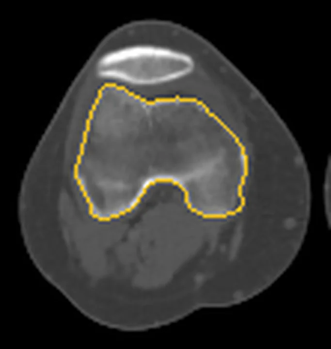
Aynı bölgenin, kaynak veri setinde patoloji içermediği belirtilen bir BT incelemesini kesit kesit kaydırın; organ sınırlarını açarak anatomiyi hasta görüntüsüyle karşılaştırın. Küçük rastlantısal bulgular tümüyle dışlanmamıştır.
Kaynak: TotalSegmentator v2.01 veri seti (Wasserthal ve ark., Radiology: Artificial Intelligence 2023), CC BY 4.0, vaka s0685; organ sınırları veri setinin otomatik segmentasyonundan, birleştirilerek sadeleştirildi. Görüntüler 2,5 mm kesit aralığına yeniden örneklendi.
Ayırıcı tanı
- Sekonder dejeneratif eklem içi cisimler
- osteoartrit gibi altta yatan bir eklem hastalığıyla ilişkili olabilir; primer forma göre daha az sayıda ve daha büyük olabilir.
- Osteokondritis dissekans fragmanı
- subkondral lezyondan ayrılan osteokondral fragman eklem içinde serbest cisme dönüşebilir; eklem yüzündeki defektle birlikte değerlendirilir.
- Pigmente villonodüler sinovit/tenosinovyal dev hücreli tümör
- MRG’de anterior ve posterior kompartımanlarda yaygın nodüler kitleler ve heterojen düşük sinyal görülebilir; mineralize olmayan sinovyal kitleler ayırıcı tanıya girer. Çok sayıda halka-yay kalsifik cisim sinovyal kondromatozis lehinedir.
- Eklem içi osteokondral kırık fragmanları
- patella çıkığı veya eklem içi kırıkla ilişkili osteokondral yüzey hasarı ve ayrılmış fragman görülebilir.
- Kalsiyum pirofosfat birikimi
- menisküs gibi fibro-kıkırdak ve hiyalin kıkırdakta noktasal/çizgisel kondrokalsinozis yapabilir; bu kalsifikasyon eklem içi serbest cisimlerle aynı bulgu değildir.
Tuzaklar
- Kıkırdak nodülleri mineralize değilse BT'de efüzyondan ayrılamayabilir; kalsifikasyon veya serbest cisim yokluğu erken sinovyal hastalığı dışlamaz ve MRG yumuşak doku yayılımını daha iyi gösterir.
- Tek veya sınırlı sayıda osteokondral fragman varsa eşleşen subkondral eklem yüzü defektini arayın; bu görünüm osteokondritis dissekansla uyumlu olabilir.
- Osteoartrit zeminindeki sekonder cisimler primer kondromatozise göre daha büyük ve boyut/biçim açısından daha değişken olabilir; dejeneratif eklem bulgularını birlikte değerlendirin.
Kendini dene
Diz BT'sinde anterior ve posterior eklem bölümlerine dağılmış, benzer boyutlu çok sayıda kalsifiye nodül görülüyor. En olası tanı nedir?
Cevabı göster
Sinovyal kondromatozis. Eklem boyunca yaygın ve benzer boyutlu çok sayıda cisim sinovyal kondromatozisi destekler. CPPD'de hiyalin veya fibro-kıkırdakta noktasal ve çizgisel kalsifikasyonlar görülebilir.
Dizde subkondral kemik lezyonundan fragman ayrılması hangi tanıyla uyumludur? En olası tanı nedir?
Cevabı göster
Osteokondritis dissekans. Osteokondritis dissekans subkondral kemik lezyonu ve olası fragman ayrılmasıyla karakterizedir. Sinovyal kondromatoziste sinovyumda kıkırdak nodülleri oluşur; bazıları eklem boşluğuna ayrılarak serbest cisimleri oluşturabilir.
İlgili konular
Kaynaklar
Bu sayfadaki 39 cümle ve 2 quiz sorusu aşağıdaki kaynaklardan alıntılarla otomatik olarak karşılaştırıldı; 13 cümle bu karşılaştırmada düzeltildi (2026-10-09).
- Synovial chondromatosis: Novel advances in understanding the pathogenesis and in diagnostic strategies (Review)pmc.ncbi.nlm.nih.gov
- Diagnostic Imaging and Management of Common Intra-articular and Peri-articular Soft Tissue Tumors and Tumorlike Conditions of the Kneepmc.ncbi.nlm.nih.gov
- Imaging of Synovial Chondromatosis with Radiologic-Pathologic Correlationpubs.rsna.org
- Unusual lesions that distend the knee joint: pictorial essayscielo.br
- Diagnosis and Treatment of Calcium Pyrophosphate Deposition (CPPD) Disease: A Reviewpmc.ncbi.nlm.nih.gov
- Osteochondritis dissecans of the knee: Imaging, instability concept, and criteriajournals.sagepub.com
- Pigmented Villonodular Synovitis: A Critical Reviewpubmed.ncbi.nlm.nih.gov
- Imaging of patellar fracturespmc.ncbi.nlm.nih.gov
- Imaging Approach to Temporomandibular Joint Disorders.pubmed.ncbi.nlm.nih.gov
- Benign synovial tumors and proliferative processes.pubmed.ncbi.nlm.nih.gov
- Primary synovial chondromatosis of the talonavicular joint: A case report.pubmed.ncbi.nlm.nih.gov
- [Diagnosis and therapy of benign intraarticular tumors].pubmed.ncbi.nlm.nih.gov
- Synovial chondromatosis of the upper extremity.pubmed.ncbi.nlm.nih.gov
- MR imaging assessment of arthritis of the knee.pubmed.ncbi.nlm.nih.gov
- Synovial Chondromatosis Surrounding the First Metatarsophalangeal Joint: A Case Report.pubmed.ncbi.nlm.nih.gov
- Cone-Beam Computed Tomography for Temporomandibular Joint Imaging.pubmed.ncbi.nlm.nih.gov
- Evaluation and Management of Intra-Articular Tumors of the Knee.pubmed.ncbi.nlm.nih.gov
- Synovial chondromatosis of the temporomandibular joint: a case report and literature review.pubmed.ncbi.nlm.nih.gov
- Synovial chondromatosis of the temporomandibular joint: a case description with systematic literature review.pubmed.ncbi.nlm.nih.gov
- Tenosynovial giant cell tumor and its differential diagnosis in children.pubmed.ncbi.nlm.nih.gov
- Innovative Strategies for Hip Synovial Chondromatosis and Its Medico-Legal Implications.pubmed.ncbi.nlm.nih.gov
- Synovial chondromatosis in the temporomandibular joint: case report with review of the literature.pubmed.ncbi.nlm.nih.gov
- Advanced arthroscopic management of diffuse knee synovial chondromatosis with large tricompartmental nodules using trans-notch visualization and a safe posteromedial portal.pubmed.ncbi.nlm.nih.gov
- Giant subpatellar synovial osteochondromatosis complicated by osteoarthritis of the knee: A case report.pubmed.ncbi.nlm.nih.gov
- Synovial Chondromatosis: A Narrative Review of Current Evidence on Diagnosis, Differential Diagnosis, and Management.pubmed.ncbi.nlm.nih.gov
- Case Report: Arthroscopic management of a giant Baker cyst containing numerous rice body-like loose bodies in a patient with rheumatoid arthritis.pubmed.ncbi.nlm.nih.gov
- Osteochondritis Dissecans of the Medial Femoral Condyle With Intra-Articular Meniscal Calcification: A Rare Case and Literature Review.pubmed.ncbi.nlm.nih.gov
- <i>In Vitro</i> Evaluation of Lateral Femur Condyle Free Chondral Fragment Swelling after Soaking in Normal Saline.pubmed.ncbi.nlm.nih.gov
- Comparing MRI and arthroscopic appearances of common knee pathologies: A pictorial review.pubmed.ncbi.nlm.nih.gov
- Total Hip Arthroplasty for Giant Hip Synovial Chondromatosis With Acetabular Involvement Complicated by Osteoarthritis: A 12-Year Follow-Up of a Case and Literature Review.pubmed.ncbi.nlm.nih.gov
- Synovial Chondromatosis of the Elbow: A Case Report and Discussion of Feared Complications.pubmed.ncbi.nlm.nih.gov
- Calcified Chondroid Mesenchymal Neoplasm of the Temporomandibular Joint: A Rare and Recently Described Benign Tumor.pubmed.ncbi.nlm.nih.gov
- Extensive Synovial Osteochondromatosis Secondary to Avascular Necrosis of the Femoral Head: A Case of Multifocal Extra-articular Involvement.pubmed.ncbi.nlm.nih.gov
- The CT knee arthrogram revisited.pubmed.ncbi.nlm.nih.gov
- Extra-articular tenosynovial chondromatosis of the right fifth digit in a 59-year-old man: A case report and literature review.pubmed.ncbi.nlm.nih.gov
- Candida parapsilosis -Caused Arthritis with Rice Body Formation: A Case Presentation and Literature Review.pubmed.ncbi.nlm.nih.gov
- Calcified or ossified benign soft tissue lesions that may simulate malignancy.pubmed.ncbi.nlm.nih.gov
- MRI imaging of soft tissue tumours of the foot and ankle.pubmed.ncbi.nlm.nih.gov
- Imaging of the temporomandibular joint: An update.pubmed.ncbi.nlm.nih.gov
- Soft-tissue masses and masslike conditions: what does CT add to diagnosis and management?pubmed.ncbi.nlm.nih.gov
- Standardized Image-Based Arthroscopic Technique for Hip Synovial Chondromatosis: Long-Term Patient Outcomes.pubmed.ncbi.nlm.nih.gov
- Primary Synovial Chondromatosis of the Elbow Joint Presenting with Ulnar Nerve Compression and Restricted Range of Motion: A Case Report.pubmed.ncbi.nlm.nih.gov
- Bilateral Knee Synovial Chondromatosis in a 2-Year-Old Girl: First Reported Case and Literature Review.pubmed.ncbi.nlm.nih.gov
- Concurrent arboreal lipoma and synovial chondromatosis in an osteoarthritic knee: Insights from a rare case study - A surgical case report.pubmed.ncbi.nlm.nih.gov
- MRI of diffuse-type tenosynovial giant cell tumour in the knee: a guide for diagnosis and treatment response assessment.pubmed.ncbi.nlm.nih.gov
- Synovial chondromatosis of the temporomandibular joint: a case report with an associated systematic review update of the literature.pubmed.ncbi.nlm.nih.gov
- A multidisciplinary approach to treatment for osteochondroma of the mandibular condyle and oral rehabilitation: A case report.pubmed.ncbi.nlm.nih.gov
- LOCALIZED PIGMENTED VILLONODULAR SYNOVITIS OF THE KNEE: A CONSECUTIVE CASE SERIES AND REVIEW OF THE LITERATURE.pubmed.ncbi.nlm.nih.gov
- Recurrent knee pain and effusion in a child: synovial chondromatosis.pubmed.ncbi.nlm.nih.gov
- Secondary Synovial Chondromatosis of the Shoulder Associated with Rotator Cuff Tear: Diagnostic Challenge and Arthroscopic Management - A Case Report.pubmed.ncbi.nlm.nih.gov
- Imaging findings of intra-articular tumor/tumor-like lesions based on pathologic correlation.pubmed.ncbi.nlm.nih.gov
- Pictorial Review of Soft Tissue Lesions with Calcification.pubmed.ncbi.nlm.nih.gov
- Prevalence and Radiographic Features of Head and Neck Soft Tissue Calcifications on Digital Panoramic Radiographs: A Retrospective Study.pubmed.ncbi.nlm.nih.gov
- Total Joint Replacement for Immediate Reconstruction following Ablative Surgery for Primary Tumors of the Temporo-Mandibular Joint.pubmed.ncbi.nlm.nih.gov
- Stem cell injections for osteoarthritis of the knee.pubmed.ncbi.nlm.nih.gov
- Diagnostic accuracy of synovial chondromatosis of the temporomandibular joint on magnetic resonance imaging.pubmed.ncbi.nlm.nih.gov
- Arthroscopic Management of Primary Synovial Chondromatosis of the Ankle: A Case Report and Review of the Literature.pubmed.ncbi.nlm.nih.gov
- A Missed Case of Synovial Osteochondromatosis.pubmed.ncbi.nlm.nih.gov
- Arthroscopic Fixation of Unstable Adolescent Knee Osteochondritis Dissecans With Structural Tibial Cancellous Bone Autograft.pubmed.ncbi.nlm.nih.gov
- Femoral Osteochondritis Dissecans and Tibial Osteochondral Defect in an Adult Revealed by Bone SPECT/CT.pubmed.ncbi.nlm.nih.gov
- Management and Outcomes of Osteochondritis Dissecans of the Knee in the Pediatric and Adolescent Population.pubmed.ncbi.nlm.nih.gov
- Bilateral Localized-Tenosynovial Giant Cell Tumor of the Knee: Case Report and Review.pubmed.ncbi.nlm.nih.gov
- Multifocal tenosynovial giant cell tumor: A case report.pubmed.ncbi.nlm.nih.gov
- Arthroscopic-assisted Suture-based Fixation of Patellar Osteochondral Fractures using the DG-lock Suturing: A Novel Technique.pubmed.ncbi.nlm.nih.gov
- High prevalence of generalized ligamentous laxity in patellar dislocation with posterior weight-bearing lateral femoral condyle osteochondral fractures: an observational study and treatment outcomes.pubmed.ncbi.nlm.nih.gov
- Risk Factor Evaluation for Loose Body Following First-Time Patellar Dislocation in Pediatric Patients.pubmed.ncbi.nlm.nih.gov
- Characterization of calcifications in posterior horn of human meniscus using micro-computed tomography.pubmed.ncbi.nlm.nih.gov
- The association of CT-detected intra-articular calcification with MRI-detected effusion-synovitis and Hoffa-synovitis in knee OA: The Multicenter Osteoarthritis study.pubmed.ncbi.nlm.nih.gov
- The current state of computed tomography in osteoarthritis research.pubmed.ncbi.nlm.nih.gov
- Mutation in the COL2A1 gene is associated with acetabular dysplasia.pubmed.ncbi.nlm.nih.gov
- Algorithm-Based Surgical Decision-Making for Focal Knee Cartilage Defects: A Pragmatic Clinical Framework with Illustrative Cases.pubmed.ncbi.nlm.nih.gov
- Arthroscopic Technique for Autologous Minced Cartilage Implantation for Focal Lesions of the Capitellum Humeri of the Elbow: A Technical Note.pubmed.ncbi.nlm.nih.gov
- Surgical Options for Primary Synovial Chondromatosis of the Knee: A Systematic Review.pubmed.ncbi.nlm.nih.gov
- Non-neoplastic Orthopedic Pathology - A Systematic Review.pubmed.ncbi.nlm.nih.gov
- Transient lateral patellar dislocation resulting in osteochondral lesion of the lateral femoral condyle: a case series of findings on cone beam CT.pubmed.ncbi.nlm.nih.gov
- Weight-bearing computed tomography in knee pathologies: current evidence and future perspectives.pubmed.ncbi.nlm.nih.gov
- The distribution in joint recesses and adjacent synovial compartments of loose bodies determined on MR and CT arthrographies of ankle joint.pubmed.ncbi.nlm.nih.gov
- Primary Synovial Chondromatosis Displacing Popliteal Neurovascular Structures: A Case Report.pubmed.ncbi.nlm.nih.gov
- The Boba Tea Sign: A Novel Magnetic Resonance Imaging Finding in Synovial Chondromatosis of the Knee - A Case Report.pubmed.ncbi.nlm.nih.gov
- Glenoid osteochondritis dissecans in 9 baseball players: arthroscopic management and radiographic outcomes.pubmed.ncbi.nlm.nih.gov
- Extraarticular Knee Joint Resection: Indications, Results, and Complications in a Series of 30 Patients.pubmed.ncbi.nlm.nih.gov
- Evaluation of the knee joint with ultrasound and magnetic resonance imaging.pubmed.ncbi.nlm.nih.gov
- Diffuse-Type Tenosynovial Giant Cell Tumor: What Are the Important Findings on the Initial and Follow-Up MRI?pubmed.ncbi.nlm.nih.gov
- Articular abnormalities that may mimic arthritis.pubmed.ncbi.nlm.nih.gov
- Natural Biomaterials for Osteochondral Repair: From Source to Strategy.pubmed.ncbi.nlm.nih.gov
- Glossary of terms for musculoskeletal radiology.pubmed.ncbi.nlm.nih.gov
Eğitim amaçlıdır; hasta başında tanı ve tedavi kararının yerine geçmez, tıbbi tavsiye değildir. Metinler yapay zekâ desteğiyle yazıldı ve otomatik bir adımda kaynak alıntılarıyla karşılaştırıldı; bu bir tıbbi doğrulama değildir ve içerik uzman radyolog incelemesinden geçmedi. Klinik kararlarda güncel kılavuzları, kurum protokollerini ve radyoloji raporunu esas alın.