Özet
Bel ağrısı, yürüme kısıtlılığı veya alt ekstremite yakınması olan erişkinde santral kanal darlığı dejeneratif disk, faset ve ligament değişikliklerinin toplam etkisini yansıtabilir. Kontrastsız BT kemik kanalın şeklini, faset hipertrofisini ve osteofitleri iyi gösterir; yumuşak doku katkısı ve kauda ekuina basısı için MR daha açıklayıcıdır. Kanal çapı ve alanı tanımlayıcı ölçümlerdir, ancak yaş, düzey ve klinik tabloyu dışarıda bırakan tek bir BT eşiği semptomatik stenozu kanıtlamaz. İleri daralma ve olası acil nöral bası atlanırsa önemli klinik bulgular görüntü raporuna yansımayabilir.
Okuma sırası
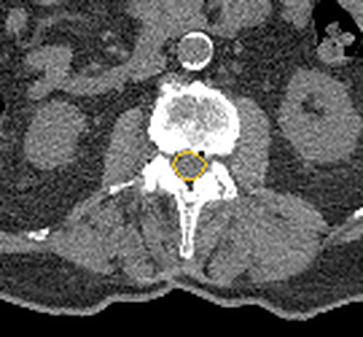
- L4-L5 santral kemik kanal. Aksiyel L4-L5 disk kesitinde santral kemik kanalın ön duvarı ile posterior elemanları arasındaki açıklığı bul; bir üst ve alt disk seviyesindeki şekille karşılaştır.
- Posterior korpus kenarı. Ön sınırdaki disk taşması veya son plak osteofitinin kanala uzanımını takip et ve kemik çıkıntıyla yumuşak disk bileşenini ayır.
- Fasetler ve ligamentum flavum. Posterolateral köşelerde fasetlerin içe kabarıklığını, lamina önünde ligament kalınlaşması ya da ossifikasyonunu ara.
- Lomber kanalın sagittal seyri. Sagittal MPR'de stenozun tek seviyeli mi çok seviyeli mi olduğunu ve eşlik eden kayma/osteofitleri kontrol et; sinir liflerinin bası derecesini BT ölçüsünden çıkarsama.
Faz ve pencere
- Kontrastsız tanısal
- BT, kemik spinal kanalın anatomik yapılarının ve kanal daralmasının değerlendirilmesinde kullanılabilir. Rutin dejeneratif kanal darlığını göstermek için intravenöz kontrast gerekmez; thekal kese ve sinir demetlerinin derecelendirilmesi BT'de sınırlıdır.
- Kemik algoritması tanısal
- Diskin santral kanala doğru bombeleşmesi, vertebral osteofitler ve ligamentum flavum hipertrofisi dejeneratif santral kanal stenozuna katkıda bulunabilir.
Önerilen pencereler G genişlik, M merkez; değerler Hounsfield birimi (HU)
- KemikG 1800 / M +400
- Yumuşak dokuG 400 / M +40
BT bulguları
- Santral kanal daralması — kemik kanalın ön-arka ya da transvers açıklığı komşu seviyelere göre azalır.
- Posterior disk-osteofit kompleksi — vertebra arka kenarından kanala uzanan disk ve kemik çıkıntısı ön sınırı daraltır.
- Faset hipertrofisi — kalınlaşmış superior/inferior artiküler çıkıntılar kanalın posterolateral köşelerini içe iter.
- Ligamentum flavum kalınlaşması/ossifikasyonu — lamina önündeki yumuşak doku veya kemik yoğunluklu bant posterior kanalı daraltır.
- Kombine daralma — disk, faset ve posterior eleman katkıları aynı seviyede birlikte bulunabilir.
- Gelişimsel dar kanal — konjenital olarak dar spinal kanal, erken yaşta nörojenik semptomlara yol açabilir ve disk herniasyonu veya spondilolistezis gibi dejeneratif süreçlerle ilişkilidir.
- Eşlik eden spondilolistezis — vertebra dizilimindeki basamaklanma kanal geometrisini ve darlığın dinamik olasılığını değiştirir.
- Lateral reses darlığında faset hipertrofisi ve osteofitler lateral resesi daraltabilir; bu daralma sinir köküne bası yapabilir.
Ölçütler ve sınıflamalar
- Literatürde en sık bildirilen nicel lomber santral stenoz ölçütleri (evrensel eşik değildir)
- Bir aksiyel yüklemeli MR çalışmasında dural kese kesit alanının <75 mm² olması şiddetli santral kanal stenozu ölçütü olarak kullanılmıştır. Lomber santral stenozun nicel değerlendirmesinde farklı radyolojik parametreler kullanılmıştır; verilen kaynaklar BT'de kemik kanal ön-arka çapı için <10 mm'lik mutlak darlık eşiğini doğrulamamaktadır.
- Lee ve ark. lomber santral kanal stenoz derecelendirmesi (aksiyel T2 ağırlıklı MR)
- Lee derecelendirme sistemi lomber santral kanal stenozunu MRI bulgularına göre sınıflandırır.
Normalde
BT, kemik spinal kanalın anatomik değerlendirilmesini sağlar; ancak normal kanalın vertebra korpusu, lamina ve fasetler arasındaki sınırları verilen kaynaklarda tanımlanmamıştır. Kıyasta en dar düzeyin komşu disk seviyelerine göre ne kadar değiştiğine, posterior disk konturuna ve iki fasetin simetrisine bak; kemik kanal ölçüsü tek başına thekal kese içindeki sinir basısını göstermez.
Normal BT ile kıyasla
Aynı bölgenin, kaynak veri setinde patoloji içermediği belirtilen bir BT incelemesini kesit kesit kaydırın; organ sınırlarını açarak anatomiyi hasta görüntüsüyle karşılaştırın. Küçük rastlantısal bulgular tümüyle dışlanmamıştır.
Kaynak: TotalSegmentator v2.01 veri seti (Wasserthal ve ark., Radiology: Artificial Intelligence 2023), CC BY 4.0, vaka s1338; organ sınırları veri setinin otomatik segmentasyonundan, birleştirilerek sadeleştirildi. Görüntüler 2,5 mm kesit aralığına yeniden örneklendi.
Ayırıcı tanı
- Lateral reses stenozu
- daralma santral thekal kese yerine posterolateral subartiküler kanalda ve geçen kökün koridorunda belirgindir.
- Nöral foramen stenozu
- daralma vertebra pedikülleri arasındaki kök çıkış tünelindedir; santral kanal açıklığı korunabilir.
- Gelişimsel dar kanal
- çok seviyeli, benzer biçimde küçük kanal çapı vardır; fokal osteofit/faset değişikliği tek başına açıklamaz.
- Epidural kitle
- kemik sınırlardan bağımsız yumuşak doku etkisi veya kemik destrüksiyonu olabilir; karakterizasyon için MR/kontrastlı inceleme gerekir.
- Konjenital kanal varyantı
- vertebral ark ve pedikül morfolojisi gelişimsel farklılık gösterir; tek seviyeli dejeneratif çıkıntıyla açıklanmaz.
Tuzaklar
- Kemik kanal darlığı ile klinik/soft-tissue stenoz eş anlamlı değildir; sayı veriliyorsa kullanılan yöntem ve düzey belirtilmeli, sinir basısı BT'de varsayılmamalıdır.
- Kanal çapı ve alan ölçümleri ayırıcı tanıda değer taşır ve karşılaştırılabilir olması için belirlenmiş disk düzeylerinde, aynı anatomik referanslarla yapılmalıdır.
- Osteofit ve kalsifiye ligament yüksek atenüasyon nedeniyle dikkat çeker; eşlik eden yumuşak disk ve ligament kalınlaşmasının katkısı kemik pencerede eksik kalabilir.
- Anatomik stenoz yaşlı ve yakınmasız kişilerde de görülebilir; görüntüdeki darlık klinik korelasyon olmadan semptom nedeni ilan edilmemelidir.
Kendini dene
L4-L5'te posterior osteofit ve bilateral faset hipertrofisi kemik kanalı daraltıyor. BT hangi sonucu doğrudan destekler?
Cevabı göster
Santral kemik kanal daralması. BT kemik kanal konturunu ve daralmaya katkı veren osteofit/fasetleri gösterir. Kauda ekuina basısı ve kök ödemi yumuşak doku/nöral değerlendirmedir; enfeksiyon için destrüktif son plak ve çevre bulgular aranır.
Gelişimsel lomber kanal stenozunun temel anatomik özelliği nedir?
Cevabı göster
Doğuştan küçük spinal kanal. Gelişimsel lomber stenoz, doğuştan küçük spinal kanal ile tanımlanır; erken başlangıçlı nörojenik belirtilerle ilişkili olabilir.
İlgili konular
- İntervertebral disk dejenerasyonu ve vakum fenomeni
- Disk hernisi: protrüzyon ve ekstrüzyon
- Lateral reses stenozu
- Nöral foramen stenozu
Kaynaklar
Bu sayfadaki 41 cümle ve 2 quiz sorusu aşağıdaki kaynaklardan alıntılarla otomatik olarak karşılaştırıldı; 9 cümle bu karşılaştırmada düzeltildi (2026-10-09).
- Dual Aetiology of Lower Limb Claudication: A Rare Case of Coexisting Lumbar Facet Joint Synovial Cyst and Critical Lower Limb Arterial Stenosespmc.ncbi.nlm.nih.gov
- Efficacy and safety of percutaneous transforaminal endoscopic surgery (PTES) compared with MIS-TLIF for surgical treatment of lumbar degenerative disease in elderly patients: A retrospective cohort studypmc.ncbi.nlm.nih.gov
- Spontaneous Spinal Subdural Hematoma Concurrent with Cranial Subdural Hematomapmc.ncbi.nlm.nih.gov
- Cervical Stenosis in a Patient with Arthrogryposis: Case Reportpmc.ncbi.nlm.nih.gov
- Assessment of Effectiveness of Percutaneous Adhesiolysis in Managing Chronic Low Back Pain Secondary to Lumbar Central Spinal Canal Stenosispmc.ncbi.nlm.nih.gov
- Role of Diffusion Tensor MR Imaging in Degenerative Cervical Spine Disease: a Review of the Literature.pubmed.ncbi.nlm.nih.gov
- Cervical stenosis in a patient with arthrogryposis: case report.pubmed.ncbi.nlm.nih.gov
- Magnetic Resonance Imaging Findings in Lumbar Disc Degeneration in Symptomatic Patients.pubmed.ncbi.nlm.nih.gov
- Clinics in diagnostic imaging (192). Flexion teardrop fracture.pubmed.ncbi.nlm.nih.gov
- Clinics in diagnostic imaging (161). Cervical OPLL with cord compression.pubmed.ncbi.nlm.nih.gov
- Decompression of Lumbar Central Spinal Canal Stenosis Following Minimally Invasive Transforaminal Lumbar Interbody Fusion.pubmed.ncbi.nlm.nih.gov
- Spontaneous spinal subdural hematoma concurrent with cranial subdural hematoma.pubmed.ncbi.nlm.nih.gov
- Magnetic Resonance Evaluation of Lumbar Disc Degenerative Disease as an Implication of Low Back Pain: A Prospective Analysis.pubmed.ncbi.nlm.nih.gov
- Diagnostic advancement of axial loaded lumbar spine MRI in patients with clinically suspected central spinal canal stenosis.pubmed.ncbi.nlm.nih.gov
- [Early-effectiveness of unilateral biportal endoscopic laminectomy in treatment of two-level lumbar spinal stenosis].pubmed.ncbi.nlm.nih.gov
- Dual Aetiology of Lower Limb Claudication: A Rare Case of Coexisting Lumbar Facet Joint Synovial Cyst and Critical Lower Limb Arterial Stenoses.pubmed.ncbi.nlm.nih.gov
- Spinal pain.pubmed.ncbi.nlm.nih.gov
- Comparative Clinical Effectiveness of Nonsurgical Treatment Methods in Patients With Lumbar Spinal Stenosis: A Randomized Clinical Trial.pubmed.ncbi.nlm.nih.gov
- Neuroimaging of Spinal Canal Stenosis.pubmed.ncbi.nlm.nih.gov
- Clinically Meaningful Improvement Across Ages 50-95 Years After Decompression Without Fusion for Central Lumbar Spinal Stenosis Without Spondylolisthesis.pubmed.ncbi.nlm.nih.gov
- Physical activity barriers and facilitators in patients awaiting spine surgery: A qualitative study.pubmed.ncbi.nlm.nih.gov
- Clinical management of a Veteran with chronic scan-negative cauda equina syndrome: a case report.pubmed.ncbi.nlm.nih.gov
- Clinical and radiological predictors of adverse outcomes after unilateral biportal endoscopic surgery for short-segment degenerative lumbar spinal stenosis.pubmed.ncbi.nlm.nih.gov
- The Role of CT in Cervical Spine Imaging for Suspected Cervical Myelopathy.pubmed.ncbi.nlm.nih.gov
- Case Report: Segment-specific minimally invasive surgical management of lumbar spinal epidural lipomatosis with adjacent-level degenerative stenosis.pubmed.ncbi.nlm.nih.gov
- Efficacy of Indirect Decompression by Posterior Longitudinal Ligament Ligamentotaxis in Minimally Invasive Oblique Lateral Interbody Fusion (MIS-OLIF) Without Posterior Decompression for Degenerative Lumbar Disease.pubmed.ncbi.nlm.nih.gov
- Degenerative spinal stenosis may influence Normal Pressure Hydrocephalus (NPH)-related imaging characteristics.pubmed.ncbi.nlm.nih.gov
- Acute Cauda Equina Syndrome With Clinicoradiological Discordance Treated via Unilateral Biportal Endoscopy: A Case Report.pubmed.ncbi.nlm.nih.gov
- Minimally Invasive Surgery for the Treatment of Lumbar Spinal Stenosis: A Comprehensive Review.pubmed.ncbi.nlm.nih.gov
- A rare case of multi-segmental lumbar intradural ependymoma: clinical presentation and surgical management: a case report.pubmed.ncbi.nlm.nih.gov
- Synthesizing lumbar computed tomography myelography from plain computed tomography images using a cycle-consistent generative adversarial network.pubmed.ncbi.nlm.nih.gov
- Lessons in balancing function with oncological control in a destructive lumbosacral tumor of uncertain pathology: illustrative case.pubmed.ncbi.nlm.nih.gov
- Minimally invasive percutaneous cannulated polyaxial screw and rod fixation thoracolumbar and lumbosacral vertebrae: cadaveric evaluation of accuracy and safety of a fluoroscopic-guided technique, in large breed dogs.pubmed.ncbi.nlm.nih.gov
- Successful Treatment of Functional Neurologic Symptom Disorder (Conversion Disorder) With Subdissociative Dose Ketamine in the Emergency Department.pubmed.ncbi.nlm.nih.gov
- Imaging the brain: diagnosis aided by structural features on neuroimaging studies.pubmed.ncbi.nlm.nih.gov
- Pain Management in Autosomal Dominant Polycystic Kidney Disease: Clinical Challenges and a Stepwise Algorithmic Approach.pubmed.ncbi.nlm.nih.gov
- Comparative efficacy of unilateral biportal endoscopy versus endoscopic foraminoplasty decompression for single-level lumbar spinal stenosis: a retrospective cohort study.pubmed.ncbi.nlm.nih.gov
- Comparison of clinical and radiological outcomes between microscope-assisted tubular unilateral laminectomy for bilateral decompression and bilateral laminotomy for crossover decompression in single-level lumbar spinal stenosis.pubmed.ncbi.nlm.nih.gov
- Laminectomy alone <i>vs</i> laminectomy with posterior fusion in lumbar spinal stenosis: The role of instability.pubmed.ncbi.nlm.nih.gov
- Is bilateral laminectomy and fusion superior to total laminectomy and fusion in two-level lumbar spinal stenosis?pubmed.ncbi.nlm.nih.gov
- Five-year outcomes from a multi-center prospective randomized controlled trial of minimally invasive interspinous fixation versus open direct decompression for lumbar spinal stenosis.pubmed.ncbi.nlm.nih.gov
- Bispecific antibody engineered extracellular vesicles redirect T cells to prevent postoperative epidural fibrosis.pubmed.ncbi.nlm.nih.gov
- Automatic segmentation of dura for quantitative analysis of lumbar stenosis: A deep learning study with 518 CT myelograms.pubmed.ncbi.nlm.nih.gov
- Congenital and Breed-Associated Vertebral Anomalies in Dogs and Cats: Developmental Anatomy, Genetic Determinants, and Biological Significance.pubmed.ncbi.nlm.nih.gov
- Anatomy-guided context-aware deep learning for lumbar degenerative disease grading and burden-aware risk assessment on MRI.pubmed.ncbi.nlm.nih.gov
- Benchmarking Multimodal Vision Frontier Models With Lumbar Spine MRIs for Grading Lumbar Spinal Stenosis.pubmed.ncbi.nlm.nih.gov
- Important postoperative magnetic resonance imaging findings correlated with clinical outcomes to evaluate adequacy and success of decompression surgery-what radiologists need to know: a systematic review.pubmed.ncbi.nlm.nih.gov
- Association Between Radiological Stenosis Level and Patient-Reported Outcomes in Patients with Lumbar Spinal Stenosis: A Cross-Sectional Study.pubmed.ncbi.nlm.nih.gov
- Postoperative radiculopathy following the use of a flowable gelatin-based hemostatic matrix in endoscopic lumbar spine surgery: two case reports of uniportal and biportal decompression procedures.pubmed.ncbi.nlm.nih.gov
- Correlation of Clinical and MRI Findings With Intraoperative Findings in Lumbar Intervertebral Disc Prolapse: A Prospective Observational Study.pubmed.ncbi.nlm.nih.gov
- Challenges in diagnosing malignant lymphoma involving the spine: illustrative cases.pubmed.ncbi.nlm.nih.gov
- Idiopathic Syringomyelia: A Systematic Scoping Review.pubmed.ncbi.nlm.nih.gov
- Diagnostic guide to hypochondroplasia.pubmed.ncbi.nlm.nih.gov
- Orthopedic preoperative evaluation and surgical strategies of hereditary multiple osteochondromas involving the spinal region.pubmed.ncbi.nlm.nih.gov
- Hemorrhagic Lumbar Synovial Cyst: Rare Mimic for Mass Lesion Causing Acute Neurological Deficit.pubmed.ncbi.nlm.nih.gov
- Lumbar Epidural Cystic Lesion Associated With <i>Finegoldia magna</i> Discitis Presenting as Cauda Equina Syndrome: A Case Report.pubmed.ncbi.nlm.nih.gov
- Cervicothoracolumbar Multiregional Spinal Stenosis (MRSS): Outcomes of Single-Stage Surgery in a Case Series of 28 Patients With a Minimum Two-Year Follow-Up.pubmed.ncbi.nlm.nih.gov
- A patient with lumbar disc herniation complicates cauda equina syndrome after epidural steroid injection: A case report.pubmed.ncbi.nlm.nih.gov
- A case series of calcium pyrophosphate deposition in the spine: An underrecognized debilitating disease.pubmed.ncbi.nlm.nih.gov
- Association of supine apical vertebral rotation with three-dimensional deformity and global spinal alignment in adult degenerative scoliosis: a retrospective study.pubmed.ncbi.nlm.nih.gov
- Absence of lumbosacral plexus abnormalities in preschool children with tethered cord syndrome: A multiparametric MRI study.pubmed.ncbi.nlm.nih.gov
- Koala Kyphoscoliosis: Radiographic and CT Features of Abnormal Vertebral Column Curvature in Koalas (<i>Phascolarctos cinereus</i>) of the Mount Lofty Ranges, South Australia.pubmed.ncbi.nlm.nih.gov
- The advancement and utility of multimodal imaging in the diagnosis of degenerative disc disease.pubmed.ncbi.nlm.nih.gov
- A Primer on Anatomy, Biophysics, Pathology, Imaging and Treatment of the Intervertebral Disc and the Anterior Spinal Column: The Discogenic, Intervertebral, Spinal Column (DISC) ASPN Workgroup.pubmed.ncbi.nlm.nih.gov
- Computed Tomography and Magnetic Resonance Imaging Overlay in the Spine for Surgical Planning: A Technical Report.pubmed.ncbi.nlm.nih.gov
- Is extension of multi-level lumbar fusion to S1 necessary in the absence of L5-S1 instability? A comparative study of selective decompression versus TLIF with S1 pedicle screw fixation.pubmed.ncbi.nlm.nih.gov
- Artificial intelligence in spine care: A scoping review of diagnostic applications.pubmed.ncbi.nlm.nih.gov
- Delayed diagnosis of spinal tuberculosis mimicking spondyloarthritis: a case report.pubmed.ncbi.nlm.nih.gov
- Patient-Specific Virtual Reality Rehearsal for Technically Challenging Lumbar Transforaminal Epidural Steroid Injection: A Pilot Randomized Controlled Trial.pubmed.ncbi.nlm.nih.gov
- Full-endoscopic lateral laminectomy on both sides with central line preservation under local anesthesia with monitored anesthesia care for thoracic spinal stenosis in a very old patient unsuitable for general anesthesia: illustrative case.pubmed.ncbi.nlm.nih.gov
- Degenerative lumbosacral spondylolisthesis: Instability as the primary pathology and stabilization as definitive treatment. Experience with 28 cases treated by only fixation without any decompression.pubmed.ncbi.nlm.nih.gov
- Congenital lumbar spinal stenosis: current perspectives on diagnosis, imaging, and treatment.pubmed.ncbi.nlm.nih.gov
- The Anterolateral Transpedicular Transverse Process Screw Technique: Surgical Techniques, Biomechanical study and Preliminary Outcomes.pubmed.ncbi.nlm.nih.gov
- AO Spine Clinical Practice Recommendations: When to Operate in Lumbar Disc Herniation.pubmed.ncbi.nlm.nih.gov
- Lumbar epidural haematoma after acupuncture: a case report and review of the literature.pubmed.ncbi.nlm.nih.gov
- Pictorial review: imaging of the spinal manifestations of achondroplasia.pubmed.ncbi.nlm.nih.gov
- 2024 Heart Disease and Stroke Statistics: A Report of US and Global Data From the American Heart Association.pubmed.ncbi.nlm.nih.gov
- Heart Disease and Stroke Statistics-2023 Update: A Report From the American Heart Association.pubmed.ncbi.nlm.nih.gov
- The Konakondla-Telfeian-Shen (KTS) Pyramid: a safe entry zone for ventral thoracic pathologies.pubmed.ncbi.nlm.nih.gov
- Long-term clinical and radiographic outcomes of interlaminar coflex dynamic stabilization versus posterior lumbar interbody fusion for lumbar spinal stenosis: a comparative study with minimum 15-year follow-up.pubmed.ncbi.nlm.nih.gov
- Residual bone mass after TLIF treated by TELD under local anesthesia: a case report.pubmed.ncbi.nlm.nih.gov
- Routine Application of a Bioabsorbable Cellulose Hemostatic Agent (WoundClot<sup>TM</sup>) in Unilateral Biportal Endoscopic Lumbar Decompression: A Retrospective Comparative Study.pubmed.ncbi.nlm.nih.gov
- Early clinical outcomes of simultaneous biportal endoscopic decompression for tandem spinal stenosis: a single-center retrospective study.pubmed.ncbi.nlm.nih.gov
- The impact of early vs. delayed surgery on outcomes in cervical spinal cord injury without fracture or dislocation.pubmed.ncbi.nlm.nih.gov
- Bilateral abducens nerve palsy following ligamentous C1-C2 distraction.pubmed.ncbi.nlm.nih.gov
- Anatomical Variants of the Cervical Spine and Sports-Related Injury in Athletes: A Narrative Review.pubmed.ncbi.nlm.nih.gov
- High-Riding Vertebral Artery in Cervical Spine Surgery: A Review of Preoperative Identification and Surgical Implications.pubmed.ncbi.nlm.nih.gov
- Radiographic measurement of the C1 lateral mass for posterior atlantoaxial fixation in patients with a persistent first intersegmental artery.pubmed.ncbi.nlm.nih.gov
- Diagnostic challenges of spinal gout: patient series.pubmed.ncbi.nlm.nih.gov
- Choosing the right treatment for degenerative cervical myelopathy.pubmed.ncbi.nlm.nih.gov
- Pediatric Spine Trauma.pubmed.ncbi.nlm.nih.gov
- A Retrospective Comparative Study on Arthroscopic-Assisted Uni-Portal Spinal Surgery (AUSS/UNSES) Versus Unilateral Biportal Endoscopy for Double-Segment Lumbar Spinal Stenosis.pubmed.ncbi.nlm.nih.gov
- Retrospective Analysis of Imaging Influencing Factors and Early Stability of Cage Subsidence After Percutaneous Endoscopic Lumbar Interbody Fusion.pubmed.ncbi.nlm.nih.gov
- Rare Case of a Cervical Ventral Root Filament Neuroma Mimicking a Sequestrated Disc Herniation in Association with Ossification of the Posterior Longitudinal Ligament.pubmed.ncbi.nlm.nih.gov
- Anterior Controllable Antedisplacement Fusion (ACAF) in Revision Surgery for Iatrogenic Cervical Kyphosis: Technical Note and Case Series.pubmed.ncbi.nlm.nih.gov
- Single-incision uniportal endoscopic bilateral decompression for two adjacent levels of lumbar spinal stenosis: a technical report.pubmed.ncbi.nlm.nih.gov
- Correlation Between Postoperative Changes in Imaging Parameters and Clinical Outcomes After Unilateral Biportal Endoscopic Decompression for Single-Level Lumbar Spinal Stenosis.pubmed.ncbi.nlm.nih.gov
- Surgical Correction of Thoracolumbar Kyphosis in Achondroplasia: Complications, Pitfalls, and Reflections on the Pursuit of Maximal Realignment in View of Correction Leading to Functional Disability.pubmed.ncbi.nlm.nih.gov
- Impact of Cervical Spine Immobility on Carotid Endarterectomy: A Report of Two Cases.pubmed.ncbi.nlm.nih.gov
- Subacute and Chronic Low-Back Pain: From MRI Phenotype to Imaging-Guided Interventions.pubmed.ncbi.nlm.nih.gov
- Development and validation of a clinical predictive model for the risk of recurrence after unilateral biportal endoscopic diskectomy.pubmed.ncbi.nlm.nih.gov
- Which subtypes of degenerative lumbar spondylolisthesis are suitable for oblique lumbar interbody fusion? A retrospective study in China based on the clinical and radiographic degenerative spondylolisthesis classification.pubmed.ncbi.nlm.nih.gov
- Dietary inadequacies and their association with pain severity and disc degeneration in patients surgically treated for degenerative lumbosacral spine disorders.pubmed.ncbi.nlm.nih.gov
- Standalone interspinous process devices for lumbar spinal stenosis: a critical literature review.pubmed.ncbi.nlm.nih.gov
- Correction of Spinal Sagittal Alignment after Posterior Lumbar Decompression: Does Severity of Central Canal Stenosis Matter?pubmed.ncbi.nlm.nih.gov
- Dural tears from traumatic spinal fractures: an illustrative case and systematic review of its incidence, diagnosis, risk factors, and management.pubmed.ncbi.nlm.nih.gov
- Intradural lumbar disc herniation with cauda equina syndrome: Case report and recent advances.pubmed.ncbi.nlm.nih.gov
- Adult degenerative scoliosis: challenges in diagnosis, pain management, and surgical decision-making.pubmed.ncbi.nlm.nih.gov
- The state of trial registrations in the field of Orthopaedics in years 2015-2020. A meta-epidemiological study.pubmed.ncbi.nlm.nih.gov
- Massage for low-back pain.pubmed.ncbi.nlm.nih.gov
- Structural remodeling impact of osteophytes and Schmorl's nodes on lumbar vertebral morphometry: anatomical basis for spinal instrumentation.pubmed.ncbi.nlm.nih.gov
- Comparison of ultrasound and magnetic resonance imaging findings in evaluation of fetal congenital anomalies: A single-institution prospective observational study.pubmed.ncbi.nlm.nih.gov
- A blast from the past!: The value of adding single slice magnetic resonance myelography sequence to magnetic resonance imaging of the spine; a flashback to the conventional myelography of the past.pubmed.ncbi.nlm.nih.gov
- Stable low-grade degenerative spondylolisthesis does not adversely affect clinical outcome of full-endoscopic decompression in patients with spinal stenosis.pubmed.ncbi.nlm.nih.gov
- Computed tomography-derived assessment of respiratory impairment in thoracic scoliosis: Comparison between idiopathic and non-idiopathic etiologies.pubmed.ncbi.nlm.nih.gov
Eğitim amaçlıdır; hasta başında tanı ve tedavi kararının yerine geçmez, tıbbi tavsiye değildir. Metinler yapay zekâ desteğiyle yazıldı ve otomatik bir adımda kaynak alıntılarıyla karşılaştırıldı; bu bir tıbbi doğrulama değildir ve içerik uzman radyolog incelemesinden geçmedi. Klinik kararlarda güncel kılavuzları, kurum protokollerini ve radyoloji raporunu esas alın.