Özet
Lateral reses (subartiküler zon), santral kanal ile nöral foramen arasındaki geçiş alanıdır; traversan sinir kökü buradan geçerek daha kaudalde nöral foramenden çıkar. Lateral resesteki dejeneratif daralma sinir kökünü sıkıştırarak radikülopatiye yol açabilir. Kontrastsız BT kemik ve kalsifiye dejeneratif değişiklikleri gösterebilir; sinir kökleri ve dural kese gibi yumuşak dokuları değerlendirmede MR daha üstündür. Lateral reses, santral kanal ile sinir kökünün çıktığı nöral foramen arasındaki geçiş alanıdır.
Okuma sırası
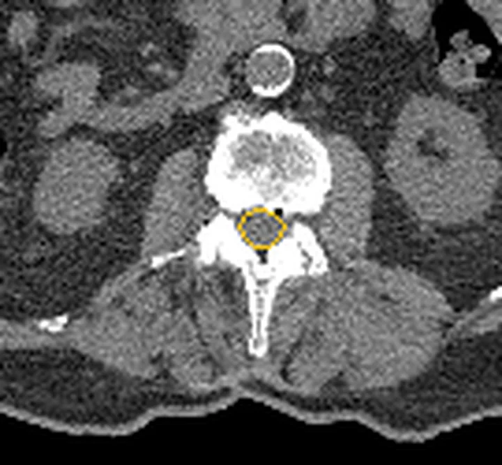
- Sağ ve sol lateral reses. L4-L5 aksiyel disk kesitinde santral kanalın iki posterolateral köşesine bak ve pediküle uzanan sağ-sol subartiküler koridorları bul.
- Posterior disk-osteofit kompleksi. Posterior disk çıkıntısının lateral reseste sinir köküne temas edip etmediğini değerlendir; posterior son plak osteofiti de daralmaya katkıda bulunabilir.
- Superior artiküler faset çıkıntıları. Medial faset çıkıntısının resese doğru büyüyüp büyümediğini kontrol et; aynı kesitte karşı tarafla açıklık ve kontur bakımından kıyasla.
- Sağ lateral reses ve nöral foramen. Son olarak traversan sinir kökünün lateral resesten nöral foramenden çıkışına kadar uzanan seyrini değerlendir; eşlik eden disk hernisi veya faset sinovyal kisti varsa belirt.
Faz ve pencere
- Kontrastsız tanısal
- Aksiyel disk düzeyi kesitinde subartiküler alanın ön sınırını vertebra korpusu, son plak ve disk kenarı; arka sınırını superior artiküler faset, dış sınırını pedikül oluşturur; bu sınırlar arasındaki yağlı/yumuşak doku koridoru ve asimetri incelenir. Kontrastsız BT kemik konturlarını ve osteofitleri gösterebilir, ancak kök ile BOS ilişkisini güvenilir biçimde derecelendiremeyebilir.
- Rekonstrüksiyon (kemik algoritması) tanısal
- BT kemik konturlarını ve osteofitleri gösterebilir; medial faset hipertrofisi ve posterior son plak osteofitleri lateral reses stenozuna katkıda bulunabilir.
Önerilen pencereler G genişlik, M merkez; değerler Hounsfield birimi (HU)
- KemikG 1800 / M +400
- Yumuşak dokuG 400 / M +40
BT bulguları
- Subartiküler kanal daralması — lateral reses, santral kanal ile nöral foramen arasındaki geçişte daralmıştır.
- Medial faset hipertrofisi — superior artiküler fasetin dejeneratif büyümesi lateral reseste traversan sinir kökünü sıkıştırabilir.
- Faset osteofiti — faset eklemindeki dejeneratif kemik büyümesi lateral resesteki sinir köküne basıya katkıda bulunabilir.
- Posterolateral disk çıkıntısı ve posterior vertebra kenarı osteofiti — lateral reses daralmasına katkıda bulunabilecek bu yapıları ayrı ayrı değerlendir.
- Tek taraflı asimetri — sağ ve sol reses genişlikleri aynı disk seviyesinde farklılaşabilir.
- Birden çok düzey tutulumu — komşu subartiküler koridorlarda ardışık daralma bulunabilir.
- Eşlik eden spondilolistezis — faset örtüşmesini ve reses şeklini değiştirerek daralmaya katkı verebilir.
Ölçütler ve sınıflamalar
- Bartynski ve Lin lateral reses kök basısı derecelendirmesi
- Bartynski sınıflamasında lateral reses stenozu 0 (normal) ile 3 (şiddetli) arasında derecelendirilir. Lateral reses stenozu derecelendirmesi MRI üzerinden yapılabilir; BT kemik ve kalsifiye değişiklikleri gösterir.
Normalde
Lateral reses, traversan sinir kökünün nöral foramenden çıkmadan önce geçtiği alandır. Lateral reses, santral kanalın yanındaki sinir kökü geçiş alanıdır; sinir kökü buradan nöral foramenden çıkmadan önce geçer.
Normal BT ile kıyasla
Aynı bölgenin, kaynak veri setinde patoloji içermediği belirtilen bir BT incelemesini kesit kesit kaydırın; organ sınırlarını açarak anatomiyi hasta görüntüsüyle karşılaştırın. Küçük rastlantısal bulgular tümüyle dışlanmamıştır.
Kaynak: TotalSegmentator v2.01 veri seti (Wasserthal ve ark., Radiology: Artificial Intelligence 2023), CC BY 4.0, vaka s1338; organ sınırları veri setinin otomatik segmentasyonundan, birleştirilerek sadeleştirildi. Görüntüler 2,5 mm kesit aralığına yeniden örneklendi.
Ayırıcı tanı
- Santral kanal stenozu
- daralma spinal kanalın merkezindeki dural kese çevresindedir; lateral reses stenozu ise daha yan taraftaki sinir kökü geçiş alanını etkiler.
- Nöral foramen stenozu
- daralma, sinir kökünün omurga kanalından çıktığı nöral foramende bulunur; lateral reses kökün bu foramene ulaşmadan geçtiği alandır.
- Fokal disk hernisi
- lateral resese uzanan herniye disk, traversan sinir kökünü sıkıştırabilir.
- Faset sinovyal kisti
- faset eklemine komşu kistik yumuşak doku odağı resesi dıştan daraltabilir; BT’deki yumuşak doku özelliği MR kadar özgül biçimde karakterize edilemeyebilir.
- Epidural kitle
- disk ve faset konturlarından ayrı yumuşak doku etkisi varsa MR ile karakterizasyon gerekebilir.
Tuzaklar
- Resesin anatomik darlığı tek başına sinir kökü basısını kanıtlamaz; kök deformasyonu veya çevresindeki BOS alanı BT’de seçilemiyorsa yalnız görülebilen anatomik daralmayı tarif et.
- Tek aksiyel kesite dayanma; komşu görüntüler ve sagittal/koronal MPR ile daralmanın düzeyini, uzanımını ve iki taraflı dağılımını doğrula.
- Kemik algoritmasında osteofitin son plakla devamlılığını izle; posterior disk materyalini kemik penceresi bulgusuyla karıştırma ve yumuşak doku penceresini de değerlendir.
- Lateral reses stenozu ile nöral foramen stenozunu aynı terimle raporlama; sinir kökü lateral resesten geçer ve daha sonra nöral foramenden çıkar.
Kendini dene
Aksiyel L4-L5 BT'de medial faset osteofiti sağ subartiküler koridoru daraltıyor; santral kanal açık. Bölge hangisidir?
Cevabı göster
Lateral reses. Santral kanal ile kök çıkış forameninin arasındaki subartiküler geçiş lateral resestir. Santral kanal merkezi kesedir; nöral foramen pediküller arasında daha lateralde, vertebra korpusu ise omur gövdesidir.
Daralma pediküller arasındaki çıkış tünelinde; subartiküler koridor korunmuş. Hangi tanı yerleşime uyar?
Cevabı göster
Foraminal stenoz. Pediküller arası kök çıkış tüneli nöral foramendir. Lateral reses stenozu daha medial geçiş alanındadır; santral stenoz thekal kese çevresini daraltır, spondiloliz pars interartikülaris defektidir.
İlgili konular
- Disk hernisi: protrüzyon ve ekstrüzyon
- Santral kanal stenozu
- Nöral foramen stenozu
- İntervertebral disk dejenerasyonu ve vakum fenomeni
Kaynaklar
Bu sayfadaki 41 cümle ve 2 quiz sorusu aşağıdaki kaynaklardan alıntılarla otomatik olarak karşılaştırıldı; 22 cümle bu karşılaştırmada düzeltildi (2026-10-09).
- ABCs of the degenerative spinepmc.ncbi.nlm.nih.gov
- Magnetic resonance imaging of the spinepmc.ncbi.nlm.nih.gov
- Natural Course and Diagnosis of Lumbar Spinal Stenosis: WFNS Spine Committee Recommendationspubmed.ncbi.nlm.nih.gov
- Lumbar Root Compression in the Lateral Recess: MR Imaging, Conventional Myelography, and CT Myelography Comparison with Surgical Confirmationscottalexander.me
- Lumbar Disc Herniation with Contralateral Symptoms: A Case‐Series of 11 Patients and Literature Reviewpmc.ncbi.nlm.nih.gov
- Predictors for second-stage posterior direct decompression after lateral lumbar interbody fusion: a review of five hundred fifty-seven patients in the past five yearspmc.ncbi.nlm.nih.gov
- Nursing Review Section of Surgical Neurology International Part 2: Lumbar Spinal Stenosispmc.ncbi.nlm.nih.gov
- State-of-the-art transforaminal percutaneous endoscopic lumbar surgery under local anesthesia: Discectomy, foraminoplasty, and ventral facetectomy.pubmed.ncbi.nlm.nih.gov
- A new concept of transforaminal ventral facetectomy including simultaneous decompression of foraminal and lateral recess stenosis: Technical considerations in a fresh cadaver model and a literature review.pubmed.ncbi.nlm.nih.gov
- Comparative Clinical Effectiveness of Nonsurgical Treatment Methods in Patients With Lumbar Spinal Stenosis: A Randomized Clinical Trial.pubmed.ncbi.nlm.nih.gov
- Microendoscopic decompression of conjoined lumbosacral nerve roots.pubmed.ncbi.nlm.nih.gov
- Facet Arthropathy Following Disc Replacement Versus Rehabilitation: A Prospective Study With 8-Year Follow-Up.pubmed.ncbi.nlm.nih.gov
- Intra- and Intersubspecialty Variability in Lumbar Spine MRI Interpretation: A Multireader Study Comparing Musculoskeletal Radiologists and Neuroradiologists.pubmed.ncbi.nlm.nih.gov
- [Treatment of senile lumbar spinal stenosis by unilateral approach and bilateral decompression with large channel endoscopy].pubmed.ncbi.nlm.nih.gov
- Sacral plexus disorder caused by a wooden toothpick in the rectum.pubmed.ncbi.nlm.nih.gov
- Lumbar Lateral Recess Decompression: 2-Dimensional Operative Video.pubmed.ncbi.nlm.nih.gov
- Diagnostic capability of low- versus high-field magnetic resonance imaging for lumbar degenerative disease.pubmed.ncbi.nlm.nih.gov
- A novel deep learning system for automated diagnosis and grading of lumbar spinal stenosis based on spine MRI: model development and validation.pubmed.ncbi.nlm.nih.gov
- Total Hip Arthroplasty Complicated by a Gluteal Hematoma Resulting in Acute Foot Drop.pubmed.ncbi.nlm.nih.gov
- Acute foot drop secondary to lumbar disc prolapse after seizure.pubmed.ncbi.nlm.nih.gov
- Posterior spinal fusion using pedicle screws.pubmed.ncbi.nlm.nih.gov
- Anatomy and Surgical Implications of the Lumbar Epidural Venous Plexus: A Risk Zone Framework.pubmed.ncbi.nlm.nih.gov
- The Infraneural Approach to Lumbar Transforaminal Epidural Steroid Injections: A Practical Review for Trainees and Interventionalists.pubmed.ncbi.nlm.nih.gov
- Morphometric analysis of lumbar nerve root takeoff in the lumbar spine.pubmed.ncbi.nlm.nih.gov
- Application of unilateral biportal endoscopy in lumbar double crush syndrome: a retrospective study and literature-based classification.pubmed.ncbi.nlm.nih.gov
- Ten Years of Lumbar Transforaminal Epidural Steroid Injections in a Tertiary Referral Center: Trends in Clinical Indications and Outcomes.pubmed.ncbi.nlm.nih.gov
- Comparative efficacy of unilateral biportal endoscopy versus endoscopic foraminoplasty decompression for single-level lumbar spinal stenosis: a retrospective cohort study.pubmed.ncbi.nlm.nih.gov
- Percutaneous Endoscopic Lumbar Decompression Using a Novel L-Shaped Impactor for Treating Lumbar Foraminal and Nerve Root Canal Stenosis in Elderly Patients With High Iliac Crests: A Retrospective Cohort Study.pubmed.ncbi.nlm.nih.gov
- Comparative efficacy and safety of unilateral biportal endoscopic posterior cervical foraminotomy versus anterior cervical discectomy and fusion for single-level cervical radiculopathy: a retrospective cohort study.pubmed.ncbi.nlm.nih.gov
- Efficacy of Indirect Decompression by Posterior Longitudinal Ligament Ligamentotaxis in Minimally Invasive Oblique Lateral Interbody Fusion (MIS-OLIF) Without Posterior Decompression for Degenerative Lumbar Disease.pubmed.ncbi.nlm.nih.gov
- Full-endoscopic lateral laminectomy on both sides with central line preservation under local anesthesia with monitored anesthesia care for thoracic spinal stenosis in a very old patient unsuitable for general anesthesia: illustrative case.pubmed.ncbi.nlm.nih.gov
- Association of supine apical vertebral rotation with three-dimensional deformity and global spinal alignment in adult degenerative scoliosis: a retrospective study.pubmed.ncbi.nlm.nih.gov
- Intervertebral Disc Diseases PART 2: A Review of the Current Diagnostic and Treatment Strategies for Intervertebral Disc Disease.pubmed.ncbi.nlm.nih.gov
- Long-term safety and effectiveness of the "OptEase" vena cava filter.pubmed.ncbi.nlm.nih.gov
- Contralateral trans-spinous base inclined approach using AUSS for two-level lumbar lateral recess and foraminal stenosis: a case report.pubmed.ncbi.nlm.nih.gov
- Correlation Between Postoperative Changes in Imaging Parameters and Clinical Outcomes After Unilateral Biportal Endoscopic Decompression for Single-Level Lumbar Spinal Stenosis.pubmed.ncbi.nlm.nih.gov
- Comparison of clinical and radiological outcomes between microscope-assisted tubular unilateral laminectomy for bilateral decompression and bilateral laminotomy for crossover decompression in single-level lumbar spinal stenosis.pubmed.ncbi.nlm.nih.gov
- Case Report: Experience in surgical management of vertebral posterior marginal osteophytes completely encasing the nerve root: a report of two cases.pubmed.ncbi.nlm.nih.gov
- Laminectomy alone <i>vs</i> laminectomy with posterior fusion in lumbar spinal stenosis: The role of instability.pubmed.ncbi.nlm.nih.gov
- The Konakondla-Telfeian-Shen (KTS) Pyramid: a safe entry zone for ventral thoracic pathologies.pubmed.ncbi.nlm.nih.gov
- A Primer on Anatomy, Biophysics, Pathology, Imaging and Treatment of the Intervertebral Disc and the Anterior Spinal Column: The Discogenic, Intervertebral, Spinal Column (DISC) ASPN Workgroup.pubmed.ncbi.nlm.nih.gov
- Percutaneous Transforaminal Endoscopic Lumbar Foraminotomy in Stable Degenerative Lumbar Isthmic Spondylolisthesis with Radicular Leg Pain: A Retrospective Study.pubmed.ncbi.nlm.nih.gov
- Nonsurgical Treatment of Radiculopathy From a Lumbar Synovial Cyst Using the Minimally Invasive Lumbar Decompression (MILD) Procedure: A Case Report.pubmed.ncbi.nlm.nih.gov
- Combined oblique lateral interbody fusion and spinal endoscopy (Endo-OLIF) for degenerative lumbar disease: early clinical outcomes of a novel minimally invasive approach.pubmed.ncbi.nlm.nih.gov
- Chiropractic management of a Veteran with persistent spinal pain syndrome-2 status-post L3-L5 laminectomy and Coflex interlaminar stabilization: a case report.pubmed.ncbi.nlm.nih.gov
- Endoscopic management of a duplicate nerve root-case report and technique.pubmed.ncbi.nlm.nih.gov
- Full Endoscopic Spinal Surgery Techniques: Advancements, Indications, and Outcomes.pubmed.ncbi.nlm.nih.gov
- Single-incision uniportal endoscopic bilateral decompression for two adjacent levels of lumbar spinal stenosis: a technical report.pubmed.ncbi.nlm.nih.gov
- Indirect Decompression Using the Keyhole Transfacet Full-Endoscopic Lumbar Interbody Fusion: Technical Note and Preliminary Clinical Application.pubmed.ncbi.nlm.nih.gov
- Interlaminar Contralateral Endoscopic Foraminotomy Reduces Postoperative Dysesthesia in Patients With Degenerative Lumbar Deformity: A Multicenter Comparative Cohort Study.pubmed.ncbi.nlm.nih.gov
- Residual bone mass after TLIF treated by TELD under local anesthesia: a case report.pubmed.ncbi.nlm.nih.gov
- Simultaneous or staged operation for tandem spinal stenosis: surgical strategy and efficacy comparison.pubmed.ncbi.nlm.nih.gov
- Robot-assisted Percutaneous Transfacet Screw Fixation Supplementing Oblique Lateral Interbody Fusion Procedure: Accuracy and Safety Evaluation of This Novel Minimally Invasive Technique.pubmed.ncbi.nlm.nih.gov
- Heart Disease and Stroke Statistics-2021 Update: A Report From the American Heart Association.pubmed.ncbi.nlm.nih.gov
- Unilateral Endo-LIF in treating central lumbar spinal stenosis.pubmed.ncbi.nlm.nih.gov
- Association Between Radiological Stenosis Level and Patient-Reported Outcomes in Patients with Lumbar Spinal Stenosis: A Cross-Sectional Study.pubmed.ncbi.nlm.nih.gov
- Dural tears from traumatic spinal fractures: an illustrative case and systematic review of its incidence, diagnosis, risk factors, and management.pubmed.ncbi.nlm.nih.gov
- Routine Application of a Bioabsorbable Cellulose Hemostatic Agent (WoundClot<sup>TM</sup>) in Unilateral Biportal Endoscopic Lumbar Decompression: A Retrospective Comparative Study.pubmed.ncbi.nlm.nih.gov
- Current Evidence and Clinical Implications of Full-Endoscopic Spine Surgery for Post-Vertebroplasty Lumbar Radiculopathy: A Comprehensive Review.pubmed.ncbi.nlm.nih.gov
- Artificial intelligence in spine care: A scoping review of diagnostic applications.pubmed.ncbi.nlm.nih.gov
- Combined Minimally Invasive Transforaminal Lumbar Interbody Fusion and Minimally Invasive Laminectomy.pubmed.ncbi.nlm.nih.gov
- Upper lumbar disc herniation presenting as chronic right lower abdominal pain in a patient with multilevel lumbar degeneration: a case report.pubmed.ncbi.nlm.nih.gov
- Dermatome-Specific Herpes Zoster Following Corticosteroid Therapy for Lumbar Disc Herniation: A Case Report Illustrating the Immunocompromised District Theory.pubmed.ncbi.nlm.nih.gov
- Full endoscopic decompression of ligamentum flavum cyst causing thoracic myelopathy: illustrative case.pubmed.ncbi.nlm.nih.gov
- Keyhole Foraminotomy Combined With Unilateral Laminotomy for Bilateral Decompression Under Biportal Endoscopic Spinal Surgery for the Treatment of Mixed-Type Cervical Spondylosis.pubmed.ncbi.nlm.nih.gov
- Unilateral Biportal Endoscopic en Bloc Resection of the Lumbosacral Ligament Following Bony Release: A Technique Aiming to Reduce Dorsal Root Ganglion Manipulation.pubmed.ncbi.nlm.nih.gov
- Acute Cauda Equina Syndrome With Clinicoradiological Discordance Treated via Unilateral Biportal Endoscopy: A Case Report.pubmed.ncbi.nlm.nih.gov
- Case Report: Segment-specific minimally invasive surgical management of lumbar spinal epidural lipomatosis with adjacent-level degenerative stenosis.pubmed.ncbi.nlm.nih.gov
- Obesity and Skull Base Manifestations: Clinical Implications and Management.pubmed.ncbi.nlm.nih.gov
- Congenital lumbar spinal stenosis: current perspectives on diagnosis, imaging, and treatment.pubmed.ncbi.nlm.nih.gov
- Navigation-Assisted Bilateral Troughing Technique for Lumbar Laminectomy: A Stepwise Approach to Safe, Teachable, and Efficient Decompression.pubmed.ncbi.nlm.nih.gov
- A Retrospective Comparative Study on Arthroscopic-Assisted Uni-Portal Spinal Surgery (AUSS/UNSES) Versus Unilateral Biportal Endoscopy for Double-Segment Lumbar Spinal Stenosis.pubmed.ncbi.nlm.nih.gov
- Standalone interspinous process devices for lumbar spinal stenosis: a critical literature review.pubmed.ncbi.nlm.nih.gov
- MRI underestimates lumbar spinal canal cross-sectional area compared to CT in patients with lumbar spinal stenosis.pubmed.ncbi.nlm.nih.gov
- Percutaneous cement discoplasty for the treatment of lumbar degenerative diseases: A system review and meta-analysis.pubmed.ncbi.nlm.nih.gov
- Radiologic and Clinical Predictors of Reoperation Following Unilateral Biportal Endoscopic Spine Surgery: A Retrospective Cohort Study.pubmed.ncbi.nlm.nih.gov
- Posterior uniportal endoscopic laminotomy for cervical ossification of posterior longitudinal ligament: a case report and technical summary.pubmed.ncbi.nlm.nih.gov
- Is extension of multi-level lumbar fusion to S1 necessary in the absence of L5-S1 instability? A comparative study of selective decompression versus TLIF with S1 pedicle screw fixation.pubmed.ncbi.nlm.nih.gov
- Subacute and Chronic Low-Back Pain: From MRI Phenotype to Imaging-Guided Interventions.pubmed.ncbi.nlm.nih.gov
- Trends in lumbar epidural injection selection: A survey of practitioner preferences and practice patterns.pubmed.ncbi.nlm.nih.gov
- Three-Dimensional Constructive Interference in Steady State (3D CISS) Imaging and Clinical Applications in Brain Pathology.pubmed.ncbi.nlm.nih.gov
- Lumbar spinal stenosis: current concept of management.pubmed.ncbi.nlm.nih.gov
- An Evaluation of Lumbar Foraminal Stenosis Radiologic Grading Systems: A Systematic Review.pubmed.ncbi.nlm.nih.gov
- Lumbar Spinal Stenosis and Minimally Invasive Lumbar Decompression: A Narrative Review.pubmed.ncbi.nlm.nih.gov
Eğitim amaçlıdır; hasta başında tanı ve tedavi kararının yerine geçmez, tıbbi tavsiye değildir. Metinler yapay zekâ desteğiyle yazıldı ve otomatik bir adımda kaynak alıntılarıyla karşılaştırıldı; bu bir tıbbi doğrulama değildir ve içerik uzman radyolog incelemesinden geçmedi. Klinik kararlarda güncel kılavuzları, kurum protokollerini ve radyoloji raporunu esas alın.