Özet
Denis üç kolon kavramı torakolomber travmada yaralanmanın vertebra ön, orta ve arka osteoligamentöz yapılarındaki dağılımını tarif eden tarihsel bir modeldir. Ön kolon ön korpus, anterior annulus ve anterior longitudinal ligamenti; orta kolon posterior korpus duvarı, posterior annulus ve posterior longitudinal ligamenti; arka kolon nöral ark ve posterior ligamentöz kompleksi kapsar. BT’de kortikal kırık hattı ve kemik kolon tutulumu çok düzlemli olarak haritalanabilir; ligamentlerin kendisi BT’de doğrudan gösterilemez. Denis’in kompresyon, burst, emniyet kemeri tipi ve kırık-çıkık örüntüleri öğretici olsa da üç kolon kuralı güncel AO Spine/TLICS sınıflamasının veya klinik değerlendirmenin yerine geçmez.
Okuma sırası
- Anterior vertebra korpusu. BT üstün kemik tanımı sağlar; vertebra korpusunun anterior ve posterior yüksekliklerini, son plaklarını ve pedikülleri çok düzlemli olarak ölçerek kırık morfolojisini ve boyut kaybını belirle.
- Posterior korpus duvarı. Posterior korpus duvarını kesintisiz izleyin; kortikal basamak veya kanala uzanan parça varsa orta kolonun kemik bileşenini etkilediğini kaydedin.
- Pediküller, fasetler ve lamina. Aksiyel kesitlerde pedikülleri, fasetleri ve lamina halkasını tarayın; posterior arkta ek kemik kesintisi olup olmadığını gösterin.
- Kolonlar arası kırık hattı. Kırık çizgisinin kolonlar arasındaki dağılımını tarif edin; basit kompresyon, burst, fleksiyon-distraksiyon veya translasyon örüntüsünü morfolojiye göre ayırın.
- Segmental hizalanma ve PLC. Vertebral hizayı ve posterior gergi bandı için dolaylı işaretleri son kez kontrol edin; bağ bütünlüğü kuşkuluysa kemik BT bulgusunu MR değerlendirmesinden ayrı tutun.
Faz ve pencere
- Kontrastsız tanısal
- İnce kesitli kontrastsız BT’de aksiyel ve sagittal kemik rekonstrüksiyonları korpusun ön ve arka korteksini, son plakları, pedikülleri ve posterior arkı göstererek kemik yaralanmasının kolonlara dağılımını ortaya koyar. Disk ve posterior ligamentöz kompleksin bütünlüğü yalnız kemik BT ile doğrudan gösterilemez; şüphe durumunda MR bulguları ayrıca değerlendirilir.
Önerilen pencereler: Kemik (G 1800 / M 400), Yumuşak doku (G 400 / M 40).
BT bulguları
- Ön kolon — vertebra korpusunun ön bölümü ile anterior annulus ve anterior longitudinal ligamentten oluşan kavramsal bir birimdir; BT’de ön korpus korteksi ve kemik kırık hattı değerlendirilir.
- Orta kolon — posterior korpus duvarı, posterior annulus ve posterior longitudinal ligamenti kapsar; BT’de posterior korteks kırığı ve retropulsiyon orta kolon yaralanmasına kemik kanıtı sağlar.
- Arka kolon — nöral ark, fasetler ve posterior ligamentöz kompleks bileşenlerini içerir; kemik BT lamina, pedikül, faset ve spinöz çıkıntı kırıklarını gösterir ancak bağ liflerini doğrudan göstermez.
- Kompresyon örüntüsü — ağırlıklı ön korpus çökmesi ve korunmuş posterior duvar görünümü Denis’in kompresyon kırığı tanımıyla uyumludur.
- Burst örüntüsü — posterior korpus duvarı ve ön kolonun birlikte etkilenmesi klasik burst morfolojisini, retropulse fragman ise kanal yönündeki kemik uzanımı gösterir.
- Fleksiyon-distraksiyon örüntüsü — emniyet kemeri tipi yaralanmada orta ve arka kolon boyunca yatay kemik ya da osteoligamentöz ayrışma aranır.
- Kırık-çıkık örüntüsü — üç kolon boyunca kırık ve segmental translasyon/rotasyon görülebilir; AO Spine tip C bileşeni ayrıca belirtilir.
Ölçütler ve sınıflamalar
- Denis üç kolon kavramı (tarihsel model)
- Ön kolon: anterior korpus, annulus ve anterior longitudinal ligament. Orta kolon: posterior korpus duvarı, posterior annulus ve posterior longitudinal ligament. Denis modeli orta kolonu stabilite açısından belirleyici olarak görürken, güncel sistemler (TLICS/AO) posterior ligamentöz kompleks bütünlüğünün ve nörolojik durumun ayrı bir bileşen olarak değerlendirilmesini öngörür. Denis’in ana yaralanma örüntüleri kompresyon, burst, emniyet kemeri tipi ve kırık-çıkıktır. Bu model güncel AO Spine veya TLICS kodlamasının yerine geçmez.
- Güncel torakolomber sınıflama çerçevesi
- AO Spine morfolojiyi A (kompresyon), B (gergi bandı) ve C (translasyon/yer değiştirme) gruplarında tanımlar. TLICS'te morfoloji, posterior ligamentöz kompleks ve nörolojik durum ayrı bileşenler olarak ele alınır. Denis kolonları betimleyici tarihsel anatomi olarak kalır; doğrudan tedavi eşiği değildir.
Normalde
Normal torakolomber segmentte vertebral boyutlar ve pedikül açıları komşu seviyelerde tutarlıdır; travma sonrası kırık hattının hangi kolon sınırlarını kestiği çok düzlemli BT ile karşılaştırılarak belirlenir. Kırık hattının bu anatomik sınırların hangilerini kestiğini aksiyel ve sagittal kesitlerde komşu sağlam seviyelerle karşılaştır; posterior bağ bütünlüğünü kemik BT’den kesin kabul etme.
Normal BT ile kıyasla
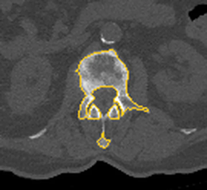
Aynı bölgenin, kaynak veri setinde patoloji içermediği belirtilen bir BT incelemesini kesit kesit kaydırın; organ sınırlarını açarak anatomiyi hasta görüntüsüyle karşılaştırın. Küçük rastlantısal bulgular tümüyle dışlanmamıştır.
Kaynak: TotalSegmentator v2.01 veri seti (Wasserthal ve ark., Radiology: Artificial Intelligence 2023), CC BY 4.0, vaka s1338; organ sınırları veri setinin otomatik segmentasyonundan, birleştirilerek sadeleştirildi. Görüntüler 2,5 mm kesit aralığına yeniden örneklendi.
Ayırıcı tanı
- Basit kompresyon kırığı
- ön korpus ve/veya son plakta çökme vardır; posterior duvar sağlam kalırsa Denis’in orta kolonunda kemik kırığı gösterilmez.
- Burst kırığı
- posterior korpus duvarı ve ön kolon birlikte etkilenir; retropulsiyon görülebilir.
- Fleksiyon-distraksiyon kırığı
- yatay kırık ya da disk/PLC ayrışması orta ve arka kolon boyunca uzanabilir; kemik Chance paterni AO B1 ile uyumlu olabilir.
- Translasyonel kırık-çıkık
- vertebra segmenti komşu segmente göre yer değiştirir; AO Spine tip C örüntüsü ayrıca değerlendirilir.
- Dejeneratif lezyonlar (osteofit, Modic değişiklikleri) uçplakta sınırlandırılmış sinyal veya kontür bozukluklarıyla izlenir; çok kolonlu akut travmatik kırık hattı oluşturmazlar.
Tuzaklar
- “İki kolon tutulduysa instabil” biçimindeki tarihsel kısa kuralı tek başına güncel stabilite ya da tedavi kararı gibi kullanma; AO Spine/TLICS morfolojisi, PLC durumu ve nörolojik değerlendirmeyi ayrı ele alır.
- Posterior duvar kırığını yalnız ön kolon kompresyonu olarak adlandırma; posterior korteks kırığı burst morfolojisini düşündürür.
- BT’de kemik kolonları haritalanabilir ancak posterior longitudinal ligament, annulus ve PLC lifleri doğrudan güvenilir biçimde değerlendirilemez.
- PLC yaralanması yalnızca kemik BT ile kesin tanı konamaz; MR'da STIR sekansında interspinöz/supraspinöz ligamanlardaki ödem ya da supraspinöz mesafe artışı gibi işaretler değerlendirilir.
- Denis’in üç kolon kavramını Denis sakral zonlarıyla karıştırma; biri torakolomber kolon modelidir, diğeri sakrum kırığının foramene göre zonlamasıdır.
Kendini dene
BT’de ön korpus çökmesine ek olarak posterior korpus duvarında kırık ve kanala retropulse parça görülüyor. Denis kolonlarına göre hangi kemik bileşen etkilenmiştir?
Cevabı göster
Ön ve orta kolon. Ön korpus çökmesi ön kolonun, posterior korpus duvarı kırığı ise orta kolonun kemik bileşeninin tutulduğunu gösterir; bu burst morfolojisidir. Arka kolon değerlendirmesinde posterior kemik elemanları ve posterior ligamentöz kompleks dikkate alınır; BT kemik yapıları gösterse de saf ligamentöz yaralanmaları değerlendirmede sınırlıdır.
Denis üç kolon modeliyle ilgili güncel ve güvenli yorum hangisidir?
Cevabı göster
Tarihsel morfolojik çerçevedir; güncel AO Spine/TLICS değerlendirmesinin yerine geçmez. Denis modeli torakolomber yaralanmayı üç anatomik kolonda betimleyen tarihsel bir çerçevedir. Güncel AO Spine morfoloji grupları ve TLICS’in PLC/nörolojik bileşenleri ayrı değerlendirilir; Denis modeli sakral zon sınıflaması değildir.
İlgili konular
- Torakolomber burst kırığı
- Kemik Chance (fleksiyon-distraksiyon) kırığı
- Ligamentöz fleksiyon-distraksiyon yaralanması
Kaynaklar
Bu sayfadaki 48 cümle ve 2 quiz sorusu aşağıdaki kaynaklardan alıntılarla tek tek karşılaştırıldı; 10 cümle bu karşılaştırmada düzeltildi (2026-10-08).
- A prospective study of anterior versus posterior approach for traumatic thoracolumbar fractures: A systemic reviewpmc.ncbi.nlm.nih.gov
- Association between pedicle screw dimensions and pedicle morphometry in computed tomography scanspmc.ncbi.nlm.nih.gov
- The Elusive Concept of Stability in Osteoporotic Vertebral Fractures: A Narrative Reviewpmc.ncbi.nlm.nih.gov
- Bone cement displacement after percutaneous vertebroplasty for a three-column fracture: a case reportpmc.ncbi.nlm.nih.gov
- Chance-Type Fracture of the Sacrum in an Elderly Patient: A Case Reportpmc.ncbi.nlm.nih.gov
- A prospective study of anterior versus posterior approach for traumatic thoracolumbar fractures: A systemic review.pubmed.ncbi.nlm.nih.gov
- Mechanical Instability After Civilian Thoracolumbar Gunshot Injuries: A Systematic Review to Support Classification Development.pubmed.ncbi.nlm.nih.gov
- Thoracolumbar Fractures: Historical Systems and Advancements With the AO Spine Classification.pubmed.ncbi.nlm.nih.gov
- A novel internal fixation system for sacral fractures: a finite element biomechanical study.pubmed.ncbi.nlm.nih.gov
- One-Stage Posterior Open Fixation with SpineJack Expansion Kyphoplasty for Unstable A4 Complete Thoracolumbar Burst Fracture with Neurological Deficit: A Proof-of-Concept Case Series.pubmed.ncbi.nlm.nih.gov
- Association between pedicle screw dimensions and pedicle morphometry in computed tomography scans.pubmed.ncbi.nlm.nih.gov
- A pictorial essay of thoracic wall diseases: multiple pathologies in the same anatomical site.pubmed.ncbi.nlm.nih.gov
- Isolated posterior stabilization in type B and C thoracolumbar fractures associated with ankylosing spine disorders: A single center experience with clinical and radiological outcomes.pubmed.ncbi.nlm.nih.gov
- Evaluation of Posterior Ligamentous Complex Injury in Thoracolumbar Burst Fractures: Correlation Analysis of CT and MRI Findings.pubmed.ncbi.nlm.nih.gov
- The Integrated Nerve, Discoligamentous Complex, and Vertebra Scoring System for Thoracolumbar Junction (TLJ) Injury.pubmed.ncbi.nlm.nih.gov
- Spine Trauma Classifications: Historical, Current, and Emerging Perspectives for Radiologists.pubmed.ncbi.nlm.nih.gov
- QCT-based geometric, densitometric, and biomechanical properties of the aging human thoracolumbar spine.pubmed.ncbi.nlm.nih.gov
- Complete abdominal aortic occlusion following seat-belt trauma: a proposal for early recognition.pubmed.ncbi.nlm.nih.gov
- Posterior-Only Reduction of High-Grade Traumatic Thoracic Spondyloptosis.pubmed.ncbi.nlm.nih.gov
- Comparative analysis of two modified TLICS systems in guiding surgical decision-making for thoracolumbar fractures.pubmed.ncbi.nlm.nih.gov
- Reliability and reproducibility of the modified thoracolumbar injury classification and severity score and the thoracolumbar AO spine injury score for guiding surgical decision-making in thoracolumbar fractures.pubmed.ncbi.nlm.nih.gov
- Agreement and discordance between the modified thoracolumbar injury classification and severity score and the thoracolumbar AOSpine injury score in guiding surgical decision-making for thoracolumbar fractures: a comparative study.pubmed.ncbi.nlm.nih.gov
- The evolution of cervical spine trauma classification: a paradigm shift from morphological description to clinical decision-making.pubmed.ncbi.nlm.nih.gov
- Thoracolumbar spine trauma: a guide for the FRCS examination.pubmed.ncbi.nlm.nih.gov
- Traumatic lumbosacral instability: part 1 - proposing a definition and identifying underlying injury patterns.pubmed.ncbi.nlm.nih.gov
- Upper Thoracic Spine (D2-D3) Fracture With Unilateral Lock Facets Without Associated Neurological Deficits: Case Report & Literature Review.pubmed.ncbi.nlm.nih.gov
- Traumatic lumbosacral instability: part 2-indications and techniques for surgical management.pubmed.ncbi.nlm.nih.gov
- What Factors Contribute to the Poor Prognosis of Conservative Treatment for Osteoporotic Vertebral Compression Fracture (OVCF): A Systematic Review.pubmed.ncbi.nlm.nih.gov
- Quantitative analysis of bone cement dispersion height in percutaneous vertebroplasty and kyphoplasty and comparison of therapeutic effects.pubmed.ncbi.nlm.nih.gov
- Return to work following traumatic fractures of the thoracolumbar spine without spinal cord injury: a scoping review.pubmed.ncbi.nlm.nih.gov
- Chance Fracture in an Unbelted Rear Seat Passenger.pubmed.ncbi.nlm.nih.gov
- Trauma of the spine and spinal cord: imaging strategies.pubmed.ncbi.nlm.nih.gov
- Surgical outcome of thoracolumbar burst fractures with flexion-distraction injury of the posterior elements.pubmed.ncbi.nlm.nih.gov
- Missed T8-T9 Translational Fracture with Intact Neurology: A Case Report.pubmed.ncbi.nlm.nih.gov
- Anatomical and biological reconstruction of traumatic complete burst fractures of the thoracolumbar vertebrae using pedicle fixation combined with stent-armed kyphoplasty and intracorporeal bone grafting: technique rationale, 7-year experience and clinical, imaging, and histological outcomes.pubmed.ncbi.nlm.nih.gov
- MDCT Morphometry of the Thoracolumbar Junction in a Turkish Cohort: Surgical, Radiological, and Forensic Implications.pubmed.ncbi.nlm.nih.gov
- Congenital and Breed-Associated Vertebral Anomalies in Dogs and Cats: Developmental Anatomy, Genetic Determinants, and Biological Significance.pubmed.ncbi.nlm.nih.gov
- SpineMAE: A bone-window self-supervised and structure-aware framework for 3D cervical vertebra segmentation and fracture classification.pubmed.ncbi.nlm.nih.gov
- Radiomics in spinal research: a narrative review.pubmed.ncbi.nlm.nih.gov
- AstroLogics: a simulation-based framework for the analysis of boolean model ensembles.pubmed.ncbi.nlm.nih.gov
- A validated CT-based scoring system for lateral compression type one pelvic ring injuries provides insight into the spectrum of injury severity and guides treatment decisions; a prospective study.pubmed.ncbi.nlm.nih.gov
- Quantification of intervertebral disc degeneration using dual-energy computed tomography collagen maps: a comparative study with MRI Pfirrmann grading.pubmed.ncbi.nlm.nih.gov
- Risk-stratified management of ankylosing spondylitis-related spinal fractures-a meta-synthesis of contemporary surgical and nonsurgical strategies: a narrative review.pubmed.ncbi.nlm.nih.gov
- Radiological Diagnosis and Advances in Imaging of Vertebral Compression Fractures.pubmed.ncbi.nlm.nih.gov
- Traumatic posterior fracture-dislocation of the fifth lumbar vertebra: report of two cases with review of literature.pubmed.ncbi.nlm.nih.gov
- The epidemiology of thoracolumbar trauma: A meta-analysis.pubmed.ncbi.nlm.nih.gov
- Occupant and crash characteristics in thoracic and lumbar spine injuries resulting from motor vehicle collisions.pubmed.ncbi.nlm.nih.gov
- Clinicodemographic features of traumatic thoracolumbar spine fractures presenting to a tertiary care center in Nepal.pubmed.ncbi.nlm.nih.gov
- Crosslink augmentation in posterior fixation of unstable thoracic and thoracolumbar fractures: A comparative cohort study of mechanical failure and sagittal alignment.pubmed.ncbi.nlm.nih.gov
- Predicting neurological recovery after thoracolumbar spine trauma: The role of injury morphology and admission American Spinal Injury Association Impairment Scale grade.pubmed.ncbi.nlm.nih.gov
- Invited review: Recommendations for reporting intervention studies on reproductive performance in dairy cattle: Improving design, analysis, and interpretation of research on reproduction.pubmed.ncbi.nlm.nih.gov
- Effect of blending Jersey and Holstein-Friesian milk on Cheddar cheese processing, composition, and quality.pubmed.ncbi.nlm.nih.gov
- Determination of cystathionine beta-synthase activity in human plasma by LC-MS/MS: potential use in diagnosis of CBS deficiency.pubmed.ncbi.nlm.nih.gov
- Medial elbow approach via a medial epicondyle osteotomy.pubmed.ncbi.nlm.nih.gov
- Is surgery always necessary? Successful closed reduction of displaced capitellum fracture: A case report.pubmed.ncbi.nlm.nih.gov
- No single best technique: trade-offs in femoral interference screw fixation for bone-patellar tendon-bone anterior cruciate ligament reconstruction.pubmed.ncbi.nlm.nih.gov
- Single-position robot-assisted lateral lumbar corpectomy with minimally invasive posterior spinal fusion for traumatic burst fracture showing MRI-demonstrated indirect decompression: illustrative case.pubmed.ncbi.nlm.nih.gov
- Inducing thoracolumbar burst fractures in human cadaveric specimens using a combined axial-flexion loading: a fracture simulation.pubmed.ncbi.nlm.nih.gov
- Efficacy and Safety of Percutaneous Pedicle Screw Fixation for Neurologically Intact Thoracolumbar Burst Fractures: A Prospective Interventional Study.pubmed.ncbi.nlm.nih.gov
- Radiologically Devastating Yet Neurologically Intact: A Rare Case of Ambulatory C6-C7 Translational Injury with Bilateral Facet Dislocation and Vertebral Artery Thrombosis.pubmed.ncbi.nlm.nih.gov
- Lumbar Intervertebral Disc and Discovertebral Segment. Part 2: An Imaging Review of Pathologic Conditions With Anatomic Correlation.pubmed.ncbi.nlm.nih.gov
- Magnetic resonance imaging of the spine.pubmed.ncbi.nlm.nih.gov
- Identifying osteoporotic vertebral endplate and cortex fractures.pubmed.ncbi.nlm.nih.gov
- Surgical Decision-Making in Thoracolumbar Fractures: A Systematic Review of Anterior and Posterior Approach.pubmed.ncbi.nlm.nih.gov
- The healing process of vertebral body fracture in Wistar rats: creation of an animal model and demonstration of the impact of anterolateral vascularization disruption on bone healing.pubmed.ncbi.nlm.nih.gov
- Microvascular anatomy and mapping of intraosseous arterioles in the human L1 vertebral body: a histological study, literature comparison, and relevance for fracture healing.pubmed.ncbi.nlm.nih.gov
- Spinal Cord Stimulation: Mechanisms of Action, Indications, Types, Complications.pubmed.ncbi.nlm.nih.gov
- Are Ceramic Cages an Option for Lumbar Interbody Fusion? A Comparative Biomechanical Numerical Study.pubmed.ncbi.nlm.nih.gov
- Posterior-Only Sagittal Correction in Degenerative Lumbar Kyphosis Using Posterior Interpedicular Osteotomy (PIO) and Bilateral Discectomy with Hyperlordotic Cages: A Retrospective Case Series.pubmed.ncbi.nlm.nih.gov
- Application of the pivot forward combined leveraging technique in the treatment of thoracolumbar fractures: a finite element analysis and clinical study.pubmed.ncbi.nlm.nih.gov
- Occipitocervical dissociation: A narrative review and case series with a novel management algorithm proposal.pubmed.ncbi.nlm.nih.gov
- Reliability of thoracolumbar burst fracture classification in the Swedish Fracture Register.pubmed.ncbi.nlm.nih.gov
- Prevalence, Implications, and Risk Factors of Traumatic Dural Tears in Thoracic and Lumbar Fractures: A Retrospective Study.pubmed.ncbi.nlm.nih.gov
- Long-term safety and effectiveness of the "OptEase" vena cava filter.pubmed.ncbi.nlm.nih.gov
- Morphological characteristics of lumbar vertebral bodies and regional distribution patterns of bone mineral density: a CT study.pubmed.ncbi.nlm.nih.gov
- Modified bone-disc-bone osteotomy (MBDBO) for the treatment of spinal kyphotic deformity secondary to old thoracolumbar vertebral fracture: a retrospective study with 2-year follow-up.pubmed.ncbi.nlm.nih.gov
- Biomaterials for vertebral body repair: interior augmentation and total replacement-a literature review of bone fusion outcomes in clinical studies.pubmed.ncbi.nlm.nih.gov
- Current Evidence and Clinical Implications of Full-Endoscopic Spine Surgery for Post-Vertebroplasty Lumbar Radiculopathy: A Comprehensive Review.pubmed.ncbi.nlm.nih.gov
- Treatment Concept and Technical Considerations of Biportal Endoscopic Spine Surgery for Lumbar Spinal Stenosis.pubmed.ncbi.nlm.nih.gov
- Traumatic Unilateral Pedicle Fracture Treated With a Partially Threaded Screw: A Case Report.pubmed.ncbi.nlm.nih.gov
- Navigation-Assisted Bilateral Troughing Technique for Lumbar Laminectomy: A Stepwise Approach to Safe, Teachable, and Efficient Decompression.pubmed.ncbi.nlm.nih.gov
- Surgical Reduction Techniques and Outcomes for Cervical Spondyloptosis: A Technical Report and Case Series.pubmed.ncbi.nlm.nih.gov
- Radiological findings of February 2023 twin earthquakes-related spine injuries.pubmed.ncbi.nlm.nih.gov
- Acute management of spinal fractures: What you need to know.pubmed.ncbi.nlm.nih.gov
- The value of magnetic resonance imaging and computed tomography in the study of spinal disorders.pubmed.ncbi.nlm.nih.gov
- Association of Chiropractic Colleges Educational Conference and Research Agenda Conference 2026: Connected Care.pubmed.ncbi.nlm.nih.gov
- Padua Days on Muscle and Mobility Medicine, March 25-29, 2025, Hotel Petrarca, Euganean Thermae, Italy: Program and Abstracts.pubmed.ncbi.nlm.nih.gov
- Redefining the applications of navigation in spine surgery.pubmed.ncbi.nlm.nih.gov
- The Prone-Transpsoas Approach for Single-Position Lateral Corpectomy: A Case Series.pubmed.ncbi.nlm.nih.gov
- Anatomy-driven automated subsegmentation of pelvic and proximal femoral CT into clinically relevant subregions and landmarks.pubmed.ncbi.nlm.nih.gov
- Laminectomy alone <i>vs</i> laminectomy with posterior fusion in lumbar spinal stenosis: The role of instability.pubmed.ncbi.nlm.nih.gov
- Microarchitecture of historic bone samples with tuberculosis.pubmed.ncbi.nlm.nih.gov
- Clinical applications and prospects of 3D printing guide templates in orthopaedics.pubmed.ncbi.nlm.nih.gov
- Comparative Biomechanical Stability of the Fixation of Different Miniplates in Restorative Laminoplasty after Laminectomy: A Finite Element Study.pubmed.ncbi.nlm.nih.gov
- Defining mechanical instability in tuberculosis of the spine: a systematic review.pubmed.ncbi.nlm.nih.gov
- Retroperitoneal glass foreign body with renal parenchymal involvement after 25 years of silent retention: a case report.pubmed.ncbi.nlm.nih.gov
- Bone cement displacement after percutaneous vertebroplasty for a three-column fracture: a case report.pubmed.ncbi.nlm.nih.gov
- Chance-Type Fracture of the Sacrum in an Elderly Patient: A Case Report.pubmed.ncbi.nlm.nih.gov
- Staged management of a Tile C3 pelvic ring injury with bilateral sacroiliac disruption and T-shaped sacral fracture.pubmed.ncbi.nlm.nih.gov
- Recognizing Sacral Insufficiency Fractures Hidden in Plain Sight: An Illustrative Case Report.pubmed.ncbi.nlm.nih.gov
- Fetus-in-Fetu: A PRISMA-Informed Narrative Review With an Illustrative Prenatal Case.pubmed.ncbi.nlm.nih.gov
- Personalized Protocol for the Dynamic Assessment of Functional Biomarkers of Vascular Stiffness: A Novel Diagnostic Tool in P4 Medicine.pubmed.ncbi.nlm.nih.gov
- Open reduction and internal fixation versus percutaneous multiple-trajectory screw fixation for unstable pelvic fractures with severe polytrauma: a prospective study.pubmed.ncbi.nlm.nih.gov
- Assessment of Clinical Outcomes and Quality of Life Following Laminectomy and Lateral Mass Screw Fixation in Patients With Cervical Myelopathy.pubmed.ncbi.nlm.nih.gov
- Extrapedicular approach for basivertebral nerve radiofrequency ablation: a conceptual review.pubmed.ncbi.nlm.nih.gov
- An innovative 2D optical navigation workflow for percutaneous pedicle screw fixation in thoracolumbar fractures: comparison with O-arm 3D navigation.pubmed.ncbi.nlm.nih.gov
- Evolutionary Specializations of the Human Vertebral Body and Intervertebral Disc in Relation to Bipedalism.pubmed.ncbi.nlm.nih.gov
- Diagnostic accuracy of deep learning vs. human raters for detecting osteoporotic vertebral compression fractures in routine CT scans.pubmed.ncbi.nlm.nih.gov
- Spinal disorders mimicking infection.pubmed.ncbi.nlm.nih.gov
- O-arm-navigated open-door laminoplasty to reduce technical complications in multilevel cervical ossification of the posterior longitudinal ligament: operative video.pubmed.ncbi.nlm.nih.gov
- Hybrid Protective Sentinel System: A Combined Technique for Safe and Controlled Open-Wedge High Tibial Osteotomy.pubmed.ncbi.nlm.nih.gov
- Uni-portal non-coaxial spinal endoscopic surgery on percutaneous hollow screw fixation for atypical hangman's fracture: a novel minimally invasive technique case report.pubmed.ncbi.nlm.nih.gov
- Augmented reality-enabled image-guided surgical navigation in minimally invasive spine surgery: application beyond screw placement in prone lateral transpsoas interbody fusion-a cadaveric surgical technique study.pubmed.ncbi.nlm.nih.gov
- Use of Intraoperative Ultrasound During Spinal Surgery.pubmed.ncbi.nlm.nih.gov
- Subaxial cervical spine trauma.pubmed.ncbi.nlm.nih.gov
Eğitim amaçlıdır; hasta başında tanı ve tedavi kararının yerine geçmez. İçerik yapay zekâ destekli bir süreçle güncel literatüre göre cümle cümle kaynaklarla karşılaştırıldı, ancak uzman radyolog incelemesinden geçmedi. Klinik kararlarda güncel kılavuzları, kurum protokollerini ve radyoloji raporunu esas alın.