
Özet
Kontrastsız baş BT’sinde rastlantısal fizyolojik mineralizasyondan enfeksiyon, metabolik hastalık, vasküler bozukluk veya kalsifiye kitleye uzanan geniş bir neden yelpazesi intrakraniyal kalsifikasyon olarak görülebilir. Kontrastsız BT’de intrakraniyal kalsifikasyonlar gösterilebilir; değerlendirmede kalsifikasyonun örüntüsü, boyutu ve yerleşimi dikkate alınır. İntrakraniyal kalsifikasyonların örüntüsünü, boyutunu ve yerleşimini tarif etmek ayırıcı tanıya yardımcı olur. İntrakraniyal kalsifikasyonlar kontrastsız BT’de sık karşılaşılan bulgulardır.
Faz ve pencere
- Kontrastsız tanısal
- Kontrastsız BT’de intrakraniyal kalsifikasyonlar görülebilir; değerlendirmede örüntü, boyut ve yerleşimleri dikkate alınır. Odakların pallidum, dentat çekirdek, ventrikül içi koroid pleksus, arter duvarı, korteks veya dura ile anatomik ilişkisi bu fazda haritalanır.
Önerilen pencereler G genişlik, M merkez; değerler Hounsfield birimi (HU)
- BeyinG 80 / M +40
- KemikG 1800 / M +400
BT bulguları
- Pineal bez ve habenula — orta hatta birleşik ya da ayrı küçük yoğunluklar fizyolojik mineralizasyonla uyumlu olabilir.
- Koroid pleksus kalsifikasyonları fizyolojik olabilir.
- Erişkinlerde bazal ganglion kalsifikasyonları rastlantısal bulunabilir; dağılımın yaygınlığı, boyutu ve yerleşimi ayırıcı tanıda dikkate alınmalıdır.
- Yaygın, iki taraflı kalsifikasyonlar bazal ganglionlar, talamus ve serebellar dentat çekirdekleri tutabilir.
- Sturge–Weber sendromunda kortikal kalsifikasyonlar beyin atrofisiyle birlikte görülebilir.
- Parankimal kalsifikasyonlar değerlendirilirken dağılımı, boyutu ve yerleşimi tarif edilir.
- İntrakraniyal arter kalsifikasyonları kontrastsız kranial BT’de saptanabilir.
- Falx ve tentoryumda dural kalsifikasyonlar görülebilir.
- İntrakraniyal kalsifikasyonlar fizyolojik/yaşa bağlı, distrofik, doğumsal, enfeksiyöz, vasküler, neoplastik, metabolik/endokrin, inflamatuvar veya toksik kategorilerde sınıflanabilir; ayırıcı tanıda örüntü ve klinik bağlam dikkate alınır.
Normalde
Pineal bölge, habenula, koroid pleksus ve dura kalsifikasyonları fizyolojik olabilir; bazal ganglion kalsifikasyonları da asemptomatik kişilerde rastlantısal saptanabilir. Kalsifikasyonların örüntüsünü, boyutunu ve yerleşimini tarif edin.
Normal BT ile kıyasla
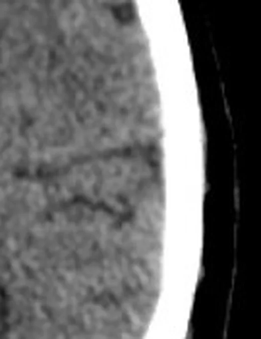
Aynı bölgenin, kaynak veri setinde patoloji içermediği belirtilen bir BT incelemesini kesit kesit kaydırın; organ sınırlarını açarak anatomiyi hasta görüntüsüyle karşılaştırın. Küçük rastlantısal bulgular tümüyle dışlanmamıştır.
Kaynak: TotalSegmentator v2.01 veri seti (Wasserthal ve ark., Radiology: Artificial Intelligence 2023), CC BY 4.0, vaka s0103; organ sınırları veri setinin otomatik segmentasyonundan, birleştirilerek sadeleştirildi. Görüntüler 2,5 mm kesit aralığına yeniden örneklendi.
Ayırıcı tanı
- Primer ailesel beyin kalsifikasyonu iki taraflı ve simetrik olarak bazal ganglionları, talamusu, subkortikal beyaz cevheri ve serebellumu tutabilir.
- Hipoparatiroidi, bazal ganglionlarda kalsifikasyonla ilişkili metabolik nedenlerden biridir.
- Sturge–Weber sendromu
- tek hemisfer korteksinde giriform tram-track kalsifikasyon, leptomeningeal anomali ve ipsilateral atrofiyle birliktedir.
Tuzaklar
- Atriumdaki koroid pleksus kalsifikasyonunu değerlendirirken odağın anatomik yerini ve yayılımını aksiyel, sagittal ve koronal yeniden format görüntülerde tarif edin.
- Kafa tabanında kemikle örtüşen damarları değerlendirmek için kemik çıkarma görüntüleri damarların daha iyi görülmesine yardımcı olabilir.
- Küçük, ara atenüasyonlu odakların niteliği belirsizse çift enerjili BT’de materyal analizi, bunların kanama, kalsifikasyon veya yabancı madde olup olmadığını ayırt etmeye yardımcı olabilir.
- İki taraflı pallidal odakları tek başına primer ailesel hastalık diye adlandırmayın; sınırlı yaş ilişkili mineralizasyon ve sekonder metabolik nedenler benzer görünebilir.
- Kalsifikasyonların örüntüsü, boyutu ve yerleşimi ayırıcı tanıya yardımcı olur; klinik bağlam da değerlendirmeye katılmalıdır.
Kendini dene
Kontrastsız BT’de pineal bölge ve koroid pleksus kalsifikasyonları görülüyor. Bu yapılardaki kalsifikasyonlar fizyolojik olabilir mi?
Cevabı göster
Sınırlı fizyolojik mineralizasyon. Pineal bölge ve koroid pleksus kalsifikasyonları fizyolojik olabilir; bilateral bazal ganglion kalsifikasyonları ise primer beyin kalsifikasyonunda görülebilir. İntrakraniyal kalsifikasyonlar fizyolojik/yaşa bağlı, enfeksiyöz, vasküler, neoplastik veya metabolik kategorilerde görülebilir; değerlendirmede örüntü, boyut ve yerleşim dikkate alınır.
Görüntülemede leptomeningeal anjiyomlar, ilerleyici beyin atrofisi ve kortikal kalsifikasyonlar birlikte bildirilmiştir. En olası ilişki nedir?
Cevabı göster
Sturge–Weber tipi kortikal vasküler malformasyon. Sturge–Weber sendromunda beyin görüntülemesinde leptomeningeal anjiyom, kortikal kalsifikasyon ve ilerleyici beyin atrofisi bildirilmiştir. Sturge–Weber sendromu beyinde ve gözlerde kapiller-venöz malformasyonlarla karakterizedir; hipoparatiroidide ise intrakraniyal kalsifikasyonlar görülebilir.
İlgili konular
- Primer ailesel beyin kalsifikasyonu (PFBC)
- Sekonder bazal ganglion kalsifikasyonu
- Sturge–Weber sendromunda giriform kalsifikasyon
- İntrakraniyal arter duvarı kalsifikasyonu
- Kalsifiye nörosistiserkoz
Kaynaklar
Bu sayfadaki 39 cümle ve 2 quiz sorusu aşağıdaki kaynaklardan alıntılarla otomatik olarak karşılaştırıldı; 33 cümle bu karşılaştırmada düzeltildi (2026-10-09).
- A novel SLC20A2 variant associated with primary brain calcification: A case reportpmc.ncbi.nlm.nih.gov
- Epistaxis and Destructive Computed Tomographic Features in Canine Nasal Transitional Carcinoma: Frequent Cribriform Plate Destruction but Uncommon CT-Detected Brain Parenchymal Involvementpmc.ncbi.nlm.nih.gov
- Circadian Blood Pressure Pattern and Microvascular and Macrovascular Cerebral Imaging Burdenpmc.ncbi.nlm.nih.gov
- Sphenoid fungal rhinosinusitis across the disease spectrum: a case series of non-invasive fungal ball and fatal acute invasive fungal rhinosinusitispmc.ncbi.nlm.nih.gov
- Intracranial Arterial Calcification on Computed Tomography and Risk of Cognitive Impairment or Dementia: A Systematic Review and Meta-Analysispmc.ncbi.nlm.nih.gov
- Infection.pubmed.ncbi.nlm.nih.gov
- Intracranial calcifications on CT: an updated review.pubmed.ncbi.nlm.nih.gov
- Bone up: craniomandibular development and hard-tissue biomineralization in neonate mice.pubmed.ncbi.nlm.nih.gov
- Asymptomatic congenital cytomegalovirus infection with neurological sequelae: A retrospective study using umbilical cord.pubmed.ncbi.nlm.nih.gov
- Perilesional edema associated with an intracranial calcifying pseudoneoplasm of the neuraxis in a child: case report and review of imaging features.pubmed.ncbi.nlm.nih.gov
- Dual-Energy CT in Emergency Neuroimaging: Added Value and Novel Applications.pubmed.ncbi.nlm.nih.gov
- [Striatopallidodentate calcinosis, hypoparathyroidism and neurological features: a case series study].pubmed.ncbi.nlm.nih.gov
- Congenital Toxoplasmosis in Tunisia: Prenatal and Neonatal Diagnosis and Postnatal Follow-up of 35 Cases.pubmed.ncbi.nlm.nih.gov
- Radiologic features of primary intracranial ectopic germinomas: Case reports and literature review.pubmed.ncbi.nlm.nih.gov
- Recognizable phenotypes associated with intracranial calcification.pubmed.ncbi.nlm.nih.gov
- Diagnostic Value of Brain Calcifications in Adult-Onset Leukoencephalopathy with Axonal Spheroids and Pigmented Glia.pubmed.ncbi.nlm.nih.gov
- Hearing brain evaluated using near-infrared spectroscopy in congenital toxoplasmosis.pubmed.ncbi.nlm.nih.gov
- Clinical data analysis of 22 cases with hypoparathyroidism misdiagnosed as epilepsy.pubmed.ncbi.nlm.nih.gov
- Vertebrobasilar artery calcification: Prevalence and risk factors in the general population.pubmed.ncbi.nlm.nih.gov
- Vertebral Artery Pseudoaneurysm in a Single-Vessel Posterior Circulation With Chronic Contralateral Occlusion and Recent Methamphetamine Use.pubmed.ncbi.nlm.nih.gov
- Tumor-mimicking sarcoid-like reaction with lymphoid infiltration showing intracranial extension from the left jugular foramen: illustrative case.pubmed.ncbi.nlm.nih.gov
- Imaging of Mobile Emboligenic Calcified Amorphous Tumor in Mitral Annular Caseous Calcification.pubmed.ncbi.nlm.nih.gov
- Statin Use and Arterial Calcification in the General Population: A Longitudinal Analysis and a Negative Control Population.pubmed.ncbi.nlm.nih.gov
- PCR-Confirmed Convexity Subarachnoid Neurocysticercosis with Negative Serology: A Case Report.pubmed.ncbi.nlm.nih.gov
- Epistaxis and Destructive Computed Tomographic Features in Canine Nasal Transitional Carcinoma: Frequent Cribriform Plate Destruction but Uncommon CT-Detected Brain Parenchymal Involvement.pubmed.ncbi.nlm.nih.gov
- Integrated Environmental Cadmium Exposure, Plaque Calcium Content, and Lifestyle Factors Improve Prediction of Carotid Plaque Echogenicity: An Exposome-Oriented Approach.pubmed.ncbi.nlm.nih.gov
- Meningioma of the foramen of Luschka choroid plexus presenting as glossopharyngeal neuralgia: A case report and review of the literature.pubmed.ncbi.nlm.nih.gov
- ABCC6 Heterozygosity as Genetic Predisposition to Cerebrovascular Disease Across Ages.pubmed.ncbi.nlm.nih.gov
- Lung adenocarcinoma dural metastasis at the petroclival region mimicking meningioma: a frozen-section diagnostic pitfall. Illustrative case.pubmed.ncbi.nlm.nih.gov
- A rare case of primary intracranial myxoma in a 10-year-old male: Case report and literature review.pubmed.ncbi.nlm.nih.gov
- Radiologist Perceptions of an AI Tool for Intracranial Hemorrhage Detection in Teleradiology: Cross-Sectional Survey Study.pubmed.ncbi.nlm.nih.gov
- Leptomeningeal Masses or Masquerades: A Spectrum of Diseases with Leptomeningeal Enhancement and their Mimics.pubmed.ncbi.nlm.nih.gov
- Uncommon Nasal Mass Presentation: A Radiological Case Series.pubmed.ncbi.nlm.nih.gov
- Pediatric Meningeal Diseases: What Radiologists Need to Know.pubmed.ncbi.nlm.nih.gov
- A Non-enhanced CT Dataset for Differential Diagnosis of Pediatric Extracranial Germ Cell Tumors.pubmed.ncbi.nlm.nih.gov
- Understanding Photon-Counting CT: Physics, Detector Technology, and Image Reconstructions.pubmed.ncbi.nlm.nih.gov
- Imaging Characteristics of Colloid Cysts: A Case Series Correlating Cyst Size and Location With Associated Acute Hydrocephalus.pubmed.ncbi.nlm.nih.gov
- Application of [<sup>18</sup>F]NaF and [<sup>18</sup>F]FDG PET/CT to Assess Molecular Calcification and Glucose Metabolism in Thyroid Cartilage.pubmed.ncbi.nlm.nih.gov
- Pineal gland senescence: an emerging ageing-related pathology?pubmed.ncbi.nlm.nih.gov
- From diagnostic marker-to-therapeutic modality: emerging paradigms in tumor biomineralization.pubmed.ncbi.nlm.nih.gov
- Age-related Vascular Alterations in the Choroid Plexus: Novel Insights from Pathophysiology and Imaging Studies.pubmed.ncbi.nlm.nih.gov
- Society for Cardiovascular Magnetic Resonance 2024 Cases of SCMR case series.pubmed.ncbi.nlm.nih.gov
- Imaging of supratentorial intraventricular masses in children:a pictorial review- part 1.pubmed.ncbi.nlm.nih.gov
- Bilateral Basal Ganglia Calcification (Fahr's Syndrome) Secondary to Hypoparathyroidism: A Case Series.pubmed.ncbi.nlm.nih.gov
- Adult-recognized 22q11.2 deletion syndrome in adult neurology practice: a brief report of two diagnostic pitfalls.pubmed.ncbi.nlm.nih.gov
- Basal Ganglia Calcifications for Nephrologists: Fahr/Primary Familial Brain Calcification (PFBC) at the Interface of Chronic Kidney Disease-Mineral and Bone Disorder (CKD-MBD) and Hypoparathyroidism.pubmed.ncbi.nlm.nih.gov
- Silent stone: Idiopathic presentation of Fahr's disease.pubmed.ncbi.nlm.nih.gov
- Rare Presentation of Homozygous S<i>LC20A2</i> Mutations Causing Intra-Arterial Cerebral Vasculopathy and Stroke in Infancy: Case Report and Review of the Literature.pubmed.ncbi.nlm.nih.gov
- Fahr's disease presenting with ischemic stroke in young adult: a case report of rare disease with unique presentation.pubmed.ncbi.nlm.nih.gov
- Longitudinal Neuroimaging of a Pediatric Patient With Sturge-Weber Syndrome: From Birth to Adolescence.pubmed.ncbi.nlm.nih.gov
- Accidental finding of Sturge-Weber syndrome presented to the emergency department with persistent seizures: an unusual case report from Somalia.pubmed.ncbi.nlm.nih.gov
- Hippocampal sclerosis in association with Sturge-weber syndrome: An intertwining of 2 epileptogenic lesions.pubmed.ncbi.nlm.nih.gov
- Clinical, Immunological, and Neuroimaging Characteristics and Outcome Profile of Neurocysticercosis in Children at a Neurology Department in Antananarivo: A Retrospective Observational Study.pubmed.ncbi.nlm.nih.gov
- Beyond Imaging: A 12-Year Clinicopathological Analysis of Surgically Excised Neurocysticercosis.pubmed.ncbi.nlm.nih.gov
- Neurocysticercosis in transition: expanding clinical spectrum, evolving diagnostics, and emerging therapies.pubmed.ncbi.nlm.nih.gov
- Aortic Arch Pseudoaneurysm with Porcelain Aortic Arch and Triple-Vessel Coronary Artery Disease: Single-Stage Total Arch Replacement with Coronary Artery Bypass Grafting.pubmed.ncbi.nlm.nih.gov
- NaF-PET Imaging for Detection of Early Arterial Microcalcification and Monitoring of Targeted Therapy: A Narrative Review.pubmed.ncbi.nlm.nih.gov
- Monckeberg's Medial Sclerosis in Athletes: Report of Two Cases and Possible Pathophysiological Mechanisms.pubmed.ncbi.nlm.nih.gov
- Molecular imaging of dural microcalcification: Detection of active mineralization with <sup>18</sup>F-NaF PET/CT.pubmed.ncbi.nlm.nih.gov
- Glioblastoma eroding through falx cerebri: a rarity or commonly seen? Illustrative case.pubmed.ncbi.nlm.nih.gov
- Gorlin-Goltz syndrome: Multidisciplinary approach for early diagnosis of rare disease for better patient outcome.pubmed.ncbi.nlm.nih.gov
- Cerebrospinal Fluid Shunts: An Updated Radiologic Review of Devices, Malfunctions, and Complications.pubmed.ncbi.nlm.nih.gov
- Fetal intracranial hemorrhage and infarct: Main sonographic and MRI characteristics: A review article.pubmed.ncbi.nlm.nih.gov
- Detection of aneurysms by 64-section multidetector CT angiography in patients acutely suspected of having an intracranial aneurysm and comparison with digital subtraction and 3D rotational angiography.pubmed.ncbi.nlm.nih.gov
- High-affinity alphavbeta3 integrin targeted optical probe as a new imaging biomarker for early atherosclerosis: initial studies in Watanabe rabbits.pubmed.ncbi.nlm.nih.gov
- Idiopathic IgG4-negative perivascular fibrosis with extensive thoracic aortic, coronary artery, and supra-aortic branch involvement: A case report.pubmed.ncbi.nlm.nih.gov
- High-Z Contrast Media for Coronary Photon-counting Detector CT Angiography: Improved Quantification of Calcified Stenoses.pubmed.ncbi.nlm.nih.gov
- Unicentric Adrenal Castleman Disease Mimicking an Adrenal Tumour.pubmed.ncbi.nlm.nih.gov
- Recurrent Tumor-Induced Osteomalacia After Surgical Resection: A Case Report Demonstrating Successful Treatment With Burosumab.pubmed.ncbi.nlm.nih.gov
- Incidental brain tumor findings in children: prevalence, natural history, management, controversies, challenges, and dilemmas.pubmed.ncbi.nlm.nih.gov
- Multinodular and Vacuolating Neuronal Tumors: Imaging Features, Diagnosis, and Management Challenges.pubmed.ncbi.nlm.nih.gov
- Management of pineal and colloid cysts.pubmed.ncbi.nlm.nih.gov
- Sturge-Weber Syndrome: A Narrative Review of Clinical Presentation and Updates on Management.pubmed.ncbi.nlm.nih.gov
- Sturge-Weber Syndrome: A Report of a Rare Case.pubmed.ncbi.nlm.nih.gov
- Emerging insights into cephalic neural crest disorders: A single center experience.pubmed.ncbi.nlm.nih.gov
- Circadian Blood Pressure Pattern and Microvascular and Macrovascular Cerebral Imaging Burden.pubmed.ncbi.nlm.nih.gov
- A novel <i>SLC20A2</i> variant associated with primary brain calcification: A case report.pubmed.ncbi.nlm.nih.gov
- SWI combined with cMRI and CT in the differentiating of intracranial Rosai-Dorfman disease from fibrous meningioma.pubmed.ncbi.nlm.nih.gov
- Physiologic pineal region, choroid plexus, and dural calcifications in the first decade of life.pubmed.ncbi.nlm.nih.gov
- Cerebroretinal Microangiopathy with Calcifications and Cysts (CRMCC): A 5-Year Diagnostic Challenge.pubmed.ncbi.nlm.nih.gov
- Low-Level Cerebrospinal Fluid β-hCG Elevation in Patients with Pineal Parenchymal Tumors: A Four-Case Series and Diagnostic Caution.pubmed.ncbi.nlm.nih.gov
- Susceptibility-weighted imaging in intracranial hemorrhage: not all bleeds are black.pubmed.ncbi.nlm.nih.gov
- Imaging of Skull Base Tumors.pubmed.ncbi.nlm.nih.gov
- Virtual non-calcium dual-energy CT: clinical applications.pubmed.ncbi.nlm.nih.gov
- Insights into neurosarcoidosis: an imaging perspective.pubmed.ncbi.nlm.nih.gov
- Non-Contrast MRI Sequences for Ischemic Stroke: A Concise Overview for Clinical Radiologists.pubmed.ncbi.nlm.nih.gov
- Electron Density and Effective Atomic Number of Normal-Appearing Adult Brain Tissues: Age-Related Changes and Correlation with Myelin Content.pubmed.ncbi.nlm.nih.gov
- Radiological Predictors of Cognitive Impairment in Paediatric Brain Tumours Using Multiparametric Magnetic Resonance Imaging: A Review of Current Practice, Challenges and Future Directions.pubmed.ncbi.nlm.nih.gov
- Case Report: Two Novel Frameshift Mutations in SLC20A2 and One Novel Splice Donor Mutation in PDGFB Associated With Primary Familial Brain Calcification.pubmed.ncbi.nlm.nih.gov
- MR Imaging of Pediatric Brain Tumors.pubmed.ncbi.nlm.nih.gov
- Clinically relevant imaging in tuberous sclerosis.pubmed.ncbi.nlm.nih.gov
- International Harmonization of Nomenclature and Diagnostic Criteria (INHAND): Non-proliferative and Proliferative Lesions of the Non-human Primate ( M. fascicularis ).pubmed.ncbi.nlm.nih.gov
- Cerebellar Mutism/Posterior Fossa Syndrome Following Resection of Posterior Fossa Tumor in Pediatric Patients: Assessing Pathophysiology, Risk Factors, and Neuroradiographic Features.pubmed.ncbi.nlm.nih.gov
- Neuroimaging of pediatric tumors of the sellar region-A review in light of the 2021 WHO classification of tumors of the central nervous system.pubmed.ncbi.nlm.nih.gov
- Bilateral lesions of the basal ganglia and thalami (central grey matter)-pictorial review.pubmed.ncbi.nlm.nih.gov
- Determination of cystathionine beta-synthase activity in human plasma by LC-MS/MS: potential use in diagnosis of CBS deficiency.pubmed.ncbi.nlm.nih.gov
- First reported case in Brazil of intraventricular pilocytic astrocytoma in an adult patient with literature review.pubmed.ncbi.nlm.nih.gov
- Primary diffuse large B-cell lymphoma of the choroid plexus: A case report and review of the literature.pubmed.ncbi.nlm.nih.gov
- Diagnostic imaging and emerging image-guided neurointervention in brain tumors: a narrative review.pubmed.ncbi.nlm.nih.gov
- Imaging for Pediatric Head and Neck Emergencies: Critical Insights.pubmed.ncbi.nlm.nih.gov
- Multimodal imaging in neuro-ophthalmology.pubmed.ncbi.nlm.nih.gov
- Imaging of the septum pellucidum: normal, variants and pathology.pubmed.ncbi.nlm.nih.gov
- Resection of a recurrent pineal region teratoma via a posterior interhemispheric transcallosal approach.pubmed.ncbi.nlm.nih.gov
- Radiological diagnosis of calcified intra- and extra-ventricular ganglioglioma involving the lateral ventricle: A case-based review of a challenging and uncommon presentation.pubmed.ncbi.nlm.nih.gov
- A systematic approach to detection of epileptogenic foci on imaging: protocols, entities, search pattern, and reporting tools.pubmed.ncbi.nlm.nih.gov
- Pediatric diencephalic tumors: a constellation of entities and management modalities.pubmed.ncbi.nlm.nih.gov
- Susceptibility-Weighted Imaging (SWI): Technical Aspects and Applications in Brain MRI for Neurodegenerative Disorders.pubmed.ncbi.nlm.nih.gov
- Neuroimaging Pearls from the MDS Congress Video Challenge. Part 1: Genetic Disorders.pubmed.ncbi.nlm.nih.gov
- Basal ganglia calcifications (Fahr's syndrome): related conditions and clinical features.pubmed.ncbi.nlm.nih.gov
- Imaging of brain tumors in children: the basics-a narrative review.pubmed.ncbi.nlm.nih.gov
- Susceptibility-weighted imaging: technical aspects and clinical applications, part 2.pubmed.ncbi.nlm.nih.gov
- Ophthalmologic assessment and intracranial pressure in children: diagnostic methods, clinical correlations, and future directions.pubmed.ncbi.nlm.nih.gov
- Etiologies of Proptosis: A review.pubmed.ncbi.nlm.nih.gov
- Patterns of injury and violence in Yaoundé Cameroon: an analysis of hospital data.pubmed.ncbi.nlm.nih.gov
- Magnetic resonance imaging in leukodystrophies: characteristic patterns and diagnostic relevance.pubmed.ncbi.nlm.nih.gov
Eğitim amaçlıdır; hasta başında tanı ve tedavi kararının yerine geçmez, tıbbi tavsiye değildir. Metinler yapay zekâ desteğiyle yazıldı ve otomatik bir adımda kaynak alıntılarıyla karşılaştırıldı; bu bir tıbbi doğrulama değildir ve içerik uzman radyolog incelemesinden geçmedi. Klinik kararlarda güncel kılavuzları, kurum protokollerini ve radyoloji raporunu esas alın.