Özet
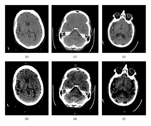
Ani fokal nörolojik defisitte erken iskemik değişiklikler, henüz belirgin infarkt hipodensitesi oluşmadan etkilenmiş dokunun yerini gösterebilir. Kontrastsız beyin BT'si öncelikle kanamayı dışlar; gri-beyaz cevher ayrımındaki silinme ve bölgesel şişme iskemi lehine ipuçlarıdır. Bulgular çok erken dönemde hafif olabilir ve normal görünen BT akut inmeyi dışlamaz. Akut dönemde yapılan erken BT'lerde iskemiye ait değişiklikler henüz oluşmamış olabilir ve ilk çekimler normal bulunabilir.
Faz ve pencere
- Kontrastsız tanısal
- İskemik parankimde karşı hemisfere göre hafif düşük atenüasyon, korteks-beyaz cevher sınırında bulanıklık, insular şeritte silinme ve sulkuslarda daralma aranır; erken bulgular tek kesitte belirsiz olabilir. İyotlu kontrast gerekmez.
Önerilen pencereler: İnme (G 40 / M 40), Beyin (G 80 / M 40).
BT bulguları
- Gri-beyaz cevher ayrım kaybı — kortikal gri cevher ile komşu subkortikal beyaz cevher arasındaki doğal kontrast azalır.
- İnsular şerit silinmesi — insular korteksin gri cevher çizgisi silikleşir; MCA yatağı iskemiğinde erken fark edilebilir.
- Lentiform çekirdek belirsizliği — putamen ve globus pallidus'un keskin sınırı karşı tarafa göre kaybolur.
- Kortikal sulkus silinmesi — ödemli giruslar arasındaki BOS aralıkları daralır.
- Fokal parankimal hipodensite — MCA veya başka arter alanına uyan gri cevher ve komşu beyaz cevherde düşük atenüasyon belirir.
- Arter alanına uyan asimetri — bir hemisferdeki gri cevher silinmesi damar dağılımını izler ve karşı tarafta benzer derecede bulunmaz.
Normalde
Normal insular korteks, Sylvian yarığı ile eksternal kapsül arasında ince ve belirgin gri şerit olarak seçilir; lentiform çekirdek de çevresindeki internal ve eksternal kapsülden ayrılır. Aynı aksiyel kesitte insular korteks, putamen ve sulkuslar gibi ASPECTS bölgeleri, karşı hemisferle kıyaslanarak gri-beyaz cevher ayrımı kaybı veya sulkus daralması açısından değerlendirilir.
Normal BT ile kıyasla
Aynı bölgenin, kaynak veri setinde patoloji içermediği belirtilen bir BT incelemesini kesit kesit kaydırın; organ sınırlarını açarak anatomiyi hasta görüntüsüyle karşılaştırın. Küçük rastlantısal bulgular tümüyle dışlanmamıştır.
Kaynak: TotalSegmentator v2.01 veri seti (Wasserthal ve ark., Radiology: Artificial Intelligence 2023), CC BY 4.0, vaka s0103; organ sınırları veri setinin otomatik segmentasyonundan, birleştirilerek sadeleştirildi. Görüntüler 2,5 mm kesit aralığına yeniden örneklendi.
Ayırıcı tanı
- Eski infarkt
- hacim kaybı, ensefalomalazi ve komşu ventrikülde çekilme akut şişmeden ayrılır.
- Kronik küçük damar hastalığı
- periventriküler ve derin beyaz cevherde lökoarayıoz izlenir; kortikal gri cevher silinmesiyle ilişkili ayırıcı tanı desteği yetersizdir.
- Postiktal değişiklik
- kortikal şişme damar alanını aşabilir ve eşlik eden fokal damar tıkanıklığı bulunmayabilir.
- Hipoglisemik ensefalopati
- derin gri çekirdek veya korteks tutulumu inmeyi taklit edebilir; bulgular klasik tek damar alanına oturmayabilir.
- İntrakraniyal kanama
- akut kan ürünleri parankimde hiperdens görünür; iskemi için tarif edilen düşük atenüasyon örüntüsünden farklıdır.
Tuzaklar
- Çok erken saatlerde parankim normal görünebilir; normal kontrastsız BT'yi iskemi yokluğu şeklinde yorumlama.
- Hareket, parsiyel hacim etkisi ve pencere ayarı insular şeridi yalancı silik gösterebilir; komşu kesitlerle ve karşı tarafla birlikte izle.
- Yaşa bağlı beyin atrofisi sulkusları genişletebilir; akut iskemi ise bölgesel ödem ve kütle etkisi ile sulkuslarda daralmaya yol açabilir.
- Eski enfarktın kalıcı hipodensitesini yeni değişiklik sanma; doku kaybı ve ex-vacuo genişlemeyi ara.
Kendini dene
Ani sağ hemiparezide sol insular şerit ve lentiform sınır silik, kanama yok. En olası erken bulgu nedir?
Cevabı göster
Akut iskemik değişiklik. Tek taraflı insula ve lentiform çekirdekte gri-beyaz ayrım kaybı, klinikle uyumlu erken MCA iskemisini destekler. Kronik kavite doku kaybı yapar; subdural hematom ekstraaksiyel koleksiyondur; postiktal ödem arter alanının dışına yayılabilir.
Kontrastsız BT'de belirgin parankim asimetrisi görülmeyen hastada akut inme şüphesi sürüyor. En doğru yorum hangisidir?
Cevabı göster
Erken iskemi dışlanmaz. Hiperakut iskemide parankim değişiklikleri henüz görünür olmayabilir; normal erken BT iskemiyi dışlamaz. Erken kontrastsız BT normal bulunabilir ve bu durum büyük damar açıklığını, tedavi endikasyonunu veya kronik lezyonları doğrudan kanıtlamaz.
Bu konunun yer aldığı turlar
İlgili konular
- ASPECTS: MCA alanı erken BT skoru
- Hiperdens arter işareti
- MCA alanı infarktı
- BT perfüzyon: temel haritalar
Kaynaklar
Bu sayfadaki 36 cümle ve 2 quiz sorusu aşağıdaki kaynaklardan alıntılarla tek tek karşılaştırıldı; 12 cümle bu karşılaştırmada düzeltildi (2026-10-08).
- Imaging in acute ischaemic stroke: assessing findings in light of evolving therapiespmc.ncbi.nlm.nih.gov
- Evolution of changes in the computed tomography scans of the brain of a patient with left middle cerebral artery infarction: a case reportpmc.ncbi.nlm.nih.gov
- Neuroimaging of Acute Stroke.pubmed.ncbi.nlm.nih.gov
- Imaging-based Selection for Endovascular Treatment in Stroke.pubmed.ncbi.nlm.nih.gov
- ASPECTS CT in Acute Ischemia: Review of Current Data.pubmed.ncbi.nlm.nih.gov
- [Pitfalls in the interpretation of central nervous system disorders from structural magnetic resonance images].pubmed.ncbi.nlm.nih.gov
- Radiological predictors of hemorrhagic transformation after acute ischemic stroke: An evidence-based analysis.pubmed.ncbi.nlm.nih.gov
- Intracerebral hemorrhage secondary to intravenous and endovascular intraarterial revascularization therapies in acute ischemic stroke: an update on risk factors, predictors, and management.pubmed.ncbi.nlm.nih.gov
- ASPECTS and other neuroimaging scores in the triage and prediction of outcome in acute stroke patients.pubmed.ncbi.nlm.nih.gov
- Current state and advancements of imaging in acute ischemic stroke: a practical review.pubmed.ncbi.nlm.nih.gov
- Searching for salvageable brain: the detection of ischemic penumbra using various imaging modalities?pubmed.ncbi.nlm.nih.gov
- Scientific Rationale for the Inclusion and Exclusion Criteria for Intravenous Alteplase in Acute Ischemic Stroke: A Statement for Healthcare Professionals From the American Heart Association/American Stroke Association.pubmed.ncbi.nlm.nih.gov
- Surfer's myelopathy--an unusual cause of acute spinal cord ischemia: a case report and review of the literature.pubmed.ncbi.nlm.nih.gov
- Deep learning in acute ischemic stroke imaging: a systematic review of CT- and MRI-based segmentation, triage, and prognostic modeling.pubmed.ncbi.nlm.nih.gov
- Diagnostic Limitations, Patient Characteristics, and Confounding Factors Impacting Neurotologic Lesion Imaging: A Systematic Review.pubmed.ncbi.nlm.nih.gov
- Advancing Stroke Diagnosis: A Comprehensive Review of Artificial Intelligence in Detecting Early Ischemic Changes on Noncontrast CT (NCCT).pubmed.ncbi.nlm.nih.gov
- Association of Brain Atrophy With Functional Outcome and Recovery Trajectories After Thrombectomy: Post Hoc Analysis of the ESCAPE-NA1 Trial.pubmed.ncbi.nlm.nih.gov
- Imaging in Acute Anterior Circulation Ischemic Stroke: Current and Future.pubmed.ncbi.nlm.nih.gov
- Predictive Value of the Alberta Stroke Program Early CT Score (ASPECTS) in the Outcome of the Acute Ischemic Stroke and Its Correlation with Stroke Subtypes, NIHSS, and Cognitive Impairment.pubmed.ncbi.nlm.nih.gov
- Pediatric Intracerebral Hemorrhage Management-Consensus Statement of the International Pediatric Stroke Organization-Part 1: Acute Phase and Workup.pubmed.ncbi.nlm.nih.gov
- EPIDEMIOLOGY, DIAGNOSIS, MANAGEMENT AND PREVENTION OF STROKE: A NARRATIVE REVIEW.pubmed.ncbi.nlm.nih.gov
- Intracerebral Hemorrhage: Advances, Knowledge Gaps, and Future Directions.pubmed.ncbi.nlm.nih.gov
- Tissue clock-guided prediction and intervention of futile recanalization: towards precision therapy in mechanical thrombectomy.pubmed.ncbi.nlm.nih.gov
- Mitochondrial dysfunction in neonatal brain injury: from molecular mechanisms to therapeutic interventions.pubmed.ncbi.nlm.nih.gov
- Acupuncture and its role in the treatment of ischemic stroke: A review.pubmed.ncbi.nlm.nih.gov
- Unraveling the mystery of alien hand syndrome: when your hand has a mind of its own.pubmed.ncbi.nlm.nih.gov
- Vascular Cognitive Impairment and Dementia.pubmed.ncbi.nlm.nih.gov
- Neuroimaging of cerebrovascular disease in the aging brain.pubmed.ncbi.nlm.nih.gov
- Alternating Hemispheric Episodes in Bilateral Rasmussen Encephalitis: The Crucial Role of Sequential Multimodal Neuroimaging.pubmed.ncbi.nlm.nih.gov
- Stroke-Like Migraine Attacks After Radiation Therapy (SMART) Syndrome: A Case Report and Review of a Rare Delayed Complication of Cranial Irradiation.pubmed.ncbi.nlm.nih.gov
- Computed tomography perfusion stroke mimics on RAPID commercial software: A case-based review.pubmed.ncbi.nlm.nih.gov
- Non-diabetic hypoglycemia: bridging gaps in diagnosis and treatment: a position statement report by the Arabic Association for the Study of Diabetes and Metabolism (AASD).pubmed.ncbi.nlm.nih.gov
- Diabetic striatopathy and concurrent peri-ictal cortical diffusion restriction in nonketotic hyperglycemia: A rare stroke mimic.pubmed.ncbi.nlm.nih.gov
- Bilateral Vein of Trolard Thrombosis Presenting with Seizure and Minimal Deficits: A Rare Case Report.pubmed.ncbi.nlm.nih.gov
- Polycythemia, a radiologic mimic in plain CT head: A case report.pubmed.ncbi.nlm.nih.gov
- Deep learning algorithm for automatic detection of acute ischemic stroke on noncontrast brain CT.pubmed.ncbi.nlm.nih.gov
- Limitations and Blind Spots of Diffusion-Weighted Imaging in the Evaluation of Acute Brain Ischemia: A Narrative Review.pubmed.ncbi.nlm.nih.gov
- CT Perfusion in lacunar stroke: a powerful tool to enhance lacunar stroke detection and prediction of patients' outcome.pubmed.ncbi.nlm.nih.gov
- The role of brain MR and FDG-PET in the diagnosis of neurodegenerative disease.pubmed.ncbi.nlm.nih.gov
- Diabetic striatopathy presenting as stroke-mimicking hemiparesis without involuntary movements: A case report.pubmed.ncbi.nlm.nih.gov
- The early fogging effect in cerebral infarction - a case report and review of the literature.pubmed.ncbi.nlm.nih.gov
- Pathophysiology-driven neuroimaging: decoding brain injury in sepsis.pubmed.ncbi.nlm.nih.gov
- Direct balloon dilatation angioplasty for intracranial atherosclerotic stenosis-related acute ischemic stroke: a single-arm retrospective study of technical feasibility and safety.pubmed.ncbi.nlm.nih.gov
- Multiterritory Acute Ischemic Stroke with Bilateral Middle Cerebellar Peduncle T2/FLAIR Hyperintensities in the Setting of a Fatal Systemic Arterial Thrombotic Process: A Case Report.pubmed.ncbi.nlm.nih.gov
- Endovascular Stent-Assisted Intracranial Lysis for Acute Middle Cerebral Artery Occlusion in a Young Woman with Primary Sjögren's Syndrome: Case Report.pubmed.ncbi.nlm.nih.gov
- StrokeCT-2C5K: A Two-Center Cranial CT Dataset for Four-Class Stroke Classification Using SE-Attention-Enhanced Deep Learning Models.pubmed.ncbi.nlm.nih.gov
- A Fatal Herpes Simplex Encephalitis with a Normocellular Cerebrospinal Fluid: A Case Report.pubmed.ncbi.nlm.nih.gov
- Rapid spontaneous regression of a massive post-thrombolysis intracerebral hemorrhage: A case report.pubmed.ncbi.nlm.nih.gov
- Stroke or Seizure? Diagnostic Role of Neuroimaging in Acute Neurologic Mimics.pubmed.ncbi.nlm.nih.gov
- Amyloid-related imaging abnormalities (ARIA) in anti-amyloid therapies for Alzheimer's disease: An update from the Alzheimer's Association ARIA workgroup.pubmed.ncbi.nlm.nih.gov
- Cribriform Brain Unraveling Virchow-Robin Space Dilation.pubmed.ncbi.nlm.nih.gov
- Hypoglycemic Encephalopathy Caused by Insulinoma With Reversible High Signals in the White Matter on Diffusion-Weighted MRI: A Case Report.pubmed.ncbi.nlm.nih.gov
- Severe hypoglycemic coma with radiological evidence of hypoglycemic encephalopathy due to impaired awareness to hypoglycemia: a case report.pubmed.ncbi.nlm.nih.gov
- An Atypical Case of Marchiafava-Bignami Disease in a Young Chronic Alcoholic: Challenges in Diagnosis and Prognosis.pubmed.ncbi.nlm.nih.gov
- Brain Frailty on Neuroimaging Beyond Chronological Age Is Associated with Functional Outcome After Endovascular Thrombectomy in Patients with Anterior Large Vessel Occlusion.pubmed.ncbi.nlm.nih.gov
- Clinicoradiologic Criteria for the Diagnosis of Stroke-like Episodes in MELAS.pubmed.ncbi.nlm.nih.gov
- Neuroimaging findings in children with retinopathy-confirmed cerebral malaria.pubmed.ncbi.nlm.nih.gov
- Automated ASPECTS Segmentation and Scoring Tool: a Method Tailored for a Colombian Telestroke Network.pubmed.ncbi.nlm.nih.gov
- The robust UCATR algorithm enhances the specificity and sensitivity to detect the infarct of acute ischaemic stroke within 6 hours of onset via non-contrast computed tomography images.pubmed.ncbi.nlm.nih.gov
- An interpretable machine learning model for predicting prognosis in acute ischemic stroke with large vessel occlusion.pubmed.ncbi.nlm.nih.gov
- Insights into the pathophysiology of stroke-heart syndrome: a review of recent literature and clinical trials to illuminate promising therapeutic strategies.pubmed.ncbi.nlm.nih.gov
- Association of serum magnesium levels with acute ischaemic stroke in patients with type 2 diabetes mellitus: a propensity score-matched case-control study.pubmed.ncbi.nlm.nih.gov
- Ischemic infarct detection, localization, and segmentation in noncontrast CT human brain scans: review of automated methods.pubmed.ncbi.nlm.nih.gov
- Contributing factors to pediatric COVID-19 and MIS-C during the initial waves: A systematic review of 92 case reports.pubmed.ncbi.nlm.nih.gov
- Malignant middle cerebral artery infarction revealing atrial fibrillation: A Multimodal imaging case of M1 occlusion treated with decompressive craniectomy.pubmed.ncbi.nlm.nih.gov
- A case of contrast-induced encephalopathy after thoracic endovascular aortic repair for a ruptured thoracic aortic aneurysm.pubmed.ncbi.nlm.nih.gov
- Cortical Spreading Depression Mimicking Ischemic Stroke: A Case Report of Complex Hemiplegic Migraine with Reversible Cortical Magnetic Resonance Imaging Changes.pubmed.ncbi.nlm.nih.gov
- Mechanical stress from the hyoid bone as a potential etiology of bony-related ischemic stroke secondary to carotid artery dissection: A case report and systematic review.pubmed.ncbi.nlm.nih.gov
- Disseminated Tuberculosis With Cutaneous, Cerebrovascular, and Thromboembolic Manifestations in a Young Immunocompetent Male Patient: A Case Report.pubmed.ncbi.nlm.nih.gov
- Real-World Comparison of Human and Software Image Assessment in Acute Ischemic Stroke Patients' Qualification for Reperfusion Treatment.pubmed.ncbi.nlm.nih.gov
- Gray-Matter Volume Estimate Score: A Novel Semi-Automatic Method Measuring Early Ischemic Change on CT.pubmed.ncbi.nlm.nih.gov
- Gastric Juice miR-106a-5p as a Non-Invasive Biomarker of Neuroinflammation and Neurodegeneration: A Prospective Observational Study.pubmed.ncbi.nlm.nih.gov
- Modified small vessel disease score as the top predictor of stroke outcome after thrombectomy: a CT-based machine learning study.pubmed.ncbi.nlm.nih.gov
- When headache meets visual disturbance: An evidence-based review of common and critical entities.pubmed.ncbi.nlm.nih.gov
- Minimal Imaging Requirements.pubmed.ncbi.nlm.nih.gov
- Effect of blending Jersey and Holstein-Friesian milk on Cheddar cheese processing, composition, and quality.pubmed.ncbi.nlm.nih.gov
- Comparative CT Ventricular Morphometrics in Hydrocephalus, Stroke, and Traumatic Brain Injury: A Distortion-Controlled Analysis.pubmed.ncbi.nlm.nih.gov
- Hydrocephalus: Molecular and Neuroimaging Biomarkers in Diagnosis and Management.pubmed.ncbi.nlm.nih.gov
- Radiological assessment of dementia: the Italian inter-society consensus for a practical and clinically oriented guide to image acquisition, evaluation, and reporting.pubmed.ncbi.nlm.nih.gov
- Continuum of Irreversible and Potentially Reversible Traumatic Brain Injury Sequelae: Axonal Injury and Hydrocephalus.pubmed.ncbi.nlm.nih.gov
- Paediatric cranial ultrasound: abnormalities of the brain in term neonates and young infants.pubmed.ncbi.nlm.nih.gov
- Hidden in the Rash: A Sneddon Syndrome Case Report.pubmed.ncbi.nlm.nih.gov
- The Central Variant of Posterior Reversible Encephalopathy Syndrome: A Systematic Review and Meta-Analysis.pubmed.ncbi.nlm.nih.gov
- A Case-Based Review of Cerebral Venous Infarcts With Perfusion Imaging and Comparison to Arterial Ischemic Stroke.pubmed.ncbi.nlm.nih.gov
Eğitim amaçlıdır; hasta başında tanı ve tedavi kararının yerine geçmez. İçerik yapay zekâ destekli bir süreçle güncel literatüre göre cümle cümle kaynaklarla karşılaştırıldı, ancak uzman radyolog incelemesinden geçmedi. Klinik kararlarda güncel kılavuzları, kurum protokollerini ve radyoloji raporunu esas alın.