
Özet
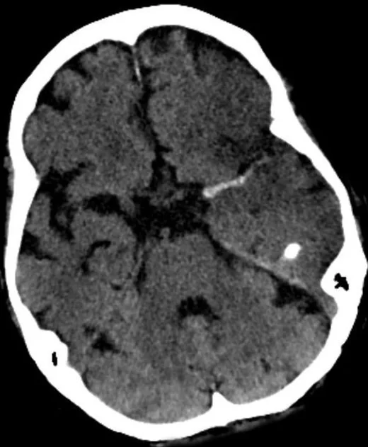
Hiperdens arter işareti, kontrastsız BT'de pıhtıyla tıkanmış bir intrakraniyal damarın normal kan havuzundan daha yoğun görünmesidir. Kontrastsız BT'de proksimal intrakraniyal arterlerde intraluminal pıhtının oluşturduğu hiperdensite oklüzyonu gösterir; kesin tanı ve oklüzyon düzeyinin belirlenmesi için CTA altın standarttır. İşaretin olmaması oklüzyonu dışlamaz, varlığı da kalsifikasyon ve hematokrit etkileri dışlanmadan kesin pıhtı kabul edilmemelidir. Hiperdens arter işareti, CTA'nın yerine geçmez; olguları tek başına elemek amacıyla kullanılmamalıdır.
Faz ve pencere
- Kontrastsız tanısal
- Kontrastsız BT'de intraluminal trombüs, MCA M1 ve baziler arterde yüksek ate nüasyonlu bir yapı olarak hiperdens arter işareti biçiminde belirir ve tanıda kontralateral sağlıklı damarla karşılaştırma esastır. Bu işaret kontrastsız fazda aranır.
- BTA tanısal
- Kontrastlı BT anjiyografi (CTA), büyük damar oklüzyonunu tespit etmek için referans yöntemdir ve oklüzyonun yerini doğrulayarak kontrastsız BT'deki hiperdens alanla anatomik olarak örtüşür.
Önerilen pencereler: İnme (G 40 / M 40), Anjiyo (G 600 / M 150), Beyin (G 80 / M 40).
BT bulguları
- Hiperdens MCA işareti — M1 segmenti Sylvian fissürde karşı taraftaki MCA'den belirgin yoğun ve tübüler görünür.
- MCA'nın Sylvian fissürdeki distal kesitsel görüntülerinde, komşu dokuya göre daha yüksek atenüasyonda nokta şeklinde odak izlenebilir (MCA nokta işareti).
- Prepontin sistern içindeki baziler arterin, komşu yapılarla kıyaslandığında artmış Hounsfield birimi değerleriyle yüksek atenüasyonlu görünmesi oklüzyonu düşündürür.
- Etkilenen bölgedeki damarın Hounsfield birimi (HU) değerinin, kontralateral sağlıklı damarla karşılaştırılarak tek taraflı artışın saptanması, hiperdensitenin özgüllüğünü artırır.
- CTA lümen eşleşmesi — hiperdens görünen segmentteki dolum kesilmesi, işaretin pıhtı ile uyumunu artırır.
- Damarın anatomik seyri — hiperdensite damar yolunu izler; çevre parankimde noktasal veya çizgisel kalsifikasyonla karıştırılmamalıdır.
Normalde
Karşılaştırma amacıyla kontrastsız BT'de normal intrakraniyal arterler homojen ve düşük ate nüasyona sahiptir; hiperdensiteden şüphelenildiğinde her iki hemisferdeki eş düzeydeki damarların yoğunlukları karşılaştırılmalıdır. İlgili anatomik düzeyde etkilenen ve sağlam taraftaki arterlerin Hounsfield birimi değerleri karşılaştırılarak tek taraflı artış saptanmalı ve hiperdensitenin damar seyrini izleyip izlemediği ardışık kesitlerde gözden geçirilmelidir.
Normal BT ile kıyasla
Aynı bölgenin, kaynak veri setinde patoloji içermediği belirtilen bir BT incelemesini kesit kesit kaydırın; organ sınırlarını açarak anatomiyi hasta görüntüsüyle karşılaştırın. Küçük rastlantısal bulgular tümüyle dışlanmamıştır.
Kaynak: TotalSegmentator v2.01 veri seti (Wasserthal ve ark., Radiology: Artificial Intelligence 2023), CC BY 4.0, vaka s0103; organ sınırları veri setinin otomatik segmentasyonundan, birleştirilerek sadeleştirildi. Görüntüler 2,5 mm kesit aralığına yeniden örneklendi.
Ayırıcı tanı
- Kronik vasküler kalsifikasyonlar da hiperdensite oluşturabilir; çift enerjili BT veya sanal kalsiyum kaldırma görüntüleme ile akut trombüsten ayrılabilir.
- Hemokonsantrasyon
- yüksek hematokrit iki taraftaki damarları ve dural sinüsleri yaygın biçimde yoğunlaştırabilir.
- Kısmi hacim etkisi
- kafa tabanı düzeyindeki kalın kesitlerde damar komşuluğundaki yoğun kemik yapının yalancı hiperdensiteye neden olması mümkündür.
- Kontrast kalıntısı veya yakın zamanda kontrast uygulanması
- spontan damar yoğunluğunu taklit edebilir; çekim zamanı ve önceki inceleme sorgulanır.
Tuzaklar
- Farklı çalışmalar ve cihazlarda farklı Hounsfield birimi (HU) eşikleri bildirilmektedir; sabit bir HU değerini tek başına kesin tanı sınırı kabul etmek yanıltıcı olabilir, görsel değerlendirme ve CTA bulgularıyla birlikte yorumlanmalıdır.
- İki taraflı simetrik damar yoğunluğunu pıhtı kabul etme; hemokonsantrasyon, yaş ve teknik parametreler damar atenüasyonunu etkileyebilir.
- Bazal gangliyonlardın kronik kalsifikasyonları, akut iskemi veya trombüs bulgusuyla karıştırılarak yanlış tanıya veya uygun tedavi gecikmesine yol açabilir.
- Hiperdens arter görülmemesi distal veya daha düşük atenüasyonlu trombüsü dışlamaz; damar açıklığı CTA'da değerlendirilir.
Kendini dene
Kontrastsız BT'de sol M1, sağ M1'den belirgin yoğun; CTA'da aynı yerde dolum kesiliyor. En olası açıklama nedir?
Cevabı göster
Akut intraluminal trombüs. Tek taraflı tübüler damar hiperdensitesi ve aynı segmentte CTA dolum kesilmesi akut trombüsü destekler. Kalsifikasyon damar duvarında bulunurken, yüksek hematokrit genellikle bilateral ve simetrik arteriyel yoğunluk artışına neden olur.
Her iki MCA'nın ve dural sinüslerin benzer biçimde yüksek atenüasyonlu görünmesi öncelikle hangi taklit nedenini düşündürür?
Cevabı göster
Hemokonsantrasyon (polisitemi). Yaygın ve simetrik damar-sinüs yoğunluğu, polisitemi veya yüksek hematokrit durumlarında görülebilir; bu durum fokal ve tek taraflı embolik olaylardan farklıdır.
Kaynaklar
Bu sayfadaki 34 cümle ve 2 quiz sorusu aşağıdaki kaynaklardan alıntılarla tek tek karşılaştırıldı; 20 cümle bu karşılaştırmada düzeltildi (2026-10-08).
- Unmet Needs in Acute Ischemic Stroke: Overcoming Recalcitrant Clotspmc.ncbi.nlm.nih.gov
- Risk factors of hemorrhagic transformation in acute ischaemic stroke: A systematic review and meta-analysispmc.ncbi.nlm.nih.gov
- Computed Tomography-Based Thrombus Imaging for the Prediction of Recanalization after Reperfusion Therapy in Strokepmc.ncbi.nlm.nih.gov
- Automated detection of hyperdense artery sign on non-contrast CT for rapid identification of large vessel occlusion: a multicenter validation studypmc.ncbi.nlm.nih.gov
- Segmentation of the Hyperdense Artery Sign on Noncontrast CT in Ischemic Stroke Using Artificial Intelligencepmc.ncbi.nlm.nih.gov
- Computed Tomography-Based Thrombus Imaging for the Prediction of Recanalization after Reperfusion Therapy in Stroke.pubmed.ncbi.nlm.nih.gov
- Sensitivity and specificity of the hyperdense artery sign for arterial obstruction in acute ischemic stroke.pubmed.ncbi.nlm.nih.gov
- Associations between early ischemic signs on non-contrast CT and time since acute ischemic stroke onset: A scoping review.pubmed.ncbi.nlm.nih.gov
- Correlation of imaging and histopathology of thrombi in acute ischemic stroke with etiology and outcome: a systematic review.pubmed.ncbi.nlm.nih.gov
- Risk factors of hemorrhagic transformation in acute ischaemic stroke: A systematic review and meta-analysis.pubmed.ncbi.nlm.nih.gov
- Smartphone teleradiology application is successfully incorporated into a telestroke network environment.pubmed.ncbi.nlm.nih.gov
- Histological composition behind CT-based thrombus density and perviousness in acute ischemic stroke.pubmed.ncbi.nlm.nih.gov
- Hyperdense artery sign, symptomatic infarct swelling and effect of alteplase in acute ischaemic stroke.pubmed.ncbi.nlm.nih.gov
- Development and Validation of a Predictive Model for Spontaneous Hemorrhagic Transformation After Ischemic Stroke.pubmed.ncbi.nlm.nih.gov
- Predictive Value of Computed Tomography Angiography-Determined Occlusion Type in Stent Retriever Thrombectomy.pubmed.ncbi.nlm.nih.gov
- Usefulness of deep learning-assisted identification of hyperdense MCA sign in acute ischemic stroke: comparison with readers' performance.pubmed.ncbi.nlm.nih.gov
- Thrombus Histology as It Relates to Mechanical Thrombectomy: A Meta-Analysis and Systematic Review.pubmed.ncbi.nlm.nih.gov
- Dual-Energy Computed Tomography Virtual Noncalcium Imaging of Intracranial Arteries in Acute Ischemic Stroke: Differentiation Between Acute Thrombus and Calcification.pubmed.ncbi.nlm.nih.gov
- Predictors for Cerebral Edema in Acute Ischemic Stroke Treated With Intravenous Thrombolysis.pubmed.ncbi.nlm.nih.gov
- 'Susceptibility sign' on susceptibility-weighted imaging in acute ischemic stroke.pubmed.ncbi.nlm.nih.gov
- Noncontrast CT-based habitat analysis for differentiating hemorrhagic transformation from contrast extravasation after endovascular therapy of acute ischemic stroke.pubmed.ncbi.nlm.nih.gov
- Effects of Computed Tomography Hyperdense Middle Cerebral Artery Sign on the Efficacy of Endovascular Therapy in Acute Ischemic Stroke With Large Infarcts: A Subgroup Analysis of the ANGEL-ASPECT Trial.pubmed.ncbi.nlm.nih.gov
- Middle cerebral artery occlusion due to paradoxical embolism diagnosed by pulmonary artery thrombosis in the absence of deep vein thrombosis: A case report.pubmed.ncbi.nlm.nih.gov
- Using Multimodal Imaging to Correlate Link Between Intracranial Aneurysms and Acute Ischemic Stroke.pubmed.ncbi.nlm.nih.gov
- Stroke or Seizure? Diagnostic Role of Neuroimaging in Acute Neurologic Mimics.pubmed.ncbi.nlm.nih.gov
- Current state and advancements of imaging in acute ischemic stroke: a practical review.pubmed.ncbi.nlm.nih.gov
- Segmentation of the Hyperdense Artery Sign on Noncontrast CT in Ischemic Stroke Using Artificial Intelligence.pubmed.ncbi.nlm.nih.gov
- Diagnostic Accuracy of Hyperdense Artery Sign in Early Detection of Middle Cerebral Artery Infarction: A Cross-Sectional Validation Study.pubmed.ncbi.nlm.nih.gov
- MDCT Imaging of Non-Traumatic Thoracic Aortic Emergencies and Its Impact on Diagnosis and Management-A Reappraisal.pubmed.ncbi.nlm.nih.gov
- The Importance of Predicting Bowel Necrosis in Acute Mesenteric Ischemia: Narrative Review.pubmed.ncbi.nlm.nih.gov
- Delayed diagnosis of basilar artery occlusion in a 28-year-old postpartum woman.pubmed.ncbi.nlm.nih.gov
- True Spontaneous Splenic Rupture Mimicking Peptic Ulcer Disease: A Case Report.pubmed.ncbi.nlm.nih.gov
- Early Detection of Hyperdense Basilar Artery Signs Through Comparison With Previous Images.pubmed.ncbi.nlm.nih.gov
- Insights into Angiographic Recanalization of Large Arterial Occlusion: Institutional Experience with Mechanical Thrombectomy for Acute Ischemic Stroke.pubmed.ncbi.nlm.nih.gov
- Imaging in Acute Anterior Circulation Ischemic Stroke: Current and Future.pubmed.ncbi.nlm.nih.gov
- Efficacy of Thrombus Density on Noninvasive Computed Tomography Neuroimaging for Predicting Thrombus Pathology and Patient Outcome after Mechanical Thrombectomy in Acute Ischemic Stroke.pubmed.ncbi.nlm.nih.gov
- Mechanical Thrombectomy for Large-Vessel Occlusion after Fontan Procedure in an Adult.pubmed.ncbi.nlm.nih.gov
- Automated detection of hyperdense artery sign on non-contrast CT for rapid identification of large vessel occlusion: a multicenter validation study.pubmed.ncbi.nlm.nih.gov
- Endovascular Treatment Strategies for Acute Intracranial Atherosclerotic Stenosis-Related Large Vessel Occlusion.pubmed.ncbi.nlm.nih.gov
- Reversible cerebral vasoconstriction syndrome: A narrative review.pubmed.ncbi.nlm.nih.gov
- The Quantitative Evaluation of the Density of the Segmental Branches of the MCA in Acute Ischemic Stroke Patients.pubmed.ncbi.nlm.nih.gov
- A multi-center study of the MicroNET-covered stent in consecutive patients with acute carotid-related stroke: SAFEGUARD-STROKE.pubmed.ncbi.nlm.nih.gov
- Predictors of Mortality in Posterior Circulation Ischemic Stroke.pubmed.ncbi.nlm.nih.gov
- Computed tomography perfusion-defined ischemic core predicts functional outcome after basilar artery thrombectomy.pubmed.ncbi.nlm.nih.gov
- Intracranial venous thrombosis imaging by anatomical location: an educational review with mimics and diagnostic pitfalls.pubmed.ncbi.nlm.nih.gov
- Hyperintense acute reperfusion marker (HARM) and thrombus analysis in acute ischemic stroke.pubmed.ncbi.nlm.nih.gov
- Association of the C-reactive protein-albumin-lymphocyte index with clinical outcomes after mechanical thrombectomy for acute ischemic stroke.pubmed.ncbi.nlm.nih.gov
- Integrated Radiology-Biochemistry Diagnostic Flow Framework for Emergency Clinical Decision Support: A Simulation-Based Educational Model.pubmed.ncbi.nlm.nih.gov
- Splenic pathologies: A pictorial review of common, wandering, twisting and rare presentations.pubmed.ncbi.nlm.nih.gov
- When Cancer Clots: An Extensive Radiologic Analysis of Cancer-Associated Thromboembolism.pubmed.ncbi.nlm.nih.gov
- Endovascular treatment for isolated cervical internal carotid artery occlusion: ETIICA study.pubmed.ncbi.nlm.nih.gov
- Utility of Non-Contrast CT and MRA Fusion Images for Depicting Occluded Vessels in Mechanical Thrombectomy.pubmed.ncbi.nlm.nih.gov
- Neutrophil extracellular traps in retrieved thrombi and functional outcome after stroke thrombectomy.pubmed.ncbi.nlm.nih.gov
- Analysis of 90-day risk factors for poor prognosis in patients with postoperative CT with hyperdense signs after thrombectomy for acute ischemic stroke.pubmed.ncbi.nlm.nih.gov
- Effect Modification of Rescue Stenting in ICAS-Related Middle Cerebral Artery Occlusion: The Role of Thrombectomy Failure, Stenosis Severity, and ASPECTS.pubmed.ncbi.nlm.nih.gov
- Artificial Intelligence in Acute Neuroimaging Pathways: Diagnostic Accuracy, Workflow Performance, and Clinical Outcomes Across CT and MRI in Stroke and Trauma.pubmed.ncbi.nlm.nih.gov
- Imaging features of lacrimal gland disease.pubmed.ncbi.nlm.nih.gov
- Application of postmortem imaging modalities in cases of sudden death due to cardiovascular diseases-current achievements and limitations from a pathology perspective : Endorsed by the Association for European Cardiovascular Pathology and by the International Society of Forensic Radiology and Imaging.pubmed.ncbi.nlm.nih.gov
- Artificial intelligence in computed tomography imaging for pulmonary embolism: a narrative review from computed tomography pulmonary angiography to non-contrast computed tomography.pubmed.ncbi.nlm.nih.gov
- Spontaneous Rectus Sheath Hematoma Presenting As Acute Abdomen in an Anticoagulated Patient: A Case Report.pubmed.ncbi.nlm.nih.gov
- Postpartum ovarian vein thrombosis: plain CT with clinical indicators-diagnostic performance and clinical application.pubmed.ncbi.nlm.nih.gov
- Diagnostic Value and Operational Recommendations for Late Iodine Enhancement and ECV Quantification in Single-Energy Computed Tomography: A Narrative Review.pubmed.ncbi.nlm.nih.gov
- A Forgotten Rare Cause of Unilateral Basal Ganglia Calcinosis Due to Venous Angioma and Complicating Acute Stroke Management: A Case Report.pubmed.ncbi.nlm.nih.gov
- Pediatric Meningeal Diseases: What Radiologists Need to Know.pubmed.ncbi.nlm.nih.gov
- Invasive Mediastinal Aspergillosis in an 18-Year-Old Immunocompetent Female Leading to Stroke.pubmed.ncbi.nlm.nih.gov
- Computed tomography of renal emergencies: A comprehensive diagnostic guide for radiology residents.pubmed.ncbi.nlm.nih.gov
- An Atlas of Neonatal Neurovascular Imaging Anatomy as Depicted with Microvascular Imaging: The Intracranial Arteries.pubmed.ncbi.nlm.nih.gov
- Leptomeninges: Anatomy, Mechanisms of Disease and Neuroimaging.pubmed.ncbi.nlm.nih.gov
- The ABCs of Subarachnoid Hemorrhage Blood Volume Measurement: A Simplified Quantitative Method Predicts Outcomes and Delayed Cerebral Ischemia.pubmed.ncbi.nlm.nih.gov
- Prognostic value of the hyperdense middle cerebral artery sign in patients undergoing mechanical thrombectomy.pubmed.ncbi.nlm.nih.gov
- Neuroimaging of hemorrhage and vascular defects.pubmed.ncbi.nlm.nih.gov
- Neuroimaging for neurovascular complications of traumatic brain injury.pubmed.ncbi.nlm.nih.gov
- Management of spontaneous abdominal wall hematomas: a narrative review with a focus on CTA-negative endovascular cases.pubmed.ncbi.nlm.nih.gov
- Middle cerebral artery dissection: pathophysiology, diagnosis, and therapeutic options.pubmed.ncbi.nlm.nih.gov
- Update on cerebrovascular manifestations of COVID-19.pubmed.ncbi.nlm.nih.gov
- Pediatric Stroke Imaging.pubmed.ncbi.nlm.nih.gov
- Imaging differentiation of solid pseudopapillary neoplasms and neuroendocrine neoplasms of the pancreas.pubmed.ncbi.nlm.nih.gov
- Infarcts and ischemia in the abdomen: an imaging perspective with an emphasis on cross-sectional imaging findings.pubmed.ncbi.nlm.nih.gov
- Signs in Neuroradiology: A Pictorial Review.pubmed.ncbi.nlm.nih.gov
- Spreading depolarization and angiographic spasm are separate mediators of delayed infarcts.pubmed.ncbi.nlm.nih.gov
- The risk and outcome of malignant brain edema in post-mechanical thrombectomy: acute ischemic stroke by anterior circulation occlusion.pubmed.ncbi.nlm.nih.gov
- Retrieval of an atherosclerotic plaque-vessel wall complex during mechanical thrombectomy: illustrative case.pubmed.ncbi.nlm.nih.gov
- Multicenter Validation of Artificial Intelligence Predicting Anterior Circulation Large Vessel Occlusion Using Noncontrast Head CT.pubmed.ncbi.nlm.nih.gov
- Associations of nine composite inflammatory indices with clinical outcomes after mechanical thrombectomy for acute ischemic stroke: a single-center retrospective study.pubmed.ncbi.nlm.nih.gov
- Association between hyperglycemia and unfavorable outcome in patients with successful thrombectomy for acute ischemic stroke: a single-center study.pubmed.ncbi.nlm.nih.gov
- Neurological complications following cerebral angiography: a case of contrast-induced encephalopathy.pubmed.ncbi.nlm.nih.gov
- Multimodal Imaging Reveals Labbé Vein Thrombosis Mimicking Subdural Hematoma: A Diagnostic Pitfall in Emergency Neuroimaging.pubmed.ncbi.nlm.nih.gov
- Brain computerized tomography reading in suspected acute ischemic stroke patients: what are essentials for medical students?pubmed.ncbi.nlm.nih.gov
- Polycythemia, a radiologic mimic in plain CT head: A case report.pubmed.ncbi.nlm.nih.gov
- Modified Alberta Stroke Program Early CT Score (ASPECTS) of Contrast Extravasation on Dual-Energy CT Predicts Haemorrhagic Transformation and Poor Outcome After Endovascular Thrombectomy.pubmed.ncbi.nlm.nih.gov
- Net water uptake and ASPECTS in predicting futile recanalization for acute large vessel occlusion stroke: insights from time window stratification.pubmed.ncbi.nlm.nih.gov
- Analysis of the baseline multiphase computed tomographic angiography findings to predict clinical outcomes in patients with middle cerebral artery M1 occlusion treated with mechanical thrombectomy.pubmed.ncbi.nlm.nih.gov
- Multiple Saccular Aneurysms of the Posterior Cerebral Artery With Mass Effect: A Case Report.pubmed.ncbi.nlm.nih.gov
- Influence of deep learning image reconstruction and adaptive statistical iterative reconstruction-V on automated Alberta stroke program early CT score- evaluation.pubmed.ncbi.nlm.nih.gov
- Attenuation measurements on non-contrast-enhanced CT brain: a re-visit of their role in the diagnosis of cerebral venous thrombosis.pubmed.ncbi.nlm.nih.gov
- Vertebrobasilar dolichoectasia and deep sylvian territory hemorrhage: Any causality? One case report of young man.pubmed.ncbi.nlm.nih.gov
- Thrombus migration in intracranial large vessel occlusion: course, predictors, and impact on endovascular thrombectomy.pubmed.ncbi.nlm.nih.gov
- The diagnostic efficacy of unenhanced CT for detecting acute central pulmonary embolism.pubmed.ncbi.nlm.nih.gov
- Natural language processing for triage of cerebral large-vessel occlusion.pubmed.ncbi.nlm.nih.gov
- Usefulness of maximum intensity projection images of non-enhanced CT for detection of hyperdense middle cerebral artery sign in acute thromboembolic ischemic stroke.pubmed.ncbi.nlm.nih.gov
- Bilateral hyperdense middle cerebral arteries: Stroke sign or not?pubmed.ncbi.nlm.nih.gov
- Distal Medium Vessel Occlusion Strokes: Understanding the Present and Paving the Way for a Better Future.pubmed.ncbi.nlm.nih.gov
- The middle cerebral artery (MCA) dot sign.pubmed.ncbi.nlm.nih.gov
- Mechanical thrombectomy for tandem internal carotid artery-middle cerebral artery occlusion in a patient with a persistent hypoglossal artery: illustrative case.pubmed.ncbi.nlm.nih.gov
- Selecting the Appropriate First-Line Strategy Based on Hyperdense Vessel Sign in Acute Ischemic Stroke Increases First Pass Recanalization: A Tertiary Center Experience.pubmed.ncbi.nlm.nih.gov
- Clot composition in retrieved thrombi after mechanical thrombectomy in strokes due to carotid web.pubmed.ncbi.nlm.nih.gov
- Dual-Energy Heart CT: Beyond Better Angiography-Review.pubmed.ncbi.nlm.nih.gov
- A wide-detector CT protocol for one-stop cardio-cerebrovascular angiography: a technical feasibility study comparing an integrated protocol with conventional two-scan approach.pubmed.ncbi.nlm.nih.gov
- Perforator-Sparing Microsurgical Clipping of Tandem Dominant-Hemisphere Middle Cerebral Artery Aneurysms: Geometry-Guided Reconstruction of a Wide-Neck Bifurcation and Dorsal M1 Fusiform Lesion.pubmed.ncbi.nlm.nih.gov
- Predictive value of thrombus enhancement sign for stroke subtype and recanalization in acute basilar-artery occlusion.pubmed.ncbi.nlm.nih.gov
- Ischemic Stroke at a Tertiary Academic Hospital in Tanzania: A Prospective Cohort Study With a Focus on Presumed Large Vessel Occlusion.pubmed.ncbi.nlm.nih.gov
- A Case-Based Review of Cerebral Venous Infarcts With Perfusion Imaging and Comparison to Arterial Ischemic Stroke.pubmed.ncbi.nlm.nih.gov
- Spontaneous calcified cerebral emboli: a comprehensive review and proposed diagnostic criteria.pubmed.ncbi.nlm.nih.gov
- Imaging diagnosis of intracranial atherosclerosis stenosis-related large vessel occlusion before and during endovascular therapy.pubmed.ncbi.nlm.nih.gov
- Patchy profile sign in RAPID software: a specific marker for intracranial atherosclerotic stenosis in acute ischemic stroke.pubmed.ncbi.nlm.nih.gov
Eğitim amaçlıdır; hasta başında tanı ve tedavi kararının yerine geçmez. İçerik yapay zekâ destekli bir süreçle güncel literatüre göre cümle cümle kaynaklarla karşılaştırıldı, ancak uzman radyolog incelemesinden geçmedi. Klinik kararlarda güncel kılavuzları, kurum protokollerini ve radyoloji raporunu esas alın.