Özet
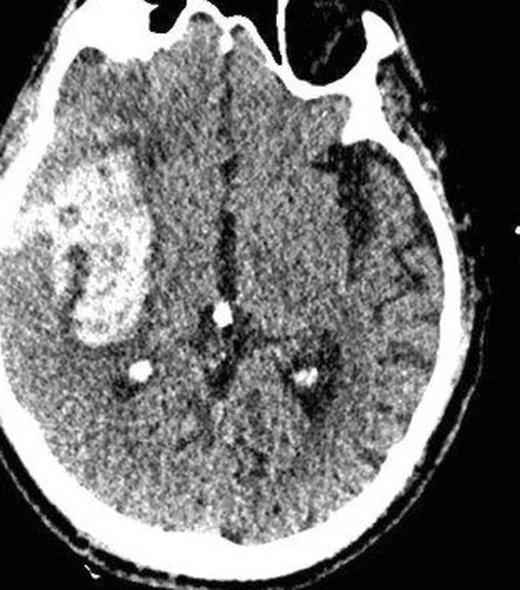
İntraparenkimal hematom, kanın beyin dokusu içinde birikmesidir; ani nörolojik defisit veya bilinç bozukluğunda hızlı tanı ve yayılım değerlendirmesi kritik önem taşır. Kontrastsız BT, akut hematomu ve buna bağlı kitle etkisini değerlendirmede birincil incelemedir; ventriküle uzanım ve hidrosefali de bu seride izlenebilir. Seçilmiş hastada BTA aktif kontrast ekstravazasyonunu ve altta yatan makrovasküler nedeni araştırmaya yardımcı olur; derin, lobar, infratentoryal veya travmatik yerleşim etiyoloji yorumunu değiştirir. Hematom hacmi, beyin sapı/serebellum basısı ya da ventrikül tıkanmasının atlanması hızlı kötüleşmenin ve cerrahi açıdan önemli anatomik etkilerin gözden kaçmasına neden olabilir.
Faz ve pencere
- Kontrastsız tanısal
- Akut pıhtı BT'de tipik olarak hiperdens odak oluşturur; zaman, anemi, pıhtılaşma durumu ve hematom içi serum nedeniyle yoğunluk değişkenlik gösterebilir. Çevresel düşük atenüasyonlu ödem, ventrikül içine açılım, sistern basısı ve orta hat kayması aynı seride incelenir.
- BTA tanısal
- Seçilmiş olguda hematom içinde damar lümenine bağlı olmayan kontrast yoğun odağı (spot sign/ekstravazasyon) aktif kanama ve hematom genişlemesi olasılığını düşündürür. Bu inceleme akut kanamanın saptanması için zorunlu değildir ve tüm intraparenkimal hematomlarda rutin tanısal faz olarak yorumlanmamalıdır.
Önerilen pencereler: Beyin (G 80 / M 40), Subdural (G 200 / M 75), Kemik (G 1800 / M 400), Anjiyo (G 600 / M 150).
BT bulguları
- İntraparenkimal hiperdens odak — bazal gangliyon, talamus, lobar kortiko-subkortikal alan, beyin sapı veya serebellum içinde pıhtı.
- Heterojen hematom — pıhtı içinde farklı atenüasyonlu alanlar; heterojenlik veya düşük yoğunluklu odaklar hematom genişlemesi açısından dikkate alınmalıdır.
- Çevresel ödem — hematom çevresinde parankimden daha düşük dansiteli, sınırları silikleştiren alan.
- Ventriküle açılım — hematomdan lateral, üçüncü veya dördüncü ventriküle uzanan hiperdens kan.
- Hidrosefali — ventrikül içi kanla ilişkili ventriküler genişleme ve transependimal BOS geçişi.
- Kitle etkisi — komşu sulkusların silinmesi, ventrikül deformasyonu, bazal sisternlerin basısı veya orta hat yer değiştirmesi.
- Yerleşim örüntüsü — derin gangliyon/talamik, lobar, infratentoryal ya da travmayla ilişkili dağılım olası nedeni yönlendirir.
- BTA spot sign — hematom içinde kontrastlanan küçük odaklar aktif ekstravazasyon lehine olabilir; damar devamlılığı gösteren yapıdan ayrılmalıdır.
Normalde
Normal karşılaştırmada beyin penceresi, parankimi ve ince dansite değişikliklerini değerlendirmeyi sağlar. Karşı hemisferdeki eş anatomik bölgeyle kıyaslayarak parankim yoğunluğunu, ventrikül biçimini ve orta hat konumunu inceleyin; bazal sisternlerin açık olup olmadığı da kitle etkisini gösterir.
Normal BT ile kıyasla
Aynı bölgenin, kaynak veri setinde patoloji içermediği belirtilen bir BT incelemesini kesit kesit kaydırın; organ sınırlarını açarak anatomiyi hasta görüntüsüyle karşılaştırın. Küçük rastlantısal bulgular tümüyle dışlanmamıştır.
Kaynak: TotalSegmentator v2.01 veri seti (Wasserthal ve ark., Radiology: Artificial Intelligence 2023), CC BY 4.0, vaka s0103; organ sınırları veri setinin otomatik segmentasyonundan, birleştirilerek sadeleştirildi. Görüntüler 2,5 mm kesit aralığına yeniden örneklendi.
Ayırıcı tanı
- Hemorajik tümör
- kanama çevresinde solid bileşen veya nodüler yapı düşünülmelidir; kanama nedeniyle altta yatan lezyonun görüntülenmesi zorlaşabilir.
- Hemorajik kontüzyon
- travma sonrası yüzeyel, sıklıkla inferior frontal veya temporal kutup yerleşimli düzensiz parankimal kan odakları.
- Kavernom kanaması
- akut BT'de kalsifikasyon veya kanama olarak görünüp ayırıcı tanıda yetersiz kalabilir; MR'da SWI/T2* dizilerde izlenen 'mulberry' çekirdek ve hemosiderin çeperi ayırt edicidir.
- Kalsifikasyon
- yoğunluğu yüksek olsa da kronik ve keskin sınırlıdır; önceki incelemeler, kemik penceresi ve anatomik dağılım yardımcıdır.
Tuzaklar
- Hiperdens odağın tamamını taze kan varsaymayın; kalsifikasyon, kontrast tutulumu ve damar kesiti kemik penceresi veya önceki BT ile ayırt edilebilir.
- Hematoma çevresindeki hipodens alanları otomatik olarak ödem saymayın; kaynağı belirlemek için Hounsfield birimi eşiklerini kullanın (örneğin <40–55 HU ödem lehine, >80 HU kalsifikasyon/artefakt lehine).
- Sadece en büyük aksiyel kesiti incelemek infratentoryal uzanımı veya ventrikül içi kanı kaçırabilir; tüm seri boyunca hematomun sınırını takip edin.
- Tek fazlı BTA spot sign'ı kaçırabilir; çok fazlı BTA ile geç ortaya çıkan kontrast ekstravazasyonu tespit edilerek hematom genişleme riski daha doğru değerlendirilir.
Kendini dene
Hipertansiyon öykülü hastada putamen içinde hiperdens hematom, çevresel ödem ve ipsilateral ventrikül basısı var. Kanama hangi kompartımandadır?
Cevabı göster
Beyin parankimi. Putamen içindeki odak parankimal hematomdur; çevresel ödem ve ventrikül basısı kitle etkisini gösterir. Subaraknoid kan sulkus/sisternleri doldurur; subdural ve epidural hematomlar beyin dokusu dışındadır.
Lobar hematomun içinde BTA'da damar lümenine bağlı olmayan kontrastlı küçük odak görülüyor. Bu bulgu en çok neyi düşündürür?
Cevabı göster
Aktif kontrast ekstravazasyonu. Hematoma gömülü kontrast odağı spot sign olarak adlandırılır ve aktif ekstravazasyon olasılığıyla ilişkilidir. Sinüs trombozu venöz damarda dolum kusurudur; kalsifikasyon kontrast fazında yeni odak değildir; infarkt parankim dağılımıyla tanınır.
İlgili konular
Kaynaklar
Bu sayfadaki 39 cümle ve 2 quiz sorusu aşağıdaki kaynaklardan alıntılarla tek tek karşılaştırıldı; 10 cümle bu karşılaştırmada düzeltildi (2026-10-08).
- An updated review for clinical and radiological predictors of acute intraparenchymal hematoma progression in cerebral contusionpmc.ncbi.nlm.nih.gov
- Preoperative brain shift is a prognostic factor for survival in certain neurosurgical diseases other than severe head injury: a case series and literature reviewpmc.ncbi.nlm.nih.gov
- Surgery for spontaneous intracerebral hemorrhagepmc.ncbi.nlm.nih.gov
- Primary cerebral malignant melanoma in insular region with extracranial metastasis: case report and review literaturepmc.ncbi.nlm.nih.gov
- A multi-task deep learning pipeline for classification, detection, and weakly supervised 3D segmentation of intraparenchymal hematoma on brain CTpmc.ncbi.nlm.nih.gov
- Vaccine-induced immune thrombotic thrombocytopenia after ChAdOx1 nCoV-19 vaccine in an older patient: Minireview and a case report.pubmed.ncbi.nlm.nih.gov
- Hemorrhagic giant cerebral capillary telangiectasia resulting in death: Case report and literature review.pubmed.ncbi.nlm.nih.gov
- Primary cerebral malignant melanoma in insular region with extracranial metastasis: case report and review literature.pubmed.ncbi.nlm.nih.gov
- An uncommon clinical presentation of primary pancreatic lymphoma: Bleeding. Case report and literature review.pubmed.ncbi.nlm.nih.gov
- Traumatic Pseudoaneurysm of Middle Meningeal Artery with Delayed Presentation as Intracerebral Hematoma: A Report with Review of Literature.pubmed.ncbi.nlm.nih.gov
- Ethical and medicolegal aspects in the management of neurosurgical emergencies among Jehovah's Witnesses: Clinical implications and review.pubmed.ncbi.nlm.nih.gov
- Acute subdural hematoma without subarachnoid hemorrhage or intraparenchymal hematoma caused by rupture of a posterior communicating artery aneurysm: case report and review of the literature.pubmed.ncbi.nlm.nih.gov
- Multicenter assessment of the Brain Injury Guidelines and a proposal of guideline modifications.pubmed.ncbi.nlm.nih.gov
- Management of Minor Traumatic Brain Injury in an ED Observation Unit.pubmed.ncbi.nlm.nih.gov
- Reversible Cerebral Vasoconstriction Syndrome: A Severe Neurological Complication in Postpartum Period.pubmed.ncbi.nlm.nih.gov
- Focal Neurological Deficit at Onset of Aneurysmal Subarachnoid Hemorrhage: Frequency and Causes.pubmed.ncbi.nlm.nih.gov
- Feasibility and validity of monitoring subarachnoid hemorrhage by a noninvasive MRI imaging perfusion technique: Pulsed Arterial Spin Labeling (PASL).pubmed.ncbi.nlm.nih.gov
- Blood-Brain Barrier Disruption Predicts Poor Outcome in Subarachnoid Hemorrhage: A Dynamic Contrast-Enhanced MRI Study.pubmed.ncbi.nlm.nih.gov
- Novel method to evaluate the risk of tumor adhesions and post-operative hemorrhage of meningiomas using 320 row CT-DSA: A clinical research study.pubmed.ncbi.nlm.nih.gov
- Progressively Enlarging Cerebellar Hematoma Concurrent with T-DM1 Treatment.pubmed.ncbi.nlm.nih.gov
- Age-related metabolic vulnerability of glia in intracerebral hemorrhage: Mechanisms and therapeutic opportunities.pubmed.ncbi.nlm.nih.gov
- Rapid spontaneous regression of a massive post-thrombolysis intracerebral hemorrhage: A case report.pubmed.ncbi.nlm.nih.gov
- The Anticoagulation Dilemma: Concurrent Traumatic Brain Hemorrhage and Cerebral Venous Thrombosis in a Blast Injury Patient.pubmed.ncbi.nlm.nih.gov
- Before the bolus: why anti-amyloid therapy must become part of every stroke code.pubmed.ncbi.nlm.nih.gov
- Multifocal intracranial hemorrhage on emergency brain CT in a child with end-stage nephrotic syndrome.pubmed.ncbi.nlm.nih.gov
- Explainable Artificial Intelligence in Non-Contrast Brain Computed Tomography Scan for Intracerebral Hemorrhage: A Scoping Review.pubmed.ncbi.nlm.nih.gov
- Imaging studies for predicting hematoma expansion: from traditional imaging signs to artificial intelligence-based multimodal fusion.pubmed.ncbi.nlm.nih.gov
- The "brush sign": a novel and accessible CT marker for hematoma expansion in hypertensive intracerebral hemorrhage.pubmed.ncbi.nlm.nih.gov
- A Case of Acute Massive Spinal Subdural Hematoma With a Spot Sign on CT.pubmed.ncbi.nlm.nih.gov
- Surgical intervention versus conservative care in severe hypertensive pontine hemorrhage: a retrospective analysis of clinical outcomes.pubmed.ncbi.nlm.nih.gov
- Comparative effectiveness of neuroendoscopic surgery and stereotactic aspiration for brain hemorrhage.pubmed.ncbi.nlm.nih.gov
- Early secondary brain abscess formation caused by <i>Bacteroides fragilis</i> at the intracerebral hemorrhage site: A case report.pubmed.ncbi.nlm.nih.gov
- An agentic AI framework for integrated decision support and surgical planning in intracerebral hemorrhage.pubmed.ncbi.nlm.nih.gov
- Autoregulation-Guided Blood Pressure Targets After Stroke Thrombectomy: Impact on Secondary Brain Injury and Neurologic Outcomes.pubmed.ncbi.nlm.nih.gov
- Utility of the Spot Sign on Multiphase CT Angiography for Prediction of Hematoma Expansion in Intracerebral Hemorrhage.pubmed.ncbi.nlm.nih.gov
- Hematoma expansion in intracerebral hemorrhage: Development of a preoperative risk prediction model.pubmed.ncbi.nlm.nih.gov
- Reporting Clinically Actionable Imaging Features of Intracerebral Hemorrhage: A Survey of Neuroradiologist Practice.pubmed.ncbi.nlm.nih.gov
- Multimodal Monitoring in Eclamptic Intracerebral Hemorrhage Complicated by Reversible Cerebral Vasoconstriction Syndrome (RCVS): A Case Report.pubmed.ncbi.nlm.nih.gov
- Aneurysmal subarachnoid hemorrhage: predictive score for angiographic vasospasm.pubmed.ncbi.nlm.nih.gov
- DWI-Positive Lesions in Patients With Acute Intracerebral Hemorrhage: Associations With Cerebral Small Vessel Disease and Clinical Outcome.pubmed.ncbi.nlm.nih.gov
- Cognition in Cardiovascular Disease.pubmed.ncbi.nlm.nih.gov
- Stroke caused by cerebral amyloid angiopathy: a case of difficult diagnosis.pubmed.ncbi.nlm.nih.gov
- Artificial Intelligence in Intracerebral Hemorrhage: Current Applications and Future Perspectives.pubmed.ncbi.nlm.nih.gov
- Advances in Stereotactic Hematoma Evacuation for Hypertensive Intracerebral Hemorrhage and the Value of Multimodal Monitoring.pubmed.ncbi.nlm.nih.gov
- Multiscale photoacoustic imaging of stroke in preclinical models and future directions toward clinical translation [Invited].pubmed.ncbi.nlm.nih.gov
- Intracerebral Hemorrhage in a Child with Renal Artery Stenosis and COVID-19.pubmed.ncbi.nlm.nih.gov
- Persistent refractory hypercapnia following tracheostomy decannulation in a patient with brainstem hemorrhage: a case report.pubmed.ncbi.nlm.nih.gov
- Cold-Induced Reversible Cerebral Vasoconstriction Syndrome Presenting With Intraventricular Hemorrhage in a Normotensive Patient.pubmed.ncbi.nlm.nih.gov
- Risk factor analysis of long-term outcomes in patients with large intracerebral and intraventricular hemorrhage treated primarily with combined external ventricular drainage and intraventricular thrombolysis.pubmed.ncbi.nlm.nih.gov
- Glucagon-like peptide-1 receptor agonist in large vessel occlusion treated by reperfusion therapy-a phase 2 randomized trial.pubmed.ncbi.nlm.nih.gov
- Establishment and validation of prognosis model for patients with cerebral contusion.pubmed.ncbi.nlm.nih.gov
- Neurological manifestations and neuroimaging findings in patients with SARS-CoV2-a systematic review.pubmed.ncbi.nlm.nih.gov
- Spot sign as a predictor of hematoma expansion and poor functional outcomes after intracerebral hemorrhage: a systematic review and meta-analysis.pubmed.ncbi.nlm.nih.gov
- A systematic approach to MR spectroscopy of the brain: Indications, protocol, metabolites, and spectral analysis in neurologic diseases.pubmed.ncbi.nlm.nih.gov
- Correlation of Gray-to-White matter ratio on Non-Contrast CT with neurological outcome in Post-Cardiac arrest syndrome.pubmed.ncbi.nlm.nih.gov
- Early identification of vascular cognitive impairment from a multimodal perspective: a combined diagnosis from targeted cognitive assessments, imaging biomarkers, and molecular fluid biomarkers to ecological behavioral characteristics.pubmed.ncbi.nlm.nih.gov
- Roles and therapeutic prospects of the laminin family in disorders of the nervous system.pubmed.ncbi.nlm.nih.gov
- Role of miR-181 Family Members in Stroke: Insights into Mechanisms and Therapeutic Potential.pubmed.ncbi.nlm.nih.gov
- Astrocytic LMP2 Coordinates NF-κB and TGF-β1/Smad3 Signaling to Drive Neuroinflammation after Cerebral Ischemia/Reperfusion.pubmed.ncbi.nlm.nih.gov
- Diagnostic challenges of a giant cerebral cavernous malformation mimicking a glioma in a 16-year-old male.pubmed.ncbi.nlm.nih.gov
- Controversies in Cavernous Malformation Management: A Comprehensive Review of Current Literature.pubmed.ncbi.nlm.nih.gov
- Low-flow vascular malformations without arteriovenous shunting of the central nervous system: a pictorial review.pubmed.ncbi.nlm.nih.gov
- Diagnostic neuroimaging in neuro-ophthalmic disorders.pubmed.ncbi.nlm.nih.gov
- Tumor-like Lesions in Primary Angiitis of the Central Nervous System: The Role of Magnetic Resonance Imaging in Differential Diagnosis.pubmed.ncbi.nlm.nih.gov
- Low-field MRI: Clinical promise and challenges.pubmed.ncbi.nlm.nih.gov
- Automated Detection and Screening of Traumatic Brain Injury (TBI) Using Computed Tomography Images: A Comprehensive Review and Future Perspectives.pubmed.ncbi.nlm.nih.gov
- Computed tomography angiography or magnetic resonance angiography for detection of intracranial vascular malformations in patients with intracerebral haemorrhage.pubmed.ncbi.nlm.nih.gov
- 2025 Heart Disease and Stroke Statistics: A Report of US and Global Data From the American Heart Association.pubmed.ncbi.nlm.nih.gov
- 2024 Heart Disease and Stroke Statistics: A Report of US and Global Data From the American Heart Association.pubmed.ncbi.nlm.nih.gov
- Heart Disease and Stroke Statistics-2023 Update: A Report From the American Heart Association.pubmed.ncbi.nlm.nih.gov
- Clinical Utility of Circulating Biomarkers for Diagnosis and Prognosis of Acute Stroke in the Emergency Department: A Narrative Review from Current Evidence to Clinical Implementation.pubmed.ncbi.nlm.nih.gov
- Role of Low-Field MRI in Acute Stroke.pubmed.ncbi.nlm.nih.gov
- Developmental venous anomalies and cerebral cavernous malformations: Partners in crime.pubmed.ncbi.nlm.nih.gov
- Fulminant Amyloid-β-Related Angiitis after Tenecteplase: A Biopsy-Confirmed Cause of Post-Thrombolysis Neurologic Deterioration.pubmed.ncbi.nlm.nih.gov
- CT Imaging Patterns and Clinical Outcomes of Traumatic Brain Injury: Distribution Across Injury Mechanisms.pubmed.ncbi.nlm.nih.gov
- Deep-learning reconstruction for noncontrast head computed tomography: Improved image quality and potential diagnostic impact in acute ischemic stroke.pubmed.ncbi.nlm.nih.gov
- Is gray-white matter ratio in out-of-hospital cardiac arrest patients' really early predictor of neurological outcome?pubmed.ncbi.nlm.nih.gov
- Photon-Counting CT: Technology, Current and Potential Future Clinical Applications, and Overview of Approved Systems and Those in Various Stages of Research and Development.pubmed.ncbi.nlm.nih.gov
- Cerebral fat embolism syndrome in sickle cell disease: a case highlighting the predominance of susceptibility-weighted imaging findings and literature review.pubmed.ncbi.nlm.nih.gov
- Posterior reversible encephalopathy syndrome in dengue virus infection with concurrent encephalitis and ischemic stroke: a case report.pubmed.ncbi.nlm.nih.gov
- Intracranial venous thrombosis imaging by anatomical location: an educational review with mimics and diagnostic pitfalls.pubmed.ncbi.nlm.nih.gov
- Intracranial ependymoma and secondary hydrocephalus: a rare case report in a Camel (<i>Camelus bactrianus</i>).pubmed.ncbi.nlm.nih.gov
- Diagnostic imaging and emerging image-guided neurointervention in brain tumors: a narrative review.pubmed.ncbi.nlm.nih.gov
- The critical care management of spontaneous intracranial hemorrhage: a contemporary review.pubmed.ncbi.nlm.nih.gov
- Ischemic stroke and cerebral venous sinus thrombosis in pregnancy.pubmed.ncbi.nlm.nih.gov
- Progressive multifocal leukoencephalopathy in an untreated chronic lymphocytic leukemia patient: Case report and review of the literature.pubmed.ncbi.nlm.nih.gov
- Posterior Reversible Encephalopathy Syndrome Following Massive Blood Transfusion in a Case of Pelvic Schwannoma.pubmed.ncbi.nlm.nih.gov
- Diffuse high-grade glioma with gliomatosis cerebri pattern mimicking symmetric white matter disease.pubmed.ncbi.nlm.nih.gov
- Cardiovascular Risk Factors Involved in Hemorrhagic Transformation After Intravenous Thrombolytic Therapy in Patients with Acute Ischemic Stroke.pubmed.ncbi.nlm.nih.gov
- Cerebral Amyloid Angiopathy: A Case Report.pubmed.ncbi.nlm.nih.gov
- Efficacy of rt-PA versus Urokinase in early time windows for minimally invasive intracerebral hemorrhage evacuation and exploratory TAFI pharmacogenomics.pubmed.ncbi.nlm.nih.gov
- Atrial volume reduction correlates with early improvement in hemorrhage-associated normal pressure hydrocephalus-a 3D computed tomography volumetric study.pubmed.ncbi.nlm.nih.gov
- Spectral CT and Photon-counting Detector CT: Principles and Applications in Neuroradiology.pubmed.ncbi.nlm.nih.gov
- Robotic-Assisted Navigational Bronchoscopy Complicated by Systemic Air Embolism.pubmed.ncbi.nlm.nih.gov
- Middle meningeal artery embolization of meningioma en plaque misdiagnosed as subdural hematoma: illustrative cases.pubmed.ncbi.nlm.nih.gov
- Attenuation value of non-contrast computed tomography for spontaneous intracerebral hemorrhage: an important marker for the rapid identification of hematoma expansion.pubmed.ncbi.nlm.nih.gov
- The Effect of Intensive Blood Pressure Reduction on Perihematomal Edema After Intracerebral Hemorrhage: A Systematic Review and Meta-Analysis.pubmed.ncbi.nlm.nih.gov
- Renal Clear Cell Carcinoma Masquerading As Brain Arteriovenous Malformation: A Case Report and Review of the Literature.pubmed.ncbi.nlm.nih.gov
- Reversible visual pareidolia and ictal bradykinesia associated with a left parieto-occipital cavernous malformation: illustrative case.pubmed.ncbi.nlm.nih.gov
- Spontaneous hemorrhage of cavernous malformation in the region of the cochlear nucleus: A case report.pubmed.ncbi.nlm.nih.gov
- The retrosigmoid trans-middle cerebellar peduncle approach for pontine cavernous malformations: multicenter experience.pubmed.ncbi.nlm.nih.gov
- Nucleus accumbens deep brain stimulation in adult patients suffering from severe and enduring anorexia nervosa (STIMARS): protocol for a pilot study.pubmed.ncbi.nlm.nih.gov
- Commentary: an eye on PET quantification.pubmed.ncbi.nlm.nih.gov
- High-affinity alphavbeta3 integrin targeted optical probe as a new imaging biomarker for early atherosclerosis: initial studies in Watanabe rabbits.pubmed.ncbi.nlm.nih.gov
- Intracerebral hemorrhage: toward physiological imaging of hemorrhage risk in acute and chronic bleeding.pubmed.ncbi.nlm.nih.gov
- Vessel wall MR imaging in neuroradiology.pubmed.ncbi.nlm.nih.gov
- Cerebral venous thrombosis: diagnosis dilemma.pubmed.ncbi.nlm.nih.gov
- Long-term safety and effectiveness of the "OptEase" vena cava filter.pubmed.ncbi.nlm.nih.gov
Eğitim amaçlıdır; hasta başında tanı ve tedavi kararının yerine geçmez. İçerik yapay zekâ destekli bir süreçle güncel literatüre göre cümle cümle kaynaklarla karşılaştırıldı, ancak uzman radyolog incelemesinden geçmedi. Klinik kararlarda güncel kılavuzları, kurum protokollerini ve radyoloji raporunu esas alın.