Özet
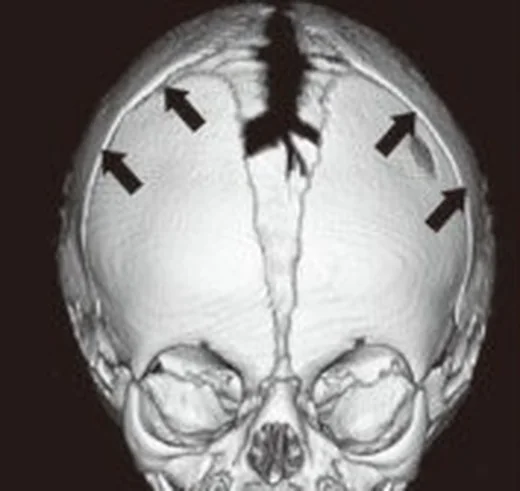
Koronal sinostoz, frontal ve parietal kemikler arasındaki sütürün tek ya da iki taraflı erken füzyonudur; ön-arka yönde büyümeyi kısıtlar. Tek taraflı füzyon ön plagiosefali, orbital asimetri ve karşı frontal bossing; iki taraflı füzyon ise kısa-geniş kafa ve bilateral orbital biçim değişikliği oluşturabilir. Kontrastsız ince kesit BT, kemik köprüsünü ve anterior kafa tabanı-orbita eşlikçilerini birlikte gösterir; koronal ve 3B rekonstrüksiyon taraf tayininde yardımcıdır. Füzyon tarafının veya karşı taraf telafi bulgularının belirtilmemesi, kafa ve orbita deformitesinin doğru paternle ilişkilendirilmesini güçleştirir.
Faz ve pencere
- Kontrastsız tanısal
- Koronal sütürlerde tırtıklı radyolüsent aralığın silinmesi, perisutural skleroz ve kemik köprüleşmesi kemik algoritmasında aranır; koronal ve 3B rekonstrüksiyon ipsilateral ön kafa çukuru sığlığı, karşı frontal bossing ve orbital çatı yükselmesini gösterir. Sütür füzyonunu değerlendirmede kemik gösterimi üstün olan BT temel görüntüleme yöntemidir.
Önerilen pencereler: Kemik (G 1800 / M 400).
BT bulguları
- Tek taraflı koronal füzyon — ilgili tarafta frontal-parietal sütür hattı kapanır ve kemik sırtı belirginleşebilir.
- İpsilateral anterior kraniyal fossa sığlığı — etkilenen frontal kemik ve ön kafa çukuru geride kalır.
- Karşı frontal bossing — karşı taraf frontal kemik dışa doğru kompansatuvar bombeleşir.
- Harlequin orbita — etkilenen tarafta orbital çatı ve lateral duvar yukarı yönelir, orbital kontur asimetrikleşir.
- Supraorbital kenar hipoplazisi — etkilenen tarafta kaş üstü kemik çıkıntısı yassı veya geride görünür.
- Ön plagiosefali ve yüz orta hat kayması — burun ucu ve çene karşı tarafa yönelmiş olabilir.
- Bilateral koronal füzyon — ön-arka kalvaryum kısalır, biparietal genişlik artar ve iki orbitada yükselmiş çatı görünümü oluşabilir.
- Kafa tabanı eşlikçisi — sfenoid kanatlar ve posterior fossa boyutu, özellikle sendromik çoklu füzyon şüphesinde incelenir.
Normalde
Normalde sağ ve sol koronal sütür, frontal kemik ile parietal kemiğin birleşiminde tırtıklı, radyolüsent ve devamlı bir çizgi oluşturur; ön kafa çukurları ve orbital çatılar karşılıklı benzer seviyededir. Tek taraflı şüphede iki koronal hattı, supraorbital kenarları ve anterior fossa derinliğini karşılaştırın; bilateral şüphede kalvaryumun ön-arka kısalığı ile enine genişliğini değerlendirin.
Normal BT ile kıyasla
Aynı bölgenin, kaynak veri setinde patoloji içermediği belirtilen bir BT incelemesini kesit kesit kaydırın; organ sınırlarını açarak anatomiyi hasta görüntüsüyle karşılaştırın. Küçük rastlantısal bulgular tümüyle dışlanmamıştır.
Kaynak: TotalSegmentator v2.01 veri seti (Wasserthal ve ark., Radiology: Artificial Intelligence 2023), CC BY 4.0, vaka s0103; organ sınırları veri setinin otomatik segmentasyonundan, birleştirilerek sadeleştirildi. Görüntüler 2,5 mm kesit aralığına yeniden örneklendi.
Ayırıcı tanı
- Pozisyonel posterior plagiosefali
- kafa asimetrisi görülebilir ancak sütürler açık kalır; kemik köprüsü ve orbital değişiklik beklenmez.
- Bikoronal sinostoz
- iki koronal sütür kapanır; tek taraflı ön plagiosefali yerine kısa-geniş kafa ve bilateral orbital şekil değişikliği gelişir.
- Lambdoid sinostozda tek taraflı erken sütür füzyonu posterior plagiosefaliye yol açar.
- Kraniyofasiyal sendrom
- iki taraflı koronal füzyona orta yüz hipoplazisi veya başka sütürlerin tutulumu eşlik edebilir; yalnız tek koronal hatla sınırlı patern değildir.
Tuzaklar
- Taraf tayininde 3B BT’nin önden görünümünde füzyonlu koronal sütürü karşı taraftaki açık sütürle karşılaştırın; sütürün izlenmemesi ve kemik sırtı füzyonu destekler.
- Harlequin benzeri orbital konturu yalnız göz çukuru şekli olarak tarif etmeyin; koronal füzyon, yükselmiş orbital çatı ve sığ ön fossa ilişkisini kurun.
- Tek taraflı koronal füzyon saptandığında diğer koronal hattı ve sagittal-lambdoid sütürleri de izleyerek eşlik eden tutulumları arayın.
Kendini dene
Sağ koronal hatta kemik köprüsü, sağ orbital çatıda yükselme ve sol frontal bossing bulunuyor. En olası tanı nedir?
Cevabı göster
Sağ koronal sinostoz. Tek taraflı koronal füzyon ipsilateral orbital ve ön fossa değişikliği ile karşı frontal bossing oluşturur. Bikoronal tutulum iki taraflıdır; sagittal sinostoz uzun-dar kafa yapar, lambdoid sinostoz posterior kalvaryumu etkiler.
İki koronal sütür de kemik köprüsüyle kapanmış; kalvaryum kısa ve enine geniş. Bu patern nasıl adlandırılır?
Cevabı göster
Bikoronal sinostoz. Bikoronal sinostozda iki koronal sütür füzyondur; ön-arka boyut kısalması genellikle brakisefaliye (kısa kafaya) yol açar. Tek taraflı koronal sinostoz oblik ön kafa asimetrisi, metopik sinostoz üçgensi alın yapar; pozisyonel deformitede sütürler açık kalır.
İlgili konular
- Kraniyosinostoz ve sütür füzyonu
- Sagittal sütür sinostozu
- Metopik sütür sinostozu
- Lambdoid sütür sinostozu
Kaynaklar
Bu sayfadaki 37 cümle ve 2 quiz sorusu aşağıdaki kaynaklardan alıntılarla otomatik olarak karşılaştırıldı; 5 cümle bu karşılaştırmada düzeltildi (2026-10-09).
- Craniosynostosis : Updates in Radiologic Diagnosispmc.ncbi.nlm.nih.gov
- Quantitative Craniofacial Analysis and Generation of Human Induced Pluripotent Stem Cells for Muenke Syndrome: A Case Reportpmc.ncbi.nlm.nih.gov
- The influence of suturectomy on age-related changes in cerebral blood flow in rabbits with familial bicoronal suture craniosynostosis: A quantitative analysispmc.ncbi.nlm.nih.gov
- A de novo substitution in BCL11B leads to loss of interaction with transcriptional complexes and craniosynostosispmc.ncbi.nlm.nih.gov
- Isolation and Characterization of Human Suture Mesenchymal Stem Cells In Vitropmc.ncbi.nlm.nih.gov
- Phenotype profile of a genetic mouse model for Muenke syndrome.pubmed.ncbi.nlm.nih.gov
- Brain phenotypes in two FGFR2 mouse models for Apert syndrome.pubmed.ncbi.nlm.nih.gov
- Benign radiographic coronal synostosis after sagittal synostosis repair.pubmed.ncbi.nlm.nih.gov
- [COMPUTED TOMOGRAPHIC EXAMINATION OF CRANIAL LESIONS, A PALEORADIOLOGICAL APPROACH].pubmed.ncbi.nlm.nih.gov
- Atypical presentation of a newborn with Apert syndrome.pubmed.ncbi.nlm.nih.gov
- The Muenke syndrome mutation (FgfR3P244R) causes cranial base shortening associated with growth plate dysfunction and premature perichondrial ossification in murine basicranial synchondroses.pubmed.ncbi.nlm.nih.gov
- The influence of surgical correction on white matter microstructural integrity in rabbits with familial coronal suture craniosynostosis.pubmed.ncbi.nlm.nih.gov
- Transforming Growth Factor-β3 Therapy Delays Postoperative Reossification and Improves Craniofacial Growth in Craniosynostotic Rabbits.pubmed.ncbi.nlm.nih.gov
- Quantitative Craniofacial Analysis and Generation of Human Induced Pluripotent Stem Cells for Muenke Syndrome: A Case Report.pubmed.ncbi.nlm.nih.gov
- Development of secondary unilateral coronal suture synostosis with a sagittal suture synostosis in a nonsyndromic patient.pubmed.ncbi.nlm.nih.gov
- Atypical scaphocephaly: a review.pubmed.ncbi.nlm.nih.gov
- Synostosis of the lambdoid suture: a spectrum.pubmed.ncbi.nlm.nih.gov
- Quantitative evaluation of facial hypoplasia and airway obstruction in infants with syndromic craniosynostosis: relationship with skull base and splanchnocranium sutural pattern.pubmed.ncbi.nlm.nih.gov
- The surgical treatment of plagiocephaly.pubmed.ncbi.nlm.nih.gov
- When Virchow's law fails: Crouzon syndrome as a test case for quantitative suture closure-endocranial shape relationships.pubmed.ncbi.nlm.nih.gov
- Clinical and genetic features of syndromic craniosynostosis in 18 Chinese probands: novel candidate genes and phenotypes of known pathogenic genes.pubmed.ncbi.nlm.nih.gov
- Neurocognitive outcomes in children with craniosynostosis after surgical correction: a narrative review.pubmed.ncbi.nlm.nih.gov
- Isolated frontosphenoidal craniosynostosis with pathogenic <i>FGFR3</i> variant: a case report and genetic insights.pubmed.ncbi.nlm.nih.gov
- Treatment strategies and prognostic outcomes in craniosynostosis with concurrent hydrocephalus: a case series.pubmed.ncbi.nlm.nih.gov
- Complications in craniosynostosis surgery in patients with rickets: illustrative case and systematic review of literature.pubmed.ncbi.nlm.nih.gov
- Impaired wound healing following cranial vault reconstruction in a patient with an atypical phenotype of Marfan syndrome: A case report.pubmed.ncbi.nlm.nih.gov
- Novel mutation detection of fibroblast growth factor receptor 1 (FGFR1) gene, FGFR2IIIa, FGFR2IIIb, FGFR2IIIc, FGFR3, FGFR4 gene for craniosynostosis: A prospective study in Asian Indian patient.pubmed.ncbi.nlm.nih.gov
- Clinical and molecular characterization of TCF12 variants in an Asian pediatric cohort with craniosynostosis.pubmed.ncbi.nlm.nih.gov
- From FGFR2 mutations to precision management: a review of prenatal diagnosis and multidisciplinary interventions in apert syndrome.pubmed.ncbi.nlm.nih.gov
- Three-dimensional Analysis of Facial Asymmetry in Unilateral Lambdoid Craniosynostosis.pubmed.ncbi.nlm.nih.gov
- Neuro-Ophthalmological Manifestations of Craniosynostosis: Current Perspectives.pubmed.ncbi.nlm.nih.gov
- Craniosynostosis of the lambdoid suture.pubmed.ncbi.nlm.nih.gov
- Craniosynostosis: molecular pathways and future pharmacologic therapy.pubmed.ncbi.nlm.nih.gov
- Cranial nerves involvement in craniosynostosis: a systematic review.pubmed.ncbi.nlm.nih.gov
- Frontofacial Features of Unilateral Lambdoid Craniosynostosis: A Multicenter Assessment.pubmed.ncbi.nlm.nih.gov
- Osteology of the Hamadryas Baboon ( Papio hamadryas ).pubmed.ncbi.nlm.nih.gov
- Craniosynostosis-4 with Heterozygous Mutation in the ERF Gene: A Case Report.pubmed.ncbi.nlm.nih.gov
- Surgery of craniosynostosis: a historical review.pubmed.ncbi.nlm.nih.gov
- Evaluation and Management of Nonsyndromic Craniosynostosis.pubmed.ncbi.nlm.nih.gov
- Pediatric craniofacial surgery for craniosynostosis: Our experience and current concepts: Part -1.pubmed.ncbi.nlm.nih.gov
- Pictorial essay: The many faces of craniosynostosis.pubmed.ncbi.nlm.nih.gov
- Neuroembryology and functional anatomy of craniofacial clefts.pubmed.ncbi.nlm.nih.gov
- Case Report: Three-dimensional characteristics of craniofacial morphology in facial asymmetry due to unilateral coronal synostosis.pubmed.ncbi.nlm.nih.gov
- Definitive reconstruction of frontal asymmetry in an adolescent with anterior plagiocephaly due to unilateral coronal craniosynostosis using a hybrid osteo-titanium technology based on an individualized approach: A case report.pubmed.ncbi.nlm.nih.gov
- Sella Turcica and Cranial Base Symmetry in Anterior Synostotic Plagiocephaly Patients: A Retrospective Case-Control Study.pubmed.ncbi.nlm.nih.gov
- Insights into craniosynostosis management in low- and middle-income countries: A narrative review of outcomes, shortcomings and paediatric neurosurgery capacity.pubmed.ncbi.nlm.nih.gov
- Surgical Correction of Unicoronal Synostosis: Fronto-Orbital Distraction versus Calvarial Switch.pubmed.ncbi.nlm.nih.gov
- Rethinking the Origin of the Primary Respiratory Mechanism.pubmed.ncbi.nlm.nih.gov
- Stem cell-associated osteogenic deficiency causes craniofacial deformities with progeroid accumulation of prelamin A.pubmed.ncbi.nlm.nih.gov
- Neuroimaging in Nonsyndromic Craniosynostosis: Key Concepts to Unlock Innovation.pubmed.ncbi.nlm.nih.gov
- Developments in diagnostic and surgical techniques in children with sagittal suture craniosynostosis: a systematic review spanning the last 30 years.pubmed.ncbi.nlm.nih.gov
- Exploring Different Management Modalities of Nonsyndromic Craniosynostosis.pubmed.ncbi.nlm.nih.gov
- Emphasis on Early Prenatal Diagnosis and Perinatal Outcomes Analysis of Apert Syndrome.pubmed.ncbi.nlm.nih.gov
- A tissue engineering approach to regenerate the cranial suture skeletal stem cell niche with a multicompartment biomaterial scaffold.pubmed.ncbi.nlm.nih.gov
- Wormian bones: expanded differential diagnosis and implications for abnormal head shape in infancy.pubmed.ncbi.nlm.nih.gov
- TGFβ-mediated dural progenitor cell migration into the coronal suture is crucial for preventing craniosynostosis.pubmed.ncbi.nlm.nih.gov
- Craniofacial Morphology and Dental Age Assessment in Pediatric Craniosynostosis: A Comparative Cross-Sectional Study.pubmed.ncbi.nlm.nih.gov
- Embryodysgenesis of the Lids and Orbit.pubmed.ncbi.nlm.nih.gov
- Visual outcome in 5-year-old children operated for non-syndromic craniosynostosis.pubmed.ncbi.nlm.nih.gov
- Prevalence of motor development delay in children with craniosynostosis: a systematic review and meta-analysis.pubmed.ncbi.nlm.nih.gov
- Fzd7 Restrains Pink1-Dependent Mitophagy in Suture Stem Cells to Maintain Cranial Suture Patency.pubmed.ncbi.nlm.nih.gov
- The Effect of Cranial Sutures Should Be Considered in Transcranial Electrical Stimulation.pubmed.ncbi.nlm.nih.gov
- A de novo substitution in BCL11B leads to loss of interaction with transcriptional complexes and craniosynostosis.pubmed.ncbi.nlm.nih.gov
- International Harmonization of Nomenclature and Diagnostic Criteria (INHAND): Nonproliferative and Proliferative Lesions of the Rabbit.pubmed.ncbi.nlm.nih.gov
- Endoscopic-assisted versus open fronto-orbital distraction for unicoronal craniosynostosis: morphometric and technique considerations.pubmed.ncbi.nlm.nih.gov
- Hominin Variability and Evolutionary Relationships at Guattari Cave During the Middle and Late Pleistocene (San Felice Circeo, Latina, Italy).pubmed.ncbi.nlm.nih.gov
- Rare Diseases of the Orbit.pubmed.ncbi.nlm.nih.gov
- Ophthalmological outcomes of unilateral coronal synostosis in young children.pubmed.ncbi.nlm.nih.gov
- Unicoronal Craniosynostosis and Plagiocephaly Correction with Fronto-orbital Bone Remodeling and Advancement.pubmed.ncbi.nlm.nih.gov
- Anterior Craniofacial Changes Following Occipital Switch Cranioplasty in Unilateral Lambdoid Craniosynostosis.pubmed.ncbi.nlm.nih.gov
- Comparison of Cranial Vault Remodeling Versus Spring Cranioplasty for Nonsyndromic Unicoronal Craniosynostosis: Analysis of Outcomes.pubmed.ncbi.nlm.nih.gov
- A Unique Case of Pure Bilateral Lambdoid Craniosynostosis and Progression to Mercedes-Benz Syndrome Following Spring Cranioplasty.pubmed.ncbi.nlm.nih.gov
- Surgical management of raised ICP in craniosynostosis: experience-based selection of posterior vault expansion techniques.pubmed.ncbi.nlm.nih.gov
- Machine Learning in Metopic Craniosynostosis: Does Phenotypic Severity Predict Long-Term Esthetic Outcome?pubmed.ncbi.nlm.nih.gov
- Posterior cranial vault distraction osteogenesis: A systematic review.pubmed.ncbi.nlm.nih.gov
- Evaluation of Endoscopic Strip Craniectomy and Orthotic Therapy for Bilateral Coronal Craniosynostosis.pubmed.ncbi.nlm.nih.gov
- Craniosynostosis: Experience From a Single Tertiary Center in India.pubmed.ncbi.nlm.nih.gov
- Customized 3D printed helmet in the treatment of metopic craniosynostosis in a 7-month-old infant, a case report.pubmed.ncbi.nlm.nih.gov
- Zero-TE MRI: principles and applications in the head and neck.pubmed.ncbi.nlm.nih.gov
- Deep learning-based craniosynostosis classification via suture segmentation and mask-weighted classification.pubmed.ncbi.nlm.nih.gov
- Extended Reality in Neurosurgery : Surgical Planning, Navigation, Education, and Patient Communication.pubmed.ncbi.nlm.nih.gov
- Initial In Vivo Analyses of Small Pore Polymer Scaffolds for Creation of an Artificial Cranial Stem Cell Niche.pubmed.ncbi.nlm.nih.gov
- Endoscope-assisted release for pancraniosynostosis with delayed single-suture refusion managed with endoscopic re-release in the same patient: illustrative case.pubmed.ncbi.nlm.nih.gov
- Preservation versus Structural Cranioplasty: A New Concept for Craniosynostosis Treatment.pubmed.ncbi.nlm.nih.gov
- Skeletal Deformity in Patients With Unilateral Coronal Craniosynostosis: Perceptions of the General Public.pubmed.ncbi.nlm.nih.gov
- Cranial Base and Posterior Cranial Vault Asymmetry After Open and Endoscopic Repair of Isolated Lambdoid Craniosynostosis.pubmed.ncbi.nlm.nih.gov
- The Temporal Region in Unilateral Coronal Craniosynostosis: Fronto-orbital Advancement Versus Endoscopy-Assisted Strip Craniectomy.pubmed.ncbi.nlm.nih.gov
- 3D-printed cranial helmet therapy for the treatment of deformational plagiocephaly.pubmed.ncbi.nlm.nih.gov
- Fully Automated AI-Based Digital Workflow for Mirroring of Healthy and Defective Craniofacial Models.pubmed.ncbi.nlm.nih.gov
- A novel RBF-based predictive tool for facial distraction surgery in growing children with syndromic craniosynostosis.pubmed.ncbi.nlm.nih.gov
- Indications for Computer-Aided Design and Manufacturing in Congenital Craniofacial Reconstruction.pubmed.ncbi.nlm.nih.gov
Eğitim amaçlıdır; hasta başında tanı ve tedavi kararının yerine geçmez, tıbbi tavsiye değildir. Metinler yapay zekâ desteğiyle yazıldı ve otomatik bir adımda kaynak alıntılarıyla karşılaştırıldı; bu bir tıbbi doğrulama değildir ve içerik uzman radyolog incelemesinden geçmedi. Klinik kararlarda güncel kılavuzları, kurum protokollerini ve radyoloji raporunu esas alın.