Özet
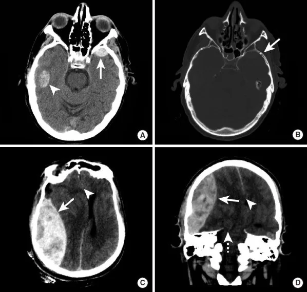
Künt kafa travmasında gelişen serebral kontüzyonlar, özellikle frontal ve temporal loblarda görülen kortikal doku hasarlarıdır ve başlangıç yaralanmasından sonra büyüme eğilimi gösterebilir. Kontrastsız beyin BT, travmatik kontüzyonların başlangıç değerlendirmesi ve tekrarlayan görüntülemelerle aradaki ilerlemenin tespiti için tercih edilen yöntemdir. Kafa tabanının pürüzlü kemik yüzeyleriyle temas nedeniyle travmatik kontüzyonlar frontotemporal bölgelerde en sık görülen yerleşimlere sahiptir. Büyüyen lezyon boyutu ve artan kütle etkisi, nörolojik bozulmaya, cerrahi ihtiyacın artmasına ve mortaliteye yol açabilir; bazal sisterna effasmanı ilerlemeyi öngörebilir.
Faz ve pencere
- Kontrastsız tanısal
- Travmatik beyin yaralanmalarının başlangıç değerlendirilmesinde ve ilerlemenin tespitinde kontrastsız BT kullanılır; lezyonlar morfoloji, anatomik yerleşim, derinlik ve hacim açısından karakterize edilir. Lezyonlar travma sonrası ilk 24 saat içinde çoğunlukla büyür ve zamanla devam eden genişlemeler de görülebilir; bu durumun başlangıç ve takip değerlendirmelerinde kontrastsız BT kullanılır.
Önerilen pencereler: Beyin (G 80 / M 40), İnme (G 40 / M 40), Kemik (G 1800 / M 400).
BT bulguları
- Kontüzyonlar frontotemporal bölgelerde en sık görülür ve lezyonun anatomik yerleşimi ile derinliği sonucu belirleyen faktörler arasındadır.
- Kontüzyonlar kontrastsız BT üzerinde morfolojileri, anatomik yerleşimleri, derinlikleri ve hacimleri açısından değerlendirilir.
- Lezyon büyüklüğü ve kütle etkisi önemli prognostik göstergelerdir; progresif kanama ve lezyon büyümesi sulkus effasmanına neden olabilir.
- Coup ve contrecoup yerleşimi — darbe noktasının altındaki korteksin yanı sıra karşı yüzeydeki beyin-kafatası temas alanında da odak bulunabilir.
- Frontal-temporal taban dağılımı — orbitofrontal giruslar, frontal kutuplar ve anterior-inferior temporal korteks sık incelenmesi gereken yüzeylerdir.
- Travmatik kontüzyonlar travmadan sonra büyüme eğilimi gösterir; başlangıç hacmine göre %30'dan fazla artış progresyon kabul edilir ve büyümenin çoğu ilk 24 saat içinde gerçekleşir.
- Bazal sisterna effasmanı ve orta hat kayması gibi kütle etkisi bulguları, lezyon ilerlemesini öngörür ve cerrahi yönetimi haklı çıkarabilir.
Normalde
Kontrastsız BT'de kontüzyonlar morfoloji, anatomik yerleşim, derinlik ve hacim açısından değerlendirilir; karşı darbe kontüzyonları klasik olarak orbitofrontal bölgelerde ve temporal kutuplarda görülebilir. Kontüzyonlar kontrastsız BT'de morfoloji, anatomik yerleşim, derinlik ve hacim açısından değerlendirilir.
Normal BT ile kıyasla
Aynı bölgenin, kaynak veri setinde patoloji içermediği belirtilen bir BT incelemesini kesit kesit kaydırın; organ sınırlarını açarak anatomiyi hasta görüntüsüyle karşılaştırın. Küçük rastlantısal bulgular tümüyle dışlanmamıştır.
Kaynak: TotalSegmentator v2.01 veri seti (Wasserthal ve ark., Radiology: Artificial Intelligence 2023), CC BY 4.0, vaka s0103; organ sınırları veri setinin otomatik segmentasyonundan, birleştirilerek sadeleştirildi. Görüntüler 2,5 mm kesit aralığına yeniden örneklendi.
Ayırıcı tanı
- Travmatik subaraknoid kanama
- yoğunluk korteks dokusunda değil, BOS ile dolu sulkus veya fissür içinde çizgisel biçimde uzanır.
- Travmatik aksonal yaralanma ile ilişkili serebral mikrokanamalar, genellikle difüzyon ağırlıklı ve manyetik duyarlılık temelli MR teknikleriyle tespit edilir; BT ince odakları algılamada yetersiz kalabilir.
- Hemorajik tümör
- travma öyküsü olmayan veya lezyonun beklenen travma yerleşiminden sapması durumunda, kısa sürede ilerleme gösteren hemoraji altında neoplazi düşünülmelidir.
- Hemorajik enfarkt
- parankim değişikliği arter sulama alanına uyar; damar dağılımı ve eşlik eden iskemik ödem travma yüzeyi yerleşiminden farklıdır.
- Kalsifikasyon
- keskin sınırlı ve çoğunlukla kronik yerleşimli yoğun odak, çevresinde akut ödem bulunmamasıyla kanamadan ayrılır.
Tuzaklar
- Tüm radyolojik ilerlemeler hemen klinik bozulmayla eşleşmeyebilir; bu nedenle seçilmiş hastalarda rutin tekrarlı görüntüleme ile lezyonların başlangıç kesitleriyle doğrudan karşılaştırılması desteklenir.
- Girus içi kanamayı subaraknoid kana katma; kontüzyonda yoğunluk kortikal dokuya gömülürken subaraknoid kan sulkusun BOS boşluğunu izler.
- Derin beyaz cevher veya korpus kallozumda yer alan ara dereceli kontüzyonlar, travmatik aksonal yaralanma ile ilişkili serebral mikrokanamalardan ayırt edilmelidir.
Kendini dene
Künt kafa travması sonrası frontal ve temporal loblarda görülebilen ve ilk yaralanmadan sonra büyüme eğiliminde olan parankimal lezyon için en uygun tanı ve özellik çifti hangisidir?
Cevabı göster
Travmatik kontüzyon — progresif hemorajik genişleme. Travmatik kontüzyonlar özellikle frontal ve temporal lobları tutar; büyümenin çoğu ilk 24 saatte görülse de sonraki günlerde de sürebilir.
Künt kafa travması sonrası izlenen küçük parankimal odak, tekrarlayan BT'de başlangıca göre %30'tan fazla hacim artışı göstermiştir. Bu radyolojik bulgu en çok aşağıdakilerden hangisiyle açıklanır?
Cevabı göster
Progresif travmatik kontüzyon. Travmatik kontüzyonların en belirgin özelliği travma sonrası büyümeye eğilimli olmasıdır; başlangıç hacmine göre %30'luk artış progresyon kabul edilir.
İlgili konular
Kaynaklar
Bu sayfadaki 33 cümle ve 2 quiz sorusu aşağıdaki kaynaklardan alıntılarla otomatik olarak karşılaştırıldı; 24 cümle bu karşılaştırmada düzeltildi (2026-10-09).
- Minimal-access exoscopic approach for caudate hemorrhage with intraventricular extension using the ViewSite Brain Access Systempmc.ncbi.nlm.nih.gov
- Correlation of Contusion Morphology and Volume With Functional Recovery and in Hospital Mortality in Traumatic Brain Injurypmc.ncbi.nlm.nih.gov
- The Anticoagulation Dilemma: Concurrent Traumatic Brain Hemorrhage and Cerebral Venous Thrombosis in a Blast Injury Patientpmc.ncbi.nlm.nih.gov
- A Rare Case of a Depressed Inner Table Fracture Without an Outer Table Fracture Following Trauma in a 20-Year-Old Male Patientpmc.ncbi.nlm.nih.gov
- AI-Based Approaches for the Identification and Quantification of Traumatic Brain Injury in Computed Tomography Imaging: Systematic Reviewpmc.ncbi.nlm.nih.gov
- Systemic Markers of Injury and Injury Response Are Not Associated with Impaired Cerebrovascular Reactivity in Adult Traumatic Brain Injury: A Collaborative European Neurotrauma Effectiveness Research in Traumatic Brain Injury (CENTER-TBI) Study.pubmed.ncbi.nlm.nih.gov
- Susceptibility-weighted angiography of intracranial blood products and calcifications compared to gradient echo sequence.pubmed.ncbi.nlm.nih.gov
- Surgical trial in traumatic intracerebral hemorrhage (STITCH(Trauma)): study protocol for a randomized controlled trial.pubmed.ncbi.nlm.nih.gov
- Radiological Parameters to Predict Hemorrhagic Progression of Traumatic Contusional Brain Injury.pubmed.ncbi.nlm.nih.gov
- Intracranial pressure monitoring and inpatient mortality in severe traumatic brain injury: A propensity score-matched analysis.pubmed.ncbi.nlm.nih.gov
- Neuroimaging findings in pediatric sports-related concussion.pubmed.ncbi.nlm.nih.gov
- Rate of Intracranial Hemorrhage After Minor Head Injury.pubmed.ncbi.nlm.nih.gov
- A cost effectiveness based safety and efficacy study of resterilized intra-parenchymal catheter based intracranial pressure monitoring in developing world.pubmed.ncbi.nlm.nih.gov
- A Clinical Predictive Nomogram for Traumatic Brain Parenchyma Hematoma Progression.pubmed.ncbi.nlm.nih.gov
- Computed tomography after decompressive craniectomy for head injury.pubmed.ncbi.nlm.nih.gov
- Isolated lower extremity monoplegia due to a traumatic intraparenchymal contusion: report of two cases.pubmed.ncbi.nlm.nih.gov
- Role of repeat CT in mild to moderate head injury: an institutional study.pubmed.ncbi.nlm.nih.gov
- Minimizing Radiation Exposure in Evaluation of Pediatric Head Trauma: Use of Rapid MR Imaging.pubmed.ncbi.nlm.nih.gov
- Risk Factors Analysis for 90-Day Mortality of Adult Patients with Mild Traumatic Brain Injury in an Italian Emergency Department.pubmed.ncbi.nlm.nih.gov
- Comparative Effectiveness of Intracranial Pressure Monitoring on 6-Month Outcomes of Critically Ill Patients With Traumatic Brain Injury.pubmed.ncbi.nlm.nih.gov
- Birth Injury: Birth Asphyxia and Birth Trauma.pubmed.ncbi.nlm.nih.gov
- Dual Impact: Coup-Contrecoup Brain Injury in a Young Cyclist-A Case Report.pubmed.ncbi.nlm.nih.gov
- Herpes simplex virus encephalitis following acute brain injury: illustrative cases.pubmed.ncbi.nlm.nih.gov
- An isolated oculomotor nerve palsy and brainstem hemorrhage in a 53-year-old male patient with mild traumatic brain injury: illustrative case.pubmed.ncbi.nlm.nih.gov
- Multi-modal lesion mapping of expressive aprosodia.pubmed.ncbi.nlm.nih.gov
- Injury Patterns in Soccer-Induced Maxillofacial Fractures and Ocular Trauma: A Narrative Review.pubmed.ncbi.nlm.nih.gov
- Multimodal PET, Diffusion Tensor Imaging, and Volumetric MRI Findings in Orbitofrontal Cortex Dysfunction: A Quantitative Single-Case Analysis.pubmed.ncbi.nlm.nih.gov
- Clinically complete spinal cord injury is not always anatomically complete.pubmed.ncbi.nlm.nih.gov
- An interpretable multimodal model for early prediction of delayed hematoma progression in frontal lobe contusion: a machine learning approach.pubmed.ncbi.nlm.nih.gov
- Lung Parenchymal and Airway Injuries: Differential Diagnosis and Severity Grading.pubmed.ncbi.nlm.nih.gov
- Cytotoxic Edema and Intra-parenchymal Hemorrhage: A Mediated Pathway to Mortality and Functional Outcome in Cerebral Venous Sinus Thrombosis- A sub-analysis of the CLOT-VENUS Registry.pubmed.ncbi.nlm.nih.gov
- Middle cerebral artery dissection: pathophysiology, diagnosis, and therapeutic options.pubmed.ncbi.nlm.nih.gov
- Computed tomography features of spontaneous acute intracranial hemorrhages in a tertiary hospital in Southern Ghana.pubmed.ncbi.nlm.nih.gov
- Neuroprotective efficacy of a proneurogenic compound after traumatic brain injury.pubmed.ncbi.nlm.nih.gov
- Commentary: an eye on PET quantification.pubmed.ncbi.nlm.nih.gov
- Herpes simplex virus infection of the central nervous system presenting as a stroke: Case report and review of the literature.pubmed.ncbi.nlm.nih.gov
- Combined Uyghur medicine, acupuncture, and rehabilitation for traumatic oculomotor nerve palsy: A case report.pubmed.ncbi.nlm.nih.gov
- Micro/Nanorobots for Combating Brain Disorders: Challenges, Advances, and Perspectives.pubmed.ncbi.nlm.nih.gov
- Correlation of Contusion Morphology and Volume With Functional Recovery and in Hospital Mortality in Traumatic Brain Injury.pubmed.ncbi.nlm.nih.gov
- An Unusual Presentation of Isolated Contrecoup Injury.pubmed.ncbi.nlm.nih.gov
- Risk factors and development of a predictive model for post-traumatic cognitive impairment in patients with frontal lobe cerebral contusion.pubmed.ncbi.nlm.nih.gov
- Ballooned neurons in semi-recent severe traumatic brain injury.pubmed.ncbi.nlm.nih.gov
- An Objective Injury Threshold for the Maximum Principal Strain Criterion for Brain Tissue in the Finite Element Head Model and Its Application.pubmed.ncbi.nlm.nih.gov
- Adjunctive middle meningeal artery embolization with low-viscosity liquid embolic agents for acute epidural hematoma: technical considerations and a case series.pubmed.ncbi.nlm.nih.gov
- Delayed Traumatic Subarachnoid Hemorrhage Following Initially Negative Computed Tomography (CT) Imaging in a Patient With a Mechanical Mitral Valve and Markedly Elevated International Normalized Ratio (INR).pubmed.ncbi.nlm.nih.gov
- Post-traumatic hydrocephalus in adults: the mechanisms of development, predictors of progression and management strategies. A narrative review and case series analysis.pubmed.ncbi.nlm.nih.gov
- Delayed Expanding Intracerebral Pneumatocele Following Severe Craniofacial Trauma: A Case Report With Serial Imaging Correlation.pubmed.ncbi.nlm.nih.gov
- Case Report: Endoscopic evacuation of acute subdural hematoma in selected young patients: a report of two cases.pubmed.ncbi.nlm.nih.gov
- Optimising 7T-fMRI for Imaging Regions of Magnetic Susceptibility.pubmed.ncbi.nlm.nih.gov
- Theta oscillations are an organizational unit of odor processing in the olfactory bulb.pubmed.ncbi.nlm.nih.gov
- Drug-resistant frontal lobe epilepsy: A review.pubmed.ncbi.nlm.nih.gov
- CSF Volumetric Analysis for Quantification of Cerebral Edema After Hemispheric Infarction.pubmed.ncbi.nlm.nih.gov
- A Review of the Effectiveness of Neuroimaging Modalities for the Detection of Traumatic Brain Injury.pubmed.ncbi.nlm.nih.gov
- Non-contrast computed tomography at the end of the acute care phase as an additional prognostic tool after aneurysmal subarachnoid hemorrhage.pubmed.ncbi.nlm.nih.gov
- Stretching the Limits: A Rare Case Report of Perimesencephalic Subarachnoid Hemorrhage During Yoga.pubmed.ncbi.nlm.nih.gov
- Size and location of intracranial hemorrhage in acute stroke presentations: correlation with baseline NIHSS and relevance to direct-to-angiography workflows.pubmed.ncbi.nlm.nih.gov
- Traumatic basal ganglia hemorrhage in the pediatric population.pubmed.ncbi.nlm.nih.gov
- From microbleeds to axonal damage: current evidence and future directions.pubmed.ncbi.nlm.nih.gov
- Fulminant cerebral fat embolism syndrome initially sparing the lungs after long-bone fractures: a case report of refractory status epilepticus.pubmed.ncbi.nlm.nih.gov
- Myositis Ossificans of the Soleus Muscle.pubmed.ncbi.nlm.nih.gov
- Atypical Presentation of a Hemorrhagic Glioblastoma Multiforme Mimicking a Cerebral Contusion.pubmed.ncbi.nlm.nih.gov
- The Dual Role of Connexins in Stroke, Neurotrauma, Neurodegenerative and Psychiatric Disorders: A Global Systematic Review.pubmed.ncbi.nlm.nih.gov
- Timing of DOAC Initiation After Atrial Fibrillation-Related Acute Ischemic Stroke: A Unified Protocol Harmonizing Clinical Safety and Imaging-Based Precision in the Post-CATALYST Era.pubmed.ncbi.nlm.nih.gov
- A Transient Left Atrial Appendage Filling Defect During Anticoagulation Interruption After Ischemic Stroke with Progressive Hemorrhagic Transformation: A Case Report.pubmed.ncbi.nlm.nih.gov
- A Diagnostic Challenge: From Suspected Ischemic Stroke to Confirmed Herpetic Encephalitis.pubmed.ncbi.nlm.nih.gov
- Imaging for Pediatric Head and Neck Emergencies: Critical Insights.pubmed.ncbi.nlm.nih.gov
- Diagnostic imaging and emerging image-guided neurointervention in brain tumors: a narrative review.pubmed.ncbi.nlm.nih.gov
- Suprasellar Desmoplastic Infantile Ganglioglioma in a 7-Month-Old Infant: A Rare Diagnostic Consideration in Infant Suprasellar Masses.pubmed.ncbi.nlm.nih.gov
- The Internal Otic Region of Oromerycids (Artiodactyla, Oromerycidae), Early Camelids (Artiodactyla, Camelidae), and the Vicuña (Artiodactyla, Camelidae), Including Notes on Intraspecific and Subadult Ontogenetic Variation.pubmed.ncbi.nlm.nih.gov
- Comparison of image quality between Deep learning image reconstruction and Iterative reconstruction technique for CT Brain- a pilot study.pubmed.ncbi.nlm.nih.gov
- CT perfusion imaging in aneurysmal subarachnoid hemorrhage. State of the art.pubmed.ncbi.nlm.nih.gov
- Computed tomography of pediatric abdominal trauma: optimizing utilization and enhancing diagnostic interpretation.pubmed.ncbi.nlm.nih.gov
- Lung parenchymal trauma biomechanics, mechanisms, and classification: a narrative review of the current knowledge.pubmed.ncbi.nlm.nih.gov
- Soft tissue hematomas on ultrasound: a case-based review and practical guide to diagnosis.pubmed.ncbi.nlm.nih.gov
- The Effectiveness Based on Optimal Dose and Administration Route, and Safety Profiles of Stem Cells and Derived Products in the Treatment of Patients With Amyotrophic Lateral Sclerosis: A Systematic Review and Meta-Analysis.pubmed.ncbi.nlm.nih.gov
- Review of imaging modalities and radiological findings of calvarial lesions.pubmed.ncbi.nlm.nih.gov
- The pathogenesis of cerebral small vessel disease and vascular cognitive impairment.pubmed.ncbi.nlm.nih.gov
- Continuum of Irreversible and Potentially Reversible Traumatic Brain Injury Sequelae: Axonal Injury and Hydrocephalus.pubmed.ncbi.nlm.nih.gov
- Striking corpus callosum lesions in immediate cerebral fat embolism: A rare clinical case.pubmed.ncbi.nlm.nih.gov
- An Atlas of Neonatal Neurovascular Imaging Anatomy as Depicted with Microvascular Imaging: The Intracranial Arteries.pubmed.ncbi.nlm.nih.gov
- Exploring optimal microscopic keyhole access to the skull base: an anatomical evaluation of transciliary supraorbital and transpalpebral orbitofrontal craniotomy approaches.pubmed.ncbi.nlm.nih.gov
- Transorbital and supraorbital uniportal multicorridor approach to the orbit, anterior, middle and posterior cranial fossa: Anatomic study.pubmed.ncbi.nlm.nih.gov
- 2024 Heart Disease and Stroke Statistics: A Report of US and Global Data From the American Heart Association.pubmed.ncbi.nlm.nih.gov
- A Comprehensive Review of the Role of the Latest Minimally Invasive Neurosurgery Techniques and Outcomes for Brain and Spinal Surgeries.pubmed.ncbi.nlm.nih.gov
- AI-Based Approaches for the Identification and Quantification of Traumatic Brain Injury in Computed Tomography Imaging: Systematic Review.pubmed.ncbi.nlm.nih.gov
- Veterans Traumatic Brain Injuries and Neurosurgical Challenges: A Narrative Review.pubmed.ncbi.nlm.nih.gov
- Case Report and Literature Review: A Severe Case of Blast-Related Traumatic Brain Injury.pubmed.ncbi.nlm.nih.gov
- Primary Blast-Induced Traumatic Brain Injury as a Risk Factor for (Cerebro)vascular Disorder: Clinical Manifestations, Blast Physics, Biomechanics, Pathobiology, and Critical Gaps.pubmed.ncbi.nlm.nih.gov
- Spectrum of Magnetic Resonance Imaging Findings in Acute Pediatric Traumatic Brain Injury - A Pictorial Essay.pubmed.ncbi.nlm.nih.gov
- Use of anti-amyloid therapies for Alzheimer's disease in Brazil: a position paper from the Scientific Department of Cognitive Neurology and Aging of the Brazilian Academy of Neurology.pubmed.ncbi.nlm.nih.gov
- Clinical manifestations, progression and treatment of post-traumatic hydrocephalus accompanied by contralateral subdural effusion after decompressive craniectomy: a single-center retrospective study.pubmed.ncbi.nlm.nih.gov
- Isolated Blunt Pancreatic Head Injury with Evolving Acute Peripancreatic Fluid Collection in a Child Successfully Managed Conservatively.pubmed.ncbi.nlm.nih.gov
- The Anticoagulation Dilemma: Concurrent Traumatic Brain Hemorrhage and Cerebral Venous Thrombosis in a Blast Injury Patient.pubmed.ncbi.nlm.nih.gov
- Association of Brain Atrophy With Functional Outcome and Recovery Trajectories After Thrombectomy: Post Hoc Analysis of the ESCAPE-NA1 Trial.pubmed.ncbi.nlm.nih.gov
- Metastasis in Adult Brain Tumors.pubmed.ncbi.nlm.nih.gov
- Neuroimaging in epilepsy.pubmed.ncbi.nlm.nih.gov
- Neurological manifestations and neuroimaging findings in patients with SARS-CoV2-a systematic review.pubmed.ncbi.nlm.nih.gov
- Clinical retrospective analysis of 14 cases of superficial siderosis of the central nervous system.pubmed.ncbi.nlm.nih.gov
- Neuroimaging for neurovascular complications of traumatic brain injury.pubmed.ncbi.nlm.nih.gov
- Traumatic Pseudo-Internuclear Ophthalmoplegia Due to Suspected Medial Rectus Injury Following Orbital Trauma: A Case Report.pubmed.ncbi.nlm.nih.gov
- Advanced multi-label brain hemorrhage segmentation using an attention-based residual U-Net model.pubmed.ncbi.nlm.nih.gov
- Seizure prophylaxis versus epilepsy prevention after acquired brain injury: a cross-sectional clinicaltrials.gov registry analysis, 2000-2026.pubmed.ncbi.nlm.nih.gov
- Case report and literature review: neuropsychiatric systemic lupus erythematosus presenting as massive intracerebral hemorrhage.pubmed.ncbi.nlm.nih.gov
- Colchicine as an Adjunctive Therapy to Improve Ischemic Stroke Reperfusion Outcomes in Mice.pubmed.ncbi.nlm.nih.gov
- Diabetic striatopathy presenting as stroke-mimicking hemiparesis without involuntary movements: A case report.pubmed.ncbi.nlm.nih.gov
- Influence of Traumatic Brain Injury on Bone Healing Rate in Mandibular Fractures: A Prospective Comparative Study Using Ultrasonographic Assessment.pubmed.ncbi.nlm.nih.gov
- Case Report: Diagnostic challenge between epileptic seizure and tetany in Albright hereditary osteodystrophy.pubmed.ncbi.nlm.nih.gov
- Characteristics of head injuries in children at King Abdullah Specialized Children's Hospital, Jeddah: a cross-sectional study.pubmed.ncbi.nlm.nih.gov
- Does the Integration of Inflammatory Markers with Clinical Scoring Systems Improve the Prediction of Neurosurgical Intervention in Emergency Department Patients Diagnosed with Traumatic Subarachnoid Hemorrhage?pubmed.ncbi.nlm.nih.gov
- The paradox of relief: how decompression sets the stage for hydrocephalus-a retrospective inquiry into its clinical determinants.pubmed.ncbi.nlm.nih.gov
- A systematic approach to detection of epileptogenic foci on imaging: protocols, entities, search pattern, and reporting tools.pubmed.ncbi.nlm.nih.gov
- Radiogenomics of congenital brain malformations: Linking embryology, genetics, and imaging.pubmed.ncbi.nlm.nih.gov
- Cranial ultrasound for beginners.pubmed.ncbi.nlm.nih.gov
- Abnormal asymmetry correlates with abnormal enlargement in a patient with chronic moderate traumatic brain injury.pubmed.ncbi.nlm.nih.gov
- Use of magnetic source imaging to assess recovery after severe traumatic brain injury-an MEG pilot study.pubmed.ncbi.nlm.nih.gov
Eğitim amaçlıdır; hasta başında tanı ve tedavi kararının yerine geçmez, tıbbi tavsiye değildir. Metinler yapay zekâ desteğiyle yazıldı ve otomatik bir adımda kaynak alıntılarıyla karşılaştırıldı; bu bir tıbbi doğrulama değildir ve içerik uzman radyolog incelemesinden geçmedi. Klinik kararlarda güncel kılavuzları, kurum protokollerini ve radyoloji raporunu esas alın.