Özet
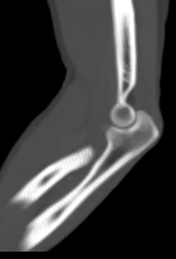
Önkol, eşsiz kemik, yumuşak doku ve eklem ilişkilerine sahip kompleks bir anatomik ve fonksiyonel ünitir; radius ve ulna şaft kırıklarında bu ilişkilerin bozulması ağrı, radioulnar eklemlerde instabilite ve azalmış hareket açıklığına yol açar. Radius malunionlarında üç boyutlu rekonstrüksiyonlar, eksenler boyunca üç yer değiştirme ve üç rotasyon parametresiyle deformiteyi değerlendirmek için kullanılabilir. Önkol kırıklarında eşlik eden yaralanmaları araştırmak için çekilen radyografilerde dirsek ve el bileği eklemleri de görüntü alanına alınmalıdır. Monteggia ve Galeazzi yaralanmaları başlangıçta gözden kaçabileceğinden, önkol kırıklarında ilgili eklem hizalanması da değerlendirilmelidir.
Okuma sırası
- Radius ve ulna diafizleri. Koronal ve transversal kesitlerde radius ve ulna kırık hatlarının proksimal, orta veya distal üçteki konumları belirlenir; kırık seviyelerinin birbirine göre uyumu değerlendirilir.
- Kırık fragmanları. Kırık fragmanlarındaki angülasyon, kayma ve uzunlamasına kısalma ölçülerek (örn. radiusun ulnaya göre mm cinsinden kısalması) kaydedilir.
- Önkol aksiyel kesitleri. Aksiyel kesitlerde radiusun ulna çevresindeki yönelimini ve kırık hattında rotasyonel basamak olup olmadığını kıyaslayın.
- Dirsek ve DRUJ. Proksimal uçta radial baş-kapitellum ve distal uçta ulnar baş-sigmoid çentik uyumunu doğrulayın.
- Önkolun iki eklem ucu. Son olarak radius başı, koronoid ve eklem yüzlerine uzanan kırık parçalarını arayıp eşlik eden kırıklı çıkığı dışlayın.
Faz ve pencere
- Kontrastsız tanısal
- Bilgisayarlı tomografi, radius ve ulna diafiz kırıklarında kallus oluşumunu ve kortikal devamlılığı değerlendirmede kullanılabilir. Radius malunionlarında üç boyutlu planlama, deformitenin altı konum parametresiyle değerlendirilmesine ve düzeltici osteotomi planlamasına yardımcı olabilir. Üç boyutlu BT rekonstrüksiyonu, karmaşık önkol yaralanmalarında eşlik eden kemik ve eklem bulgularını göstermeye yardımcı olabilir.
Önerilen pencereler: Kemik (G 1800 / M 400), Yumuşak doku (G 400 / M 40).
BT bulguları
- Radius diafiz kırığı — seviyeyi proksimal, orta veya distal şaft olarak belirtin; basit, kama veya çok parçalı morfolojiyi tarif edin.
- Radius ve ulna diafiz kırıklarının seviyeleri ayrı ayrı belirtilir; BT, kırık hattındaki kortikal devamlılığın değerlendirilmesine yardımcı olabilir.
- Kırık fragmanlarının yer değiştirmesi ve angülasyonu görüntülerde değerlendirilebilir; Galeazzi kırıklı çıkıklarında radius kısalması ulnaya göre ölçülebilir.
- Angülasyon — radius ve ulna kırıklarında apeks yönü ve iki kemiğin aks uyumu koronal-sagittal düzlemlerde incelenir.
- Rotasyonel deformite — aksiyel kemik konturlarında proksimal ve distal radius-ulna ilişkisi bozulabilir; ekstremitenin çekim rotasyonu yorumda hesaba katılır.
- Radius başı ve proksimal radioulnar eklem — radius başının kapitellumla ve ulna radial çentiğiyle ilişkisi korunmuş mu kontrol edilir.
- DRUJ ve ulnar stiloid — distal ulnar başın sigmoid çentik içindeki uyumu ve eşlik eden stiloid kırığı incelenir.
- Şaft kırığına komşu ekleme uzanan kırık çizgileri, hem metafizer hem de eklem içi tutulum gösterebilir ve stabil fiksasyonu zorlaştırabilir.
Ölçütler ve sınıflamalar
- AO/OTA 2018 önkol şaft sınıflaması
- Radius ve ulna şaft kırıkları, her iki kemiğin diafizini içeren önkol kırıklarıdır. Çift kemik önkol kırığında radius ve ulna şaftlarının ikisi de kırıktır.
Normalde
Önkolun kemik, yumuşak doku ve eklem ilişkilerinin bozulması ağrıya, radioulnar eklem instabilitesine ve hareket açıklığında azalmaya yol açabilir; ulnar yaylanma deformitesi de önkoldaki yük dağılımını değiştirebilir. Üç boyutli bilgisayarlı tomografi analizlerinde, kompleks deformitelerin doğru değerlendirilmesi ve düzeltici osteotomilerin planlanması için kontralateral sağlıklı taraf referans alınır.
Ayırıcı tanı
- Monteggia kırıklı çıkığı
- proksimal ulna kırığına radial baş çıkığı eşlik eder; radial baş hizası özellikle bozulmuştur.
- Galeazzi kırıklı çıkığı
- distal radius şaft kırığı ile DRUJ çıkığı veya subluksasyonu birliktedir.
- Radius şaft kırıklarında, özellikle Galeazi örüntüsünü dışlamak amacıyla distal radioulnar eklemdeki instabilite veya dislokasyon açısından değerlendirme yapılmalıdır.
- İzole ulna şaft kırığı
- ulna diafizinde kırık vardır; radius kırığı eşlik etmez ve distal/proksimal eklem uyumu ayrıca incelenir.
- Önkolun tek kemik kırığına eşlik eden komşu eklem çıkığı
- kırık şaftta tek kemikte görünürken radius başı ya da distal radioulnar eklem hizası bozulmuş olabilir.
Tuzaklar
- Ön kol kırıklarının tek başına değerlendirilmesi, hem proksimal hem de distal eklem yapısında eşlik edebilen patolojilerin gözden kaçmasına neden olabilir.
- Deformite analizlerinde, radius ve ulnadaki maksimum eğim yönlerinin belirlenmesi ve ideal düzeltme açılarının tayini için kontralateral sağlıklı ekstremite referans alınır.
- Önkol diafiz malunionlarında üç boyutlu kinematik analiz, kemik çarpışması veya santral bant blokajı gibi hareketi kısıtlayabilecek mekanizmaları gösterebilir.
Kendini dene
Radius şaft kırığına distal radioulnar eklem (DRUJ) çıkığı eşlik ediyor. Bu örüntü nasıl adlandırılır?
Cevabı göster
Galeazzi kırıklı çıkığı. Galeazzi yaralanması radius şaft kırığıyla DRUJ çıkığının birlikteliğidir. Monteggia yaralanmasında ulnar kırığa radial baş çıkığı eşlik eder; Essex–Lopresti yaralanması radial baş kırığı, distal radioulnar eklem çıkığı ve interosseöz membran yırtılmasıyla tanımlanır.
Önkol şaft kırığında dirsek ve el bileğinin ayrıca değerlendirilmesinin temel nedeni nedir?
Cevabı göster
Eşlik eden dirsek veya el bileği yaralanmalarını saptamak. Önkol kırıklarına başka yaralanmalar eşlik edebileceğinden dirsek ve el bileği de değerlendirilmelidir.
İlgili konular
Kaynaklar
Bu sayfadaki 40 cümle ve 2 quiz sorusu aşağıdaki kaynaklardan alıntılarla tek tek karşılaştırıldı; 22 cümle bu karşılaştırmada düzeltildi (2026-10-09).
- The Legitimacy for the Use of Splint Immobilization After Elastic Intramedullary Nailing of Both-bone Forearm Shaft Fractures in Childrenpmc.ncbi.nlm.nih.gov
- Sequential stress fractures of the ulna and radius in a patient with neurofibromatosis type 1: A case reportpmc.ncbi.nlm.nih.gov
- Complex posterolateral rotatory instability due to malunited radius fracture: diagnosis with four-dimensional computed tomography scan and corrective osteotomypmc.ncbi.nlm.nih.gov
- Complications following operative versus nonoperatively managed isolated distal third ulna shaft fractures: a retrospective single-institution cohort studypmc.ncbi.nlm.nih.gov
- Dual Fixation in Complex Long Bone Fractures: A Case Series of 30 Patientspmc.ncbi.nlm.nih.gov
- Ulnar Nerve Injury in Pediatric Midshaft Forearm Fractures: A Case Series.pubmed.ncbi.nlm.nih.gov
- Pancarpal dissociation, a very rare type of injury: A case report.pubmed.ncbi.nlm.nih.gov
- Multiple Unilateral Upper Limb Fractures in the Pediatric Setting - A Case Report.pubmed.ncbi.nlm.nih.gov
- An unprecedented pan-brachial injury: A case report of ipsilateral floating shoulder, floating elbow, and acetabular fracture following a psychosis-induced fall.pubmed.ncbi.nlm.nih.gov
- Case Report of Simultaneous Galeazzi Fracture Dislocation and Ipsilateral Scaphoid Fracture.pubmed.ncbi.nlm.nih.gov
- Emergency radiology of severe acute injury in extreme alpine summer sports: a pictorial essay.pubmed.ncbi.nlm.nih.gov
- All-Suture Anchor Fixation for Coronoid Process Fractures via a Transolecranon Fracture Approach.pubmed.ncbi.nlm.nih.gov
- Proximal humerus fractures in the elderly work up, classifications and fracture biomechanics.pubmed.ncbi.nlm.nih.gov
- High-Accuracy reduction of comminuted diaphyseal femur fractures using on-site 3-D-printed guides.pubmed.ncbi.nlm.nih.gov
- Dual Fixation in Complex Long Bone Fractures: A Case Series of 30 Patients.pubmed.ncbi.nlm.nih.gov
- The Impact of COVID-19-Related Restrictions on the Incidence of Diaphyseal and Distal Forearm Fractures: A Retrospective Analysis.pubmed.ncbi.nlm.nih.gov
- Obesity Is Independently Associated with Early Loss of Reduction After Casting in Pediatric Both-Bone Forearm Fractures: A Cohort Study of Children Aged 3-13 Years.pubmed.ncbi.nlm.nih.gov
- Glossary of terms for musculoskeletal radiology.pubmed.ncbi.nlm.nih.gov
- A hybrid technique combining intramedullary pinning with extramedullary plate fixation in unstable and comminuted radial head fractures following on-table reconstruction.pubmed.ncbi.nlm.nih.gov
- 2.5-mm articulated endoluminal forceps using a compliant mechanism.pubmed.ncbi.nlm.nih.gov
- Long-term safety and effectiveness of the "OptEase" vena cava filter.pubmed.ncbi.nlm.nih.gov
- The "Terrible Tetrad" of the Elbow: A Case Series of Two Patients with a Rare Quadruple-Component Injury and a Proposed New Classification Term.pubmed.ncbi.nlm.nih.gov
- Cementless Allograft-Prosthetic Composite Reconstruction Using an Interlocking Revision Stem for Salvage Reverse Total Shoulder Arthroplasty: A Case Report.pubmed.ncbi.nlm.nih.gov
- Conservative Treatment of Pediatric Fractures-Narrative Review of Acceptable Deformity and Remodeling Potential.pubmed.ncbi.nlm.nih.gov
- Predictors of failed closed reduction in pediatric Gartland type III and flexion-type supracondylar humerus fractures.pubmed.ncbi.nlm.nih.gov
- Remodelling in Children's Fractures and Limits of Acceptability.pubmed.ncbi.nlm.nih.gov
- Evaluation of proximal ulna morphology: a systematic review of varus angulation of the proximal ulna.pubmed.ncbi.nlm.nih.gov
- Three-wire technique for irreducible extension type Supracondylar humeral fracture in children.pubmed.ncbi.nlm.nih.gov
- The Treatment of Closed Finger and Metacarpal Fractures.pubmed.ncbi.nlm.nih.gov
- Patellar Dislocation: Workup and Decision-Making.pubmed.ncbi.nlm.nih.gov
- Computer-assisted 3D planned corrective osteotomies in eight malunited radius fractures.pubmed.ncbi.nlm.nih.gov
- Management of chronic monteggia fractures with ulnar osteotomy, external fixation and gradual closed reduction of radial head.pubmed.ncbi.nlm.nih.gov
- Efficacy of novel osteotomy techniques in the treatment of congenital radial head dislocation.pubmed.ncbi.nlm.nih.gov
- Progress in diagnosis and treatment of Essex-Lopresti injury in children.pubmed.ncbi.nlm.nih.gov
- Computed tomography-based predictors of ulnar-sided complications after distal radius fracture fixation.pubmed.ncbi.nlm.nih.gov
- Midsectional Magnetic Resonance Imaging Analysis of the Sigmoid Notch of the Distal Radioulnar Joint.pubmed.ncbi.nlm.nih.gov
- Distal Radioulnar Joint Instability and Associated Injuries: A Literature Review.pubmed.ncbi.nlm.nih.gov
- Radiographic Outcomes of Dorsal Spanning Plate for Treatment of Comminuted Distal Radius Fractures in Non-Elderly Patients.pubmed.ncbi.nlm.nih.gov
- Treatment of diaphyseal forearm fractures in children.pubmed.ncbi.nlm.nih.gov
- Nonoperative treatment of both-bone forearm shaft fractures in children: predictors of early radiographic failure.pubmed.ncbi.nlm.nih.gov
- Three-Dimensional Surgical Planning and Patient-Specific Osteotomy Guide in the Management of Chronic Monteggia Fracture: A Case Report.pubmed.ncbi.nlm.nih.gov
- Management of a Nonunion Type IIC Monteggia Fracture.pubmed.ncbi.nlm.nih.gov
- Rare pediatric Galeazzi lesion with progressive wrist deformity.pubmed.ncbi.nlm.nih.gov
- Ballistic Galeazzi Fractures: A Case Series.pubmed.ncbi.nlm.nih.gov
- Emergency Medicine Management of Distal Radius and Ulna Fractures: Narrative Review.pubmed.ncbi.nlm.nih.gov
- Elastic intramedullary nailing for recurrent radial fracture associated with persistent ulnar bowing deformity in a pediatric patient: a case report.pubmed.ncbi.nlm.nih.gov
- Sequential stress fractures of the ulna and radius in a patient with neurofibromatosis type 1: A case report.pubmed.ncbi.nlm.nih.gov
- Complications following operative versus nonoperatively managed isolated distal third ulna shaft fractures: a retrospective single-institution cohort study.pubmed.ncbi.nlm.nih.gov
- Ipsilateral concomitant fractures of the distal radius and radial head: a radial Joint-Coupled Pattern (JCP).pubmed.ncbi.nlm.nih.gov
- Ulnar-Sided Wrist Pain: Systematic Clinical Approach and Principles of Treatment.pubmed.ncbi.nlm.nih.gov
- Radiocapitellar Alignment in Suspected Pediatric Monteggia Lesions: Narrative Review and Imaging Interpretation Framework.pubmed.ncbi.nlm.nih.gov
- Case-control study of the characteristics and risk factors of hot clot artefacts on 18F-FDG PET/CT.pubmed.ncbi.nlm.nih.gov
- Molecular-informed image classification for predicting drug sensitivity in cancer therapy.pubmed.ncbi.nlm.nih.gov
- Update on elbow arthroplasties with emphasis on imaging.pubmed.ncbi.nlm.nih.gov
- High-Frequency and Ultra-High-Frequency Ultrasound in Dermatologic Diseases and Aesthetic Medicine.pubmed.ncbi.nlm.nih.gov
- Bone Phenotyping Approaches in Human, Mice and Zebrafish - Expert Overview of the EU Cost Action GEMSTONE ("GEnomics of MusculoSkeletal traits TranslatiOnal NEtwork").pubmed.ncbi.nlm.nih.gov
- Multisystem Progeroid Syndrome With Lipodystrophy, Cardiomyopathy, and Nephropathy Due to an LMNA p.R349W Variant.pubmed.ncbi.nlm.nih.gov
- Radius crossover sign: an indication of malreduced radius shaft greenstick fractures.pubmed.ncbi.nlm.nih.gov
- Can CT-Scan Measurements of Humpback Deformity, Dislocation, and the Size of Bony Cysts Predict Union after Surgery for Scaphoid Nonunion?pubmed.ncbi.nlm.nih.gov
- Fractures of distal radius: an overview.pubmed.ncbi.nlm.nih.gov
- Association of Chiropractic Colleges Educational Conference and Research Agenda Conference 2025: Transformative Trends in Healthcare.pubmed.ncbi.nlm.nih.gov
- Acute Plastic Deformation of the Forearm Involving Both Bones in an Adolescent: Distinct Deformity Vectors Requiring Individualized Three-Dimensional Correction.pubmed.ncbi.nlm.nih.gov
- Beyond Palmer: a topographic classification of non-Palmer triangular fibrocartilage complex injuries.pubmed.ncbi.nlm.nih.gov
- Osteotomies: Indications, Imaging Appearance, Surgical Techniques, and Complications.pubmed.ncbi.nlm.nih.gov
- Radial head arthroplasty: A pillar of stability in complex elbow fractures.pubmed.ncbi.nlm.nih.gov
- Outcomes and challenges in the surgical treatment of trans-olecranon fracture-dislocations: A case series study.pubmed.ncbi.nlm.nih.gov
- Subcapital Realignment of the Displaced Pediatric Radial Head-Technical Description and Early Clinical Results.pubmed.ncbi.nlm.nih.gov
- Ulnar lengthening in multiple hereditary exostosis with forearm deformity: a long-term follow-up study.pubmed.ncbi.nlm.nih.gov
- Interosseous Membrane Reconstruction with Suture Button for Essex-Lopresti Fracture and Lateral Collateral Ligament Complex Rupture Leading to Rerupture of the Ligament and Radial Head Dislocation.pubmed.ncbi.nlm.nih.gov
- Impact of coordinate system alignment on CT-based distal radius morphometry relevant to surgical planning: A bilateral comparative study.pubmed.ncbi.nlm.nih.gov
- Four-dimensional computed tomography-derived distal radioulnar joint osteokinematics during pronosupination with unilateral triangular fibrocartilage complex injuries.pubmed.ncbi.nlm.nih.gov
- Comparing Outcomes between Arthroscopic-Assisted Reduction and Fluoroscopic Reduction in AO Type C Distal Radius Fracture Treatment.pubmed.ncbi.nlm.nih.gov
- Interventions for treating proximal humeral fractures in adults.pubmed.ncbi.nlm.nih.gov
- Surgical interventions for treating extracapsular hip fractures in older adults: a network meta-analysis.pubmed.ncbi.nlm.nih.gov
- Clinical efficacy of Masquelet technique in the treatment of tibial and fibular bone defects caused by severe gunshot wounds.pubmed.ncbi.nlm.nih.gov
- Diagnostic Imaging of the Skeletal System: Overview of Applications in Human and Veterinary Medicine.pubmed.ncbi.nlm.nih.gov
- Musculoskeletal Ultrasound and Protein-Based Hydrogels: Novel Approaches for the Diagnosis and Treatment of Sports Injuries.pubmed.ncbi.nlm.nih.gov
- A Systematic Approach to the Evaluation of Acute Musculoskeletal Injuries in the Emergency Department.pubmed.ncbi.nlm.nih.gov
- 3D CT Anatomy and Morphology of the Appendicular Skeleton of European Pond Turtle (Emys orbicularis).pubmed.ncbi.nlm.nih.gov
- Reconstruction of malunited diaphyseal fractures of the forearm.pubmed.ncbi.nlm.nih.gov
- Complex forearm deformities: operative strategy in posttraumatic pathology.pubmed.ncbi.nlm.nih.gov
- Bone grafts and bone substitutes for treating distal radial fractures in adults.pubmed.ncbi.nlm.nih.gov
- External fixation versus conservative treatment for distal radial fractures in adults.pubmed.ncbi.nlm.nih.gov
- Different methods of external fixation for treating distal radial fractures in adults.pubmed.ncbi.nlm.nih.gov
- Three-Dimensional Planning and Patient-Specific Instruments in Complex Forearm Malunion.pubmed.ncbi.nlm.nih.gov
- Indications and Timing of Guided Growth Techniques for Pediatric Upper Extremity Deformities: A Literature Review.pubmed.ncbi.nlm.nih.gov
- Evidence for peripheral neuroinflammation after acute whiplash.pubmed.ncbi.nlm.nih.gov
- International Multicenter Validation of an Expanded AI Diagnostic System for 18 Pathologies in Thoracic and Musculoskeletal Radiography.pubmed.ncbi.nlm.nih.gov
- High-affinity alphavbeta3 integrin targeted optical probe as a new imaging biomarker for early atherosclerosis: initial studies in Watanabe rabbits.pubmed.ncbi.nlm.nih.gov
- ESTES recommendations regarding distal radius fractures.pubmed.ncbi.nlm.nih.gov
- Management of Pediatric Hand Injuries in the Acute Setting Part 2: Fractures and Dislocations.pubmed.ncbi.nlm.nih.gov
- Occipitocervical dissociation: A narrative review and case series with a novel management algorithm proposal.pubmed.ncbi.nlm.nih.gov
- Intramedullary Headless Screw Feasibility for Anatomical Reduction in II-V Metacarpal Fractures: A CT-Based Morphometric Study.pubmed.ncbi.nlm.nih.gov
- Analysis of characteristics and therapeutic outcomes in children with Dubberley type IIA capitellar fractures.pubmed.ncbi.nlm.nih.gov
- Variation in trabecular bone microarchitecture across rhesus macaque (Macaca mulatta) load-bearing joints.pubmed.ncbi.nlm.nih.gov
- Neotaphonomic characteristics of vertebrate site formation in underwater caves.pubmed.ncbi.nlm.nih.gov
- Impact of proximal tibio-fibular joint injury on posterolateral corner knee reconstructions, strategies for achieving knee stability in combined injuries: a systematic review.pubmed.ncbi.nlm.nih.gov
- Open Reduction and Internal Fixation of Distal Radius Fractures with Complete Intra-articular Involvement and Diaphyseal Extension.pubmed.ncbi.nlm.nih.gov
- Pediatric Chronic Monteggia Fractures: Insights From a Comprehensive Review.pubmed.ncbi.nlm.nih.gov
- Reconstructing the interosseous ligament: A technique using a single bundle synthetic tape.pubmed.ncbi.nlm.nih.gov
- Mini-Anchor Fixation vs Conservative Treatment for Hauck Type II Ulnar Styloid Fractures Associated With Distal Radius Fractures: A Retrospective Cohort Study.pubmed.ncbi.nlm.nih.gov
- Padua Days on Muscle and Mobility Medicine, March 25-29, 2025, Hotel Petrarca, Euganean Thermae, Italy: Program and Abstracts.pubmed.ncbi.nlm.nih.gov
- Evidence for deliberate burial of the dead by <i>Homo naledi</i>.pubmed.ncbi.nlm.nih.gov
- Imaging in Hip Arthroplasty Management Part 2: Postoperative Diagnostic Imaging Strategy.pubmed.ncbi.nlm.nih.gov
- Multicenter reliability study of the universal long bone nonunion classification.pubmed.ncbi.nlm.nih.gov
- Exploring the mechanisms of acupuncture in improving cognitive function in post-COVID-19 myalgic encephalomyelitis/chronic fatigue syndrome: study protocol for a randomized controlled trial using multimodal MRI.pubmed.ncbi.nlm.nih.gov
- Radiation Dose and Risk in the Radiological Investigation of Suspected Non-Accidental Injury (NAI).pubmed.ncbi.nlm.nih.gov
- Is transphyseal intramedullary fixation of the distal radius in pediatric fractures a safe procedure? An MRI study.pubmed.ncbi.nlm.nih.gov
- A Prospective Comparative Study Evaluating Functional Outcomes in Adolescents Undergoing Dynamic Compression Plating Versus Intramedullary Nailing for Both-Bone Diaphyseal Forearm Fractures.pubmed.ncbi.nlm.nih.gov
- Epidemiology and Incidence of Upper Limb Fractures: A UK Level 1 Trauma Center Perspective.pubmed.ncbi.nlm.nih.gov
- Reliable classification of children's fractures according to the comprehensive classification of long bone fractures by Müller.pubmed.ncbi.nlm.nih.gov
- Postoperative Outcomes of Anteromedial Approach in Patients with Elbow Varus Posteromedial Rotatory Instability: A Retrospective Study.pubmed.ncbi.nlm.nih.gov
- Quantifying the Toll of Disuse: A Meta-Analysis of Skeletal Muscle Mass and Strength Loss Following Upper Limb Immobilization.pubmed.ncbi.nlm.nih.gov
- Can Personalized 3D Kinematic Modeling Predict Loss of Pronation and Supination in Diaphyseal Forearm Malunions? A Clinical Validation Study.pubmed.ncbi.nlm.nih.gov
- Elbow Varus posteromedial rotatory instability: Reflections from a multidimensional perspective.pubmed.ncbi.nlm.nih.gov
Eğitim amaçlıdır; hasta başında tanı ve tedavi kararının yerine geçmez. İçerik yapay zekâ destekli bir süreçle güncel literatüre göre cümle cümle kaynaklarla karşılaştırıldı, ancak uzman radyolog incelemesinden geçmedi. Klinik kararlarda güncel kılavuzları, kurum protokollerini ve radyoloji raporunu esas alın.