Özet
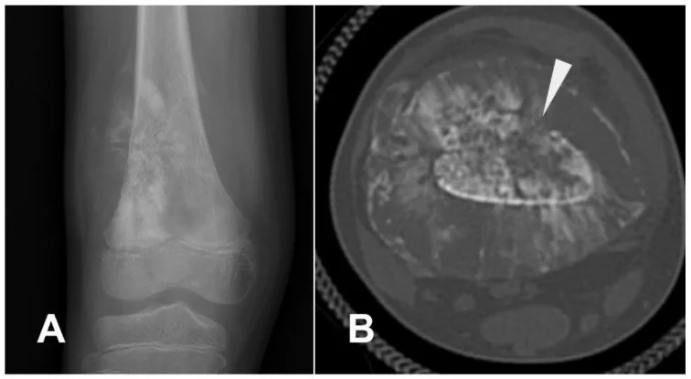
Özellikle genç hastada ilerleyici kemik ağrısı veya şişlik eşliğinde distal femur ve proksimal tibia metafizindeki agresif lezyon osteosarkom açısından dikkatle incelenir. Radyografi ilk değerlendirmede temel yöntemdir. Kontrastsız kemik algoritmalı BT seçilmiş olgularda tümör osteoidinin mineralizasyonunu, kortikal yıkımı, periostal kemik oluşumunu ve patolojik kırığı gösterir; intramedüller ve yumuşak doku yayılımı için MR temel tamamlayıcı incelemedir. Mineralize tümör matriksi BT'de bulutumsu, yoğun osteoid alanları şeklinde görülebilir ve ekstraosseöz komponent içine taşabilir. Lezyonun agresifliği veya kırığı atlanırsa patolojik kırık ve lokal uzanım eksik tarif edilir.
Faz ve pencere
- Kontrastsız tanısal
- Metafiz merkezli lezyonda litik ve sklerotik alanların karışımı olan osteoid matriks, kortikal yıkım, agresif periost reaksiyonu ve mineralize ekstraosseöz yumuşak doku komponenti incelendiğinde; ince kesit multiplanar rekonstrüksiyonlar ve uygun pencere ayarları kemik matriks detaylarını belirginleştirir.
- Kemik algoritması tanısal
- İnce kesit multiplanar rekonstrüksiyonlar osteoid mineralizasyon dağılımını, kortikal yıkımın uzunluğunu, periostal spikülleri/Codman tipi periost kalkmasını ve tümör içinden geçen patolojik kırığı gösterir.
Önerilen pencereler G genişlik, M merkez; değerler Hounsfield birimi (HU)
- KemikG 1800 / M +400
- Yumuşak dokuG 400 / M +40
- AkciğerG 1500 / M −600
BT bulguları
- Agresif metafizer lezyon — distal femur veya proksimal tibia metafizinden metadiafize uzanan, belirsiz geçiş zonlu litik-sklerotik kemik destrüksiyonu.
- Bulutumsu osteoid matriks — lezyon içinde yoğun, düzensiz ve amorf kemiksi mineralizasyon alanları.
- Kortikal yıkım — endosteal erozyonla başlayan incelme, korteks boyunca geniş destrüksiyon veya dış korteksin kesintisi.
- Spiküler periostal reaksiyon — korteksten dışa ışınsal uzanan mineralize yeni kemik.
- Codman tipi periost kalkması — tümör kenarında periostun kaldırıldığı üçgenimsi mineralize kenar.
- Ekstraosseöz tümör komponenti — osteosarkom çevre yumuşak dokulara uzanabilir; BT kemik ve yumuşak doku tümör matrikslerini göstermede ayrıntı sağlar.
- Patolojik kırık — osteosarkomda BT, kemik yapısını ve tümör matriksini değerlendirmenin yanı sıra patolojik kırıkların saptanmasına da katkı sağlar.
- Akciğer nodülleri — osteosarkom evrelemesinde BT uzak metastazları saptamada kullanılır; metastazlar en sık akciğerlerde görülür.
Normalde
Kontrastsız BT ve kemik penceresi, distal femur ve proksimal tibia metafizlerinde normal kemik yoğunluğunu ve trabeküler yapıyı gösterirken; lezyonun radyolüsant (normal kemikten daha düşük), sklerotik veya karışık alanları ile bu normal yapıdan ayrımını detaylandırır. BT'de kemik yapısını değerlendirirken, MR'da tümörün uzunlamasına yayılımını ve en yakın eklem yüzüne uzaklığını inceleyin.
Normal BT ile kıyasla
Aynı bölgenin, kaynak veri setinde patoloji içermediği belirtilen bir BT incelemesini kesit kesit kaydırın; organ sınırlarını açarak anatomiyi hasta görüntüsüyle karşılaştırın. Küçük rastlantısal bulgular tümüyle dışlanmamıştır.
Kaynak: TotalSegmentator v2.01 veri seti (Wasserthal ve ark., Radiology: Artificial Intelligence 2023), CC BY 4.0, vaka s0685; organ sınırları veri setinin otomatik segmentasyonundan, birleştirilerek sadeleştirildi. Görüntüler 2,5 mm kesit aralığına yeniden örneklendi.
Ayırıcı tanı
- Ewing sarkomu
- osteosarkomla birlikte sunburst tipi periostal reaksiyon görülebilen agresif kemik lezyonları arasındadır; bu bulgu tek başına osteosarkoma özgü değildir.
- Brodie apsesi
- distal femurda iyi kortike, sklerotik kenarlı osteolitik lezyon şeklinde görülebilir.
- Osteomiyelit
- enfeksiyon ile malign kemik tümörlerinin klinik ve görüntüleme özellikleri örtüşebileceğinden ayırıcı tanı güç olabilir.
- Telanjiektatik osteosarkom
- osteosarkomun farklı görüntüleme özellikleri gösterebilen alt tiplerinden biridir ve anevrizmal kemik kistiyle görüntüleme bulguları örtüşebilir; görüntüleme tek başına bu tümörü dışlayamaz.
- Parosteal osteosarkom
- kemik yüzeyinden gelişen osteosarkom alt tiplerinden biridir ve konvansiyonel osteosarkomdan farklı klinik ve biyolojik davranış gösterebilir.
Tuzaklar
- Sklerotik osteoid matriksin değerlendirilmesinde BT, tümörün kemik matriksini incelemek için tercih edilen yöntemdir.
- Agresif periostal reaksiyon tek başına osteosarkoma özgü değildir; matriks mineralizasyonu, metafizer yerleşim ve ekstraosseöz kitleyi birlikte değerlendirin.
- Yumuşak doku kitlesindeki mineralizasyon BT'de tümör yayılımını gösterebilir; yalnız kemik penceresindeki primer odağı tarif edip yumuşak doku penceresini atlamayın.
- BT medüller tümör uzunluğunu ve nörovasküler ilişkiyi MR kadar göstermez; kemik destrüksiyonunun tüm lokal yayılımı belirlediği izlenimini vermeyin.
- Sunburst tipi spiküler periostal reaksiyon ve Codman üçgeni agresiflik bulgularıdır; osteosarkoma özgü değildir ve tek başına tanı koydurmaz.
Kendini dene
Tümör hücrelerinin osteoid matriks üretmesiyle tanımlanan malign primer kemik tümörü hangisidir?
Cevabı göster
Osteosarkom. Osteosarkom, tümör hücrelerinin osteoid matriks üretmesiyle tanımlanan malign primer kemik tümörüdür. Diz çevresindeki uzun kemiklerin metafizleri sık yerleşim yeridir; BT kemik matriksini değerlendirmede tercih edilir.
Kemik lezyonlarında iyi sınırlı veya sklerotik kenar hangi genel davranışla daha çok ilişkilidir?
Cevabı göster
Benign davranış. İyi sınırlı veya sklerotik kenarlar benign lezyonlarda tipiktir; görüntüleme bulguları kesin histopatolojik tanının yerini tutmaz.
İlgili konular
Kaynaklar
Bu sayfadaki 38 cümle ve 2 quiz sorusu aşağıdaki kaynaklardan alıntılarla otomatik olarak karşılaştırıldı; 19 cümle bu karşılaştırmada düzeltildi (2026-10-09).
- Multimodal Imaging of Osteosarcoma: From First Diagnosis to Radiomicspmc.ncbi.nlm.nih.gov
- The quiet flame: whole-body MRI and the underestimated burden of pediatric chronic non-bacterial osteomyelitis (CNO).pubmed.ncbi.nlm.nih.gov
- Clinical and Radiological Concordance of Bone Tumours Using Histopathology as the Gold Standard: A Pilot Study.pubmed.ncbi.nlm.nih.gov
- Hybrid Prosthetic and Biological Reconstruction Using Extracorporeal Radiotherapy for Long-Segment Humeral Osteosarcoma: A Case Report.pubmed.ncbi.nlm.nih.gov
- Comparison of FRACTURE MRI, Conventional Radiography, and CT in the Evaluation of Bone Tumors.pubmed.ncbi.nlm.nih.gov
- The role of imaging in extremity sarcoma surgery.pubmed.ncbi.nlm.nih.gov
- Imaging features of primary tumors of the spine: A pictorial essay.pubmed.ncbi.nlm.nih.gov
- The solitary skull mass: An atypical presentation of anaplastic large cell lymphoma.pubmed.ncbi.nlm.nih.gov
- Imaging of Skull Base Tumors.pubmed.ncbi.nlm.nih.gov
- Proximal Tibial Giant Cell Tumor in a Skeletally Immature Child: A Diagnostic Dilemma and Review of the Literature.pubmed.ncbi.nlm.nih.gov
- Intraosseous tumours of the knee.pubmed.ncbi.nlm.nih.gov
- Beyond Chordoma: A Comprehensive Review of Sacral Lesions.pubmed.ncbi.nlm.nih.gov
- Malignant Transformation of Musculoskeletal Lesions with Imaging-Pathology Correlation-Part 1: Bone Lesions.pubmed.ncbi.nlm.nih.gov
- Myoepithelial tumor of the bone: radiologic and histopathologic correlation.pubmed.ncbi.nlm.nih.gov
- Calcified Chondroid Mesenchymal Neoplasm of the Temporomandibular Joint: A Rare and Recently Described Benign Tumor.pubmed.ncbi.nlm.nih.gov
- Multimodal Imaging of Osteosarcoma: From First Diagnosis to Radiomics.pubmed.ncbi.nlm.nih.gov
- Odontogenic myxoma with rare sunburst periosteal reaction: report of two cases and review of the literature.pubmed.ncbi.nlm.nih.gov
- Intraosseous Venous Malformation of the Rib With EWSR1-NFATC1 Fusion Mimicking Malignancy: A Pediatric Case Report.pubmed.ncbi.nlm.nih.gov
- When plasmacytoma defies expectations: An unusual maxillary presentation with mixed imaging features in a young patient.pubmed.ncbi.nlm.nih.gov
- Osteosarcoma: A Comprehensive Morphological and Molecular Review with Prognostic Implications.pubmed.ncbi.nlm.nih.gov
- A case of pelvic Ewing sarcoma misinterpreted as osteomyelitis: diagnostic and clinical implications.pubmed.ncbi.nlm.nih.gov
- Secondary distal femoral osteosarcoma in a non-irradiated survivor of bilateral retinoblastoma.pubmed.ncbi.nlm.nih.gov
- TRAF7-Mutated Fibromyxoid Spindle Cell Tumor of Bone: An Osseous Case Expanding the Spectrum of TRAF7-Mutated Tumors With Over 20 Years Clinical Follow-Up.pubmed.ncbi.nlm.nih.gov
- Risk Factors for Pathologic Fracture in Dogs With Confirmed or Suspected Appendicular Osteosarcoma Treated With Low- or Standard-Dose-Rate Palliative External Beam Radiation Therapy.pubmed.ncbi.nlm.nih.gov
- Use of the IlluminOss Photodynamic Bone Stabilisation System Following Stereotactic Body Radiation Therapy for Limb-Sparing in Six Dogs With Presumptive Appendicular Osteosarcoma.pubmed.ncbi.nlm.nih.gov
- Paget's disease of bone with superimposed osseous metastasis from lung carcinoma: A case report.pubmed.ncbi.nlm.nih.gov
- Unveiling Primary Bone Tumors of the Spine: A Review of Essential Imaging Clues.pubmed.ncbi.nlm.nih.gov
- Non-odontogenic tumors of the facial bones in children and adolescents: role of multiparametric imaging.pubmed.ncbi.nlm.nih.gov
- Brodie's abscess of the distal radius: an unusual complication after percutaneous pinning.pubmed.ncbi.nlm.nih.gov
- Imaging of lower extremity infections: predisposing conditions, atypical infections, mimics, and differentiating features.pubmed.ncbi.nlm.nih.gov
- Transphyseal Proximal Humeral Aneurysmal Bone Cyst with Pathologic Fracture in a Child: A Case Report.pubmed.ncbi.nlm.nih.gov
- Rare giant cell tumour of the left parieto-occipital skull in an 8-year-old female patient: a case report.pubmed.ncbi.nlm.nih.gov
- Case Report: Ossifying parosteal lipoma of the mandible with cortical continuity: surgical considerations from a rare case and literature review.pubmed.ncbi.nlm.nih.gov
- Extraosseous osteochondroma of the hypothenar region: a case report and literature review.pubmed.ncbi.nlm.nih.gov
- Imaging of ewing sarcoma: an updated analysis including presenting features, prognostic imaging biomarkers, and treatment response assessment.pubmed.ncbi.nlm.nih.gov
- Recurrent Fibro-Osseous Pseudotumour of the Digit Mimicking Infection: A Narrative Review and Case Report.pubmed.ncbi.nlm.nih.gov
- Dedifferentiated Central Chondrosarcoma: A Clinical, Histopathological, and Immunohistochemical Analysis of 57 Cases.pubmed.ncbi.nlm.nih.gov
- A Novel 3D-Printed Strut-Type Prosthesis Reconstructs the Hemicortical Defect After Distal Femur Parosteal Osteosarcoma Resection: The Mid-Term Follow-Up Outcome.pubmed.ncbi.nlm.nih.gov
- Human biomarker navigator.pubmed.ncbi.nlm.nih.gov
- Diagnostic and Treatment Strategies for Adult Non-Ossifying Fibromas: A Case Report and Literature Review.pubmed.ncbi.nlm.nih.gov
- Primary osseous kaposiform hemangioendothelioma confined to long bones: a retrospective imaging analysis of 12 pediatric cases.pubmed.ncbi.nlm.nih.gov
- Imaging of pediatric musculoskeletal trauma: Age-specific considerations and challenges.pubmed.ncbi.nlm.nih.gov
- Metal implantation and high-grade osteosarcoma of the femur: a case report and review of the literature.pubmed.ncbi.nlm.nih.gov
- A Rare Cause of Recurrent Left Knee Pain in an Adolescent Male: A Case Report of Distal Femoral Brodie's Abscess.pubmed.ncbi.nlm.nih.gov
- Brodie's Abscess: Case Report of an Unusual Presentation of <i>Neisseria gonorrhoeae</i>.pubmed.ncbi.nlm.nih.gov
- Unusual Sites of Brodie's Abscess and the use of Calcium Sulfate Beads: A Case Series.pubmed.ncbi.nlm.nih.gov
- High-Grade Undifferentiated Pleomorphic Sarcoma of the Distal Tibia Presenting as a Radiographically Benign-Appearing Lesion: A Case Report and Review of the Literature.pubmed.ncbi.nlm.nih.gov
- Surgically Treated Benign Bone Tumors and Tumor-like Conditions in the Pediatric Population-A 10-Year Institutional Experience.pubmed.ncbi.nlm.nih.gov
- Bone lesions of the tibia: Multimodal iconographic review and diagnostic algorithms, Part 1: Diagnostic algorithms, dysplasia and diaphyseal lesions.pubmed.ncbi.nlm.nih.gov
- Diagnosis and Current Treatment of Aneurysmal Bone Cysts.pubmed.ncbi.nlm.nih.gov
- Subcutaneous extraskeletal osteosarcoma of the gluteal region: a case report from a resource-limited setting.pubmed.ncbi.nlm.nih.gov
- Analysis and Historical Evolution of Paediatric Bone Tumours: The Importance of Early Diagnosis in the Detection of Childhood Skeletal Malignancies.pubmed.ncbi.nlm.nih.gov
- En Bloc Total Vertebrectomy of the Thoracic and Lumbar Spine.pubmed.ncbi.nlm.nih.gov
- Novel report of an osteogenic tumor in a late Jurassic Mamenchisaurid from Thailand.pubmed.ncbi.nlm.nih.gov
- Sacral intraosseous schwannoma in an adolescent patient: A case report.pubmed.ncbi.nlm.nih.gov
- CT-guided bone biopsies with non-diagnostic results in pediatric patients-a multi-institutional 10-year retrospective review.pubmed.ncbi.nlm.nih.gov
- Diagnostic challenges and therapeutic approaches in pediatric osteosarcoma: A case report.pubmed.ncbi.nlm.nih.gov
- The resection of extraosseous osteosarcoma was accompanied by the occurrence of pulmonary metastasis and distal metastasis at the primary site: a case report.pubmed.ncbi.nlm.nih.gov
- Soft Tissue Special Issue: Imaging of Bone and Soft Tissue Sarcomas in the Head and Neck.pubmed.ncbi.nlm.nih.gov
- The Neuro-Bone Axis in Metastatic Progression: Innervation, Neuro-Immune-Osteoclast Crosstalk, and Therapeutic Opportunities.pubmed.ncbi.nlm.nih.gov
- Aptamer-Functionalized Nanocarriers for Bone and Joint Diseases: Engineering Design Rules and Translational Roadmap.pubmed.ncbi.nlm.nih.gov
- International Society of Paediatric Surgical Oncology (IPSO) Surgical Practice Guidelines.pubmed.ncbi.nlm.nih.gov
- Normal Variants and Pitfalls of 18 F-FDG PET/CT Imaging in Pediatric Oncology.pubmed.ncbi.nlm.nih.gov
- Chondrosarcoma in childhood: the radiologic and clinical conundrum.pubmed.ncbi.nlm.nih.gov
- Synovial Chondromatosis of the Elbow: A Case Report and Discussion of Feared Complications.pubmed.ncbi.nlm.nih.gov
- Pharmacotherapy of Kienböck Disease and the Carpal Avascular Necrosis Family: A Narrative Review with a Four-Axis Translation Framework.pubmed.ncbi.nlm.nih.gov
- Surface osteosarcoma: Clinical features and therapeutic implications.pubmed.ncbi.nlm.nih.gov
- Patellar Non-Traumatic Pathologies: A Pictorial Review of Radiologic Findings.pubmed.ncbi.nlm.nih.gov
- Giant cell tumor of the anterior rib masquerading as a breast mass: a case report and review of current literature.pubmed.ncbi.nlm.nih.gov
- Modular Revision of a Failed Patient-Specific 3D-Printed Scapulo-Humeral Endoprosthesis in a Dog with Proximal Humeral Osteosarcoma: First Clinical Communication.pubmed.ncbi.nlm.nih.gov
- State of the Art and New Concepts in Giant Cell Tumor of Bone: Imaging Features and Tumor Characteristics.pubmed.ncbi.nlm.nih.gov
- Glossary of terms for musculoskeletal radiology.pubmed.ncbi.nlm.nih.gov
- MRI Findings of Acute on Chronic Osteomyelitis of Tibia in a 12-Year-Old Child.pubmed.ncbi.nlm.nih.gov
- Current updates in MSK infection imaging: A narrative review.pubmed.ncbi.nlm.nih.gov
- Lower extremity infections: Essential anatomy and multimodality imaging findings.pubmed.ncbi.nlm.nih.gov
- Case Report: Adult proximal humeral aneurysmal bone cyst: radical resection and reconstruction with osteoconductive allograft & reverse arthroplasty-Ecuador's first reported case and functional outcomes.pubmed.ncbi.nlm.nih.gov
- Dedifferentiated parosteal osteosarcoma with a novel low-grade leiomyosarcomatous and high-grade conventional osteosarcomatous differentiation: Case report and review of literature.pubmed.ncbi.nlm.nih.gov
- Non-Oncological Progression of an Aneurysmal Bone Cyst (ABC) in a Puppy Following Surgical Treatment.pubmed.ncbi.nlm.nih.gov
- A local vascularized scapula bone graft to treat posterior glenohumeral instability-an innovative surgical technique.pubmed.ncbi.nlm.nih.gov
- Case reports with literature review of an aneurysmal bone cyst in the maxilla and mandible of two juvenile dogs.pubmed.ncbi.nlm.nih.gov
- Spinal lesions: a comprehensive radiologic overview.pubmed.ncbi.nlm.nih.gov
- Ultrasonography in Canine Carpal Disorders.pubmed.ncbi.nlm.nih.gov
- Isolated Atypical Pedicle Stress Fractures in Patients on Prolonged Bisphosphonate Therapy: Report of Two Cases and a Literature-Based Proposal for Diagnostic Criteria.pubmed.ncbi.nlm.nih.gov
- Impaired bone healing upon neutrophil-specific adrenoreceptor beta 2 knockout in non-osteoporotic and osteoporotic mice.pubmed.ncbi.nlm.nih.gov
- MRI nomenclature for musculoskeletal infection.pubmed.ncbi.nlm.nih.gov
- MRI of Knee Joint Lesions: An Observational Study of Traumatic, Degenerative, Cystic, and Neoplastic Pathologies.pubmed.ncbi.nlm.nih.gov
Eğitim amaçlıdır; hasta başında tanı ve tedavi kararının yerine geçmez, tıbbi tavsiye değildir. Metinler yapay zekâ desteğiyle yazıldı ve otomatik bir adımda kaynak alıntılarıyla karşılaştırıldı; bu bir tıbbi doğrulama değildir ve içerik uzman radyolog incelemesinden geçmedi. Klinik kararlarda güncel kılavuzları, kurum protokollerini ve radyoloji raporunu esas alın.