Özet
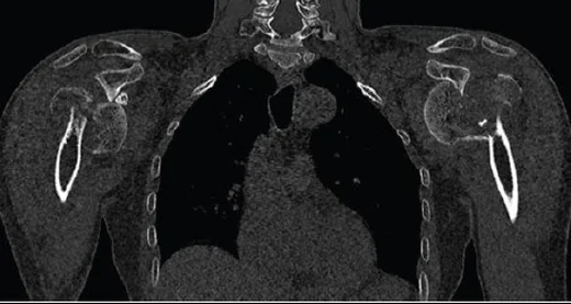
Düşme veya yüksek enerjili travma sonrası omuz ağrısı olan hastada humerus başı, büyük ve küçük tüberkül ile şaft arasındaki kırık ilişkisi birlikte haritalanır. Kontrastsız ince kesit BT, grafide üst üste binen parçaları ayırır; eklem yüzü, metafizer bağlantı ve glenoidle hizalanmayı çok düzlemli gösterir. Neer sınıflaması her anatomik parçayı kırık hattı sayısına göre değil, komşu parçaya göre yer değiştirme ve açılanmasına göre sayar. Başın glenoidden çıkması, eklem yüzü yarılması veya medial desteğin kaybı atlanırsa yaralanmanın gerçek karmaşıklığı ve başın kanlanma riski eksik tarif edilir.
Faz ve pencere
- Kontrastsız tanısal
- İnce kesit kemik rekonstrüksiyonunda kırık çizgileri, kortikal basamaklanma, parçalar arası translasyon ve impaksiyon izlenir; humerus başının glenoid ile ilişkisi ve eklem yüzündeki çökme aynı hacimde değerlendirilir. Bu kemik yaralanmasını tanımlamak için intravenöz kontrast gerekmez.
- Kemik algoritması tanısal
- Aksiyel kaynak görüntüler koronal ve sagittal rekonstrüksiyonlarla birlikte incelenerek baş, iki tüberkül ve şaftın ayrı parçalar olup olmadığı; medial kalcar ve baş-şaft kortikal temasının korunup korunmadığı belirlenir.
Önerilen pencereler G genişlik, M merkez; değerler Hounsfield birimi (HU)
- KemikG 1800 / M +400
BT bulguları
- Dört Neer parçası — eklem segmenti (humerus başı), büyük tüberkül, küçük tüberkül ve şaftın kırık hatlarıyla birbirinden ayrılıp ayrılmadığını belirleyin.
- Yer değiştirmiş parça — komşu anatomik segmente göre translasyon veya açılanma gösteren parçayı sayın; kırık çizgisi tek başına yeni bir Neer parçası oluşturmaz.
- Baş-şaft açısı — başın varus ya da valgus eğimini ve şaftla kortikal temasın sürüp sürmediğini koronal düzlemde tarif edin.
- Metafizer baş uzanımı — baş segmentine bağlı kalan medial metafiz/kalcarın uzunluğunu ve posteromedial menteşenin devamlılığını izleyin.
- Tüberkül ayrışması — büyük ve küçük tüberküllerin rotator manşet yapışma bölgelerindeki yerlerini ve baş veya şafta göre yer değiştirmelerini belirleyin.
- Eklem yüzü hasarı — başı ikiye bölen çizgi, çökme, impaksiyon ya da eklem içi basamaklanma bulunup bulunmadığını belirtin.
- Fraktür-çıkık — humerus başının glenoid fossa merkezinde olup olmadığını ve çıkığın anterior ya da posterior yönünü saptayın.
Ölçütler ve sınıflamalar
- Neer parçası için yer değiştirme
- >1 cm ayrılma veya >45° açılanma
- Neer dört parçalı sınıflama
- Humerus başı, büyük tüberkül, küçük tüberkül ve şaft dört segmenttir; eşiği geçen yer değiştirmiş segmentlerin sayısı bir ila dört parçayı belirler. Yer değiştirmemiş kırık çizgileri parça sayısını artırmaz. Neer grupları minimal yer değiştirmiş, anatomik boyun, cerrahi boyun, büyük tüberkül, küçük tüberkül ve fraktür-çıkık örüntülerini kapsar.
- AO/OTA 2018 proksimal humerus
- AO/OTA 2018’de 11A ekstraartiküler unifokal iki parçalı, 11B ekstraartiküler bifokal üç parçalı, 11C ise eklem içi veya dört parçalı kırıkları tanımlar; sınıflama Neer ölçütlerini kırık tanımına entegre eder.
Normalde
Normal omuzda humerus başı glenoid ile uyumludur; kırıkta kemik tutulumunun kapsamını, yer değiştirme ve açılanmayı ve eklem yüzünün glenoidle ilişkisini tarif edin.
Ayırıcı tanı
- Humerus başı impaksiyon lezyonu
- eklem yüzündeki sınırlı çökme çıkık yönüne göre yerleşir; tüberkül veya boyun boyunca devam eden kırık hattı olmayabilir.
- Büyük tüberkül avulsiyonu
- rotator manşet fonksiyonunu bozan avulsiyon yaralanmasıdır; izole büyük tüberkül kırıkları proksimal humerus kırıklarının yaklaşık %20'sini oluşturur.
- Glenoid kenar kırığı
- kemik parça glenoid korteksinden çıkar; humerus başı konturu ve boyun devamlılığı ayrı değerlendirilir.
- Eski kaynamış kırık
- çeşitli görüntüleme yöntemleri yaralanmadan bu yana geçen süreyi ve varsa önceden var olan durumların etkisini tahmin etmeye olanak verir.
- Normal apofiz/kemik varyantı
- omuz ekleminin anterosüperior kesimi en sık varyant ve tuzakların yerleşim yeridir; aksesuar kemikler ve varyantlar tanısal belirsizlik yaratabilir.
Tuzaklar
- Neer parça sayısını kırık çizgilerini sayarak vermeyin; her segmentin komşusuna göre eşik üstü ayrılma ya da açılanma gösterip göstermediğini ayrı kaydedin.
- Başın varus impaksiyonunu yalnızca eklem yüzü hizasıyla karıştırmayın; koronal planda medial kortikal temas ve baş-şaft yönelimini birlikte izleyin.
- Tüberkülün çok parçalı görünümünü otomatik olarak ayrı Neer segmenti saymayın; anatomik tüberkülün baş/şaftla ilişkili yer değiştirmesini değerlendirin.
- Çıkık eşlik eden kırıkta eklem yüzünün glenoid önünde ya da arkasında kalabileceğini gözden kaçırmayın; aksiyel görüntülerde gerçek baş-glenoid ilişkisini doğrulayın.
- BT kırık morfolojisini ve eklem yüzünün glenoidle ilişkisini değerlendirmeye yardımcı olur; vasküler yaralanma şüphesinde değerlendirme klinik olarak veya anjiyografik görüntülemeyle yapılabilir. Medial kalcarın sağlamlığı başın travma sonrası avasküler nekroz riskinin daha düşük olmasıyla ilişkilidir; proksimal humerus metafizinin medial bölümündeki bozulma başın arteriyel beslenmesini tehlikeye atabilir.
Kendini dene
BT'de dört anatomik segment seçiliyor; yalnız büyük tüberkül komşu başa göre eşik üstü ayrılmış, diğer çizgiler yer değiştirmemiş. Neer parça sayısı nedir?
Cevabı göster
İki parçalı. Büyük tüberkül yer değiştirmiş tek segment olduğundan iki parçalı örüntüdür. Görünen çizgilerin sayısı Neer parça sayısını belirlemez; ölçüt yer değiştirmiş segment sayısıdır.
Proksimal humerus kırığında aksiyel BT, başın glenoidin posteriorunda kaldığını ve başta ön-medial çökme bulunduğunu gösteriyor. Hangi eşlik eden yaralanma beklenir?
Cevabı göster
Posterior glenohumeral çıkık. Posterior çıkıkta başın ön-medial yüzü glenoid kenarına çarpar ve reverse Hill-Sachs impaksiyonu gelişebilir. Anterior çıkıkta tipik humerus başı impaksiyonu posterolateral olur; akromioklaviküler ayrışma farklı eklemdedir.
İlgili konular
Kaynaklar
Bu sayfadaki 46 cümle ve 2 quiz sorusu aşağıdaki kaynaklardan alıntılarla otomatik olarak karşılaştırıldı; 8 cümle bu karşılaştırmada düzeltildi (2026-10-09).
- Imaging of traumatic shoulder injuries – Understanding the surgeon’s perspective ☆pmc.ncbi.nlm.nih.gov
- Reliability and reproducibility of the new AO/OTA 2018 classification system for proximal humeral fractures: a comparison of three different classification systemspmc.ncbi.nlm.nih.gov
- Is central skeleton bone quality a predictor of the severity of proximal humeral fractures?pubmed.ncbi.nlm.nih.gov
- Predictive value of valgus head-shaft angle in identifying Neer 4-part proximal humerus fractures. A radiographic and CT-scan analysis of 120 cases.pubmed.ncbi.nlm.nih.gov
- Predicting Proximal Humerus Fracture Mechanical Complications: Are Computed Tomography Hounsfield Units the Answer?pubmed.ncbi.nlm.nih.gov
- The Impact of Three-Dimensional CT Imaging on Intraobserver and Interobserver Reliability of Proximal Humeral Fracture Classifications and Treatment Recommendations.pubmed.ncbi.nlm.nih.gov
- Biomechanical and clinical comparison of a novel dual-wing locking plate versus the PHILOS plate for neer three- and four-part proximal humeral fractures: a finite element analysis combined with a retrospective cohort study.pubmed.ncbi.nlm.nih.gov
- 3D-printed Handheld Models Do Not Improve Recognition of Specific Characteristics and Patterns of Three-part and Four-part Proximal Humerus Fractures.pubmed.ncbi.nlm.nih.gov
- Clinical and radiographic findings that impact treatment: a study of 418 proximal humerus fractures.pubmed.ncbi.nlm.nih.gov
- Locking Plate Fixation versus Nonoperative Management in Patients Aged ≥65 Years with Proximal Humerus Fractures: Clinical and Functional Outcomes from a Retrospective Cohort Study.pubmed.ncbi.nlm.nih.gov
- Neurovascular injuries associated with proximal humerus fractures: a review of the current literature.pubmed.ncbi.nlm.nih.gov
- MRI-based 3D cartilage modelling in the elbow joint : a validation study with potential for biomechanical modelling.pubmed.ncbi.nlm.nih.gov
- Transphyseal Proximal Humeral Aneurysmal Bone Cyst with Pathologic Fracture in a Child: A Case Report.pubmed.ncbi.nlm.nih.gov
- Effects of Metal Implants on Computed Tomographic Attenuation Values in the Canine Antebrachium: A Cadaveric Study.pubmed.ncbi.nlm.nih.gov
- Progress in the treatment of complex osteoporotic proximal humeral fractures in elderly patients.pubmed.ncbi.nlm.nih.gov
- Comparative analysis of clinical outcomes of different internal fixation techniques for severely displaced proximal humeral fractures in older children: A retrospective study analysis.pubmed.ncbi.nlm.nih.gov
- Management of proximal humerus fractures: a review of contemporary treatment options.pubmed.ncbi.nlm.nih.gov
- Comparing Repaired Subscapularis Tendon Integrity Using Ultrasound in Onlay Versus Inlay Reverse Shoulder Arthroplasty.pubmed.ncbi.nlm.nih.gov
- The role of sagittal axis in biceps tenodesis for superior capsular reconstruction in massive irreparable rotator cuff tears.pubmed.ncbi.nlm.nih.gov
- Proximal Humerus Fractures in the Pediatric Population.pubmed.ncbi.nlm.nih.gov
- Non-Oncological Progression of an Aneurysmal Bone Cyst (ABC) in a Puppy Following Surgical Treatment.pubmed.ncbi.nlm.nih.gov
- Reverse total shoulder replacement after traumatic acute ischemia of the upper limb due to proximal humerus fracture.pubmed.ncbi.nlm.nih.gov
- Case Report: Giant cell reparative granuloma of the humerus and femur-two rare cases with atypical skeletal involvement.pubmed.ncbi.nlm.nih.gov
- Diagnostic Imaging Analysis of Humerus Bone Structure in Long-Legged Buzzard (Buteo rufinus): Radiology and CT-Scan Insights.pubmed.ncbi.nlm.nih.gov
- Antegrade intramedullary nail and plate combination technique for proximal humeral non-union: A case report.pubmed.ncbi.nlm.nih.gov
- Paget's disease of bone with superimposed osseous metastasis from lung carcinoma: A case report.pubmed.ncbi.nlm.nih.gov
- Proximal humerus fractures in the elderly work up, classifications and fracture biomechanics.pubmed.ncbi.nlm.nih.gov
- Proximal Humerus Fractures - A Contemporary Narrative Review of Management Strategies.pubmed.ncbi.nlm.nih.gov
- Implant-specific rates of post-operative displacement and reoperation in isolated greater tuberosity fracture open reduction and internal fixation.pubmed.ncbi.nlm.nih.gov
- Delphi method consensus on radiographic characteristics influencing management decisions for proximal humerus fracture.pubmed.ncbi.nlm.nih.gov
- Conjoint tendon split approach for humeral head extraction in acute anterior-to-inferior glenoid rim-incarceration fracture-dislocation and chronic locked anterior dislocation.pubmed.ncbi.nlm.nih.gov
- Nonoperative treatment of proximal humerus fractures in the elderly.pubmed.ncbi.nlm.nih.gov
- Locking plate fixation for proximal humerus fractures: are four proximal screws adequate? A clinical and radiological analysis of 105 consecutive cases.pubmed.ncbi.nlm.nih.gov
- Radiographic outcomes of a standardized versus conventional reduction technique in proximal humerus fractures: a matched cohort study.pubmed.ncbi.nlm.nih.gov
- Internal-locking intramedullary nails (talons, claws, wings, wedge-locks, endopins): a mechanics-guided scoping review of designs, failure signatures, and technique imperatives.pubmed.ncbi.nlm.nih.gov
- Radiological Outcomes, Complications, and the Influence of Risk Factors in PHILOS Repair of Three- and Four-Part Proximal Humerus Fractures with and Without Femoral Head Allograft: Insights from a Cohort of 116 Patients.pubmed.ncbi.nlm.nih.gov
- An Arthroscopic Technique for Hybrid Double-Row Repair Using a Cancellous Screw and Washer for Enhanced Lateral-Row Fixation in Osteoporotic Massive Rotator Cuff Tears With Long-Head Biceps Augmentation.pubmed.ncbi.nlm.nih.gov
- Distal radius T-shaped locking plate with suture anchors for humeral greater tuberosity fracture with rotator cuff injury: A case report.pubmed.ncbi.nlm.nih.gov
- Surgical anatomy, biomechanics and clinical outcomes of latissimus dorsi transfer for irreparable rotator cuff tears: a review.pubmed.ncbi.nlm.nih.gov
- A Systematic Approach to the Evaluation of Acute Musculoskeletal Injuries in the Emergency Department.pubmed.ncbi.nlm.nih.gov
- The utility of locking plate fixation combined with intraosseous wiring for an intra-articular depressed fracture of the middle phalangeal base: A case report.pubmed.ncbi.nlm.nih.gov
- Imaging of pediatric musculoskeletal trauma: Age-specific considerations and challenges.pubmed.ncbi.nlm.nih.gov
- A structured three-step reduction strategy for locked posterior fracture-dislocation of the proximal humerus through a standard deltopectoral approach: A retrospective case series.pubmed.ncbi.nlm.nih.gov
- Reverse Total Shoulder Arthroplasty After Proximal Humerus Fracture Using a Novel FiberTape Cerclage Tuberosity Fixation Technique.pubmed.ncbi.nlm.nih.gov
- Anterior Elbow Dislocation Associated With Medial Epicondyle and Humeral Condyle Fractures in a Pediatric Patient: A Report of a Rare Case.pubmed.ncbi.nlm.nih.gov
- Functional Outcomes of Patients With Proximal Humerus Fractures Treated With Primary Versus Salvage Reverse Shoulder Arthroplasty: A Cohort Study of 41 Patients.pubmed.ncbi.nlm.nih.gov
- Prediction of humeral torsion from proximal humerus computed tomography is enabled by three-dimensional estimation of the transepicondylar axis using deep learning.pubmed.ncbi.nlm.nih.gov
- Corrective osteotomies of malunited proximal humerus fractures with 3D patient-specific guides: a feasibility study.pubmed.ncbi.nlm.nih.gov
- Modified McLaughlin procedure combined with proximal humeral locking plate for proximal humeral fractures with posterior shoulder dislocation and reverse Hill-Sachs lesion: a retrospective case series.pubmed.ncbi.nlm.nih.gov
- Surgical treatment of complex scapular fractures: a single centre prospective observational study.pubmed.ncbi.nlm.nih.gov
- Deep-Inspiration Breath-Hold Strategy Improves Image Quality of Transthoracic Lateral Shoulder Radiography.pubmed.ncbi.nlm.nih.gov
- Radiographic Assessment of Glenoid Morphology and Its Association with Proximal Humeral Fracture Configuration.pubmed.ncbi.nlm.nih.gov
- Proximal Humerus Fractures: A Review of Anatomy, Classification, Management Strategies, and Complications.pubmed.ncbi.nlm.nih.gov
- Interventions for treating fractures of the distal femur in adults.pubmed.ncbi.nlm.nih.gov
- Outcomes After Minimally Invasive Intramedullary Nail Fixation and Locking Plate Fixation Among Patients With Proximal Humerus Fractures.pubmed.ncbi.nlm.nih.gov
- Reverse shoulder arthroplasty for acute Neer type III and IV proximal humeral head fractures with different humeral stem inclination versus nonsurgical treatment: a prospective, single-blinded, randomized controlled trial.pubmed.ncbi.nlm.nih.gov
- Let Us Agree to Disagree on Operative Versus Nonoperative Treatment for Proximal Humerus Fractures: A Multicenter International Prospective Cohort Study of Gray-Zone, Clinical Equipoise Fractures.pubmed.ncbi.nlm.nih.gov
- Patient obesity is associated with severity of proximal humerus fractures, not outcomes.pubmed.ncbi.nlm.nih.gov
- Arthroscopic Autologous Iliac Crest Bone Augmentation for Restoring Engaging Reverse Hill-Sachs Lesion With Anterior Capsular Tightening in Posterior Shoulder Dislocation.pubmed.ncbi.nlm.nih.gov
- Clinical and Functional Outcomes of Posterior Shoulder Fracture-Dislocation: A Retrospective Case Series.pubmed.ncbi.nlm.nih.gov
- Treatment of Greater Tuberosity Fracture: Evidence-Based Strategies for Treatment and Best Practices.pubmed.ncbi.nlm.nih.gov
- Solitary Bone Plasmacytoma of the Scapula: A Rare Localization With Remarkable Radiotherapy Outcome.pubmed.ncbi.nlm.nih.gov
- Time-elapsed microstructural imaging of failure of the reverse shoulder implant.pubmed.ncbi.nlm.nih.gov
- Diagnosis and treatment of posterior shoulder instability based on the ABC classification.pubmed.ncbi.nlm.nih.gov
- Case Report: Adult proximal humeral aneurysmal bone cyst: radical resection and reconstruction with osteoconductive allograft & reverse arthroplasty-Ecuador's first reported case and functional outcomes.pubmed.ncbi.nlm.nih.gov
- Three-Dimensional Planning and Surgical Correction of Bilateral Developmental Valgus Deformity of the Manus in a Dog.pubmed.ncbi.nlm.nih.gov
- Application of Novel Biomaterials to Enhance Bone Regeneration in a Canine Non-Union Olecranon Fracture.pubmed.ncbi.nlm.nih.gov
- Ossification of the Pediatric Elbow with a Focus on Radial Head Ossification Patterns.pubmed.ncbi.nlm.nih.gov
- Multimodal Imaging of Osteosarcoma: From First Diagnosis to Radiomics.pubmed.ncbi.nlm.nih.gov
- Benign bone and soft tissue tumors of the foot.pubmed.ncbi.nlm.nih.gov
- Healthcare outcomes assessed with observational study designs compared with those assessed in randomized trials: a meta-epidemiological study.pubmed.ncbi.nlm.nih.gov
- Surgical versus conservative interventions for treating fractures of the middle third of the clavicle.pubmed.ncbi.nlm.nih.gov
- Physical tests for shoulder impingements and local lesions of bursa, tendon or labrum that may accompany impingement.pubmed.ncbi.nlm.nih.gov
- Imaging-based assessment of fracture stability does not reliably predict outcomes in patients with two-part proximal humeral fractures and may lead to unnecessary surgeries.pubmed.ncbi.nlm.nih.gov
- Multifragmentary proximal humeral fractures-precise fracture anatomy of the tuberosities identified with three-dimensional computed tomography.pubmed.ncbi.nlm.nih.gov
- Prosthetic treatment of proximal humerus fractures in the elderly.pubmed.ncbi.nlm.nih.gov
- Deep learning classification of shoulder fractures on plain radiographs of the humerus, scapula and clavicle.pubmed.ncbi.nlm.nih.gov
- Continuous clinical and radiological follow-up of isainlay reverse shoulder prostheses: results from the isa registry with a minimum of 1-year follow-up.pubmed.ncbi.nlm.nih.gov
- The Role of Arthroscopy As Minimal Invasive for Shoulder Trauma: Literature Review.pubmed.ncbi.nlm.nih.gov
- Arthroscopic Bone Block Procedure Using Iliac Crest Autograft and Labral Advancement and Repair.pubmed.ncbi.nlm.nih.gov
- Rapidly Progressive Osteonecrosis of the Humeral Head Following Arthroscopic Superior Capsule Reconstruction: A Case Report.pubmed.ncbi.nlm.nih.gov
- Case Report: Revision surgery for a missed posterior dislocation of the humeral head in a shoulder fracture-dislocation and literature review.pubmed.ncbi.nlm.nih.gov
- An Anatomic Study of Normal Humeral Shaft Bowing.pubmed.ncbi.nlm.nih.gov
- Functional outcomes and complications after locked plating of fracture-dislocations of the proximal humerus and the significance of the intertuberosity periosteal bridge.pubmed.ncbi.nlm.nih.gov
- The limited value of routine radiographs in the follow-up treatment of proximal humerus fractures.pubmed.ncbi.nlm.nih.gov
- Functional and Radiological Outcomes of Displaced Proximal Humerus Fractures Treated With the Proximal Humerus Internal Locking System (PHILOS) Plate: A Prospective Single-Center Observational Study.pubmed.ncbi.nlm.nih.gov
- osteopoikilosis and fracture healing: the bone quantity-quality paradox-a case report and systematic review with synthesis without meta-analysis.pubmed.ncbi.nlm.nih.gov
- An osteoid osteoma coexisting with a ganglion cyst in the fifth metatarsal head: A rare case report.pubmed.ncbi.nlm.nih.gov
- Radiocapitellar Alignment in Suspected Pediatric Monteggia Lesions: Narrative Review and Imaging Interpretation Framework.pubmed.ncbi.nlm.nih.gov
- Pediatric fracture classification in plain radiographs using EfficientNetV2 with proximal policy optimization fine-tuning.pubmed.ncbi.nlm.nih.gov
- Shoulder Anatomy and Normal Variants.pubmed.ncbi.nlm.nih.gov
- Retrospective analysis of open reduction and locking plate fixation in three and four part proximal humeral fractures with efficacy and complications.pubmed.ncbi.nlm.nih.gov
- Modified PHILOS system utilizing a single bone cement calcar screw achieves optimal fixation stability and promotes bone healing: a biomechanical study and corresponding case series.pubmed.ncbi.nlm.nih.gov
- The relationship between humeral head angulation and bone void within the humeral head in proximal humerus fractures.pubmed.ncbi.nlm.nih.gov
- Surgically Treated Benign Bone Tumors and Tumor-like Conditions in the Pediatric Population-A 10-Year Institutional Experience.pubmed.ncbi.nlm.nih.gov
- Os Subepicondylare Mediale in the Elbow: A Radiographic Case Report of a Sesamoid Bone.pubmed.ncbi.nlm.nih.gov
- Glossary of terms for musculoskeletal radiology.pubmed.ncbi.nlm.nih.gov
- Ultrasound and bone: a pictorial review.pubmed.ncbi.nlm.nih.gov
- Three-dimensional correction of cubitus varus deformity using patient-specific 3D-printed osteotomy guides.pubmed.ncbi.nlm.nih.gov
- The best solution is the simplest: advances in surgical and minimally invasive management of aneurysmal and simple bone cysts.pubmed.ncbi.nlm.nih.gov
- Conventional radiography for the assessment of focal bone lesions of the appendicular skeleton: fundamental concepts in the modern imaging era.pubmed.ncbi.nlm.nih.gov
- Extraosseous osteochondroma of the hypothenar region: a case report and literature review.pubmed.ncbi.nlm.nih.gov
- Brachial plexus birth injury in children and adolescents: A current concepts review.pubmed.ncbi.nlm.nih.gov
- Imaging of musculoskeletal injury: timing estimation and medico-legal issues.pubmed.ncbi.nlm.nih.gov
- Benign incidental do-not-touch bone lesions.pubmed.ncbi.nlm.nih.gov
- Avulsion injuries: an update on radiologic findings.pubmed.ncbi.nlm.nih.gov
- Magnetic resonance imaging of the wrist and hand.pubmed.ncbi.nlm.nih.gov
- Analysis of factors associated with persistent night pain after locking plate fixation of proximal humerus fractures and development of a prediction model.pubmed.ncbi.nlm.nih.gov
- What is the preferred timing for surgery after proximal humerus fractures: an e-modified Delphi Survey Study.pubmed.ncbi.nlm.nih.gov
- Outcomes of Dual Plate Fixation Without Adjunctive Grafts in Complex Proximal Humerus Fractures.pubmed.ncbi.nlm.nih.gov
- Pediatric Supracondylar Humerus Fractures: Evaluation and Management Approach in Resource-limited Settings.pubmed.ncbi.nlm.nih.gov
- Risk factors for cephalic necrosis after plate and screw osteosynthesis of 3- and 4-part proximal humerus fractures: prospective cohort study of 121 patients.pubmed.ncbi.nlm.nih.gov
Eğitim amaçlıdır; hasta başında tanı ve tedavi kararının yerine geçmez, tıbbi tavsiye değildir. Metinler yapay zekâ desteğiyle yazıldı ve otomatik bir adımda kaynak alıntılarıyla karşılaştırıldı; bu bir tıbbi doğrulama değildir ve içerik uzman radyolog incelemesinden geçmedi. Klinik kararlarda güncel kılavuzları, kurum protokollerini ve radyoloji raporunu esas alın.