Özet
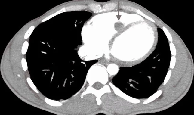
Sağ ventrikül trombüsü, pulmoner emboliyle birlikte görülebilen hareketli bir pıhtı veya düşük akım durumlarında gelişebilen duvara tutunmuş trombüs biçiminde olabilir. Pulmoner BT anjiyografi eşlik eden pulmoner emboliyi ve sağ kalp dolum kusurlarını gösterebilir; küçük intrakardiyak defektlerin fark edilmesi güç olabilir. Şüpheli dolum kusurunun çok düzlemli kesitlerde intraluminal kitle olarak görüntülenmesi ve normal kas yapılarıyla anatomik süreklilik göstermemesi trombüsü destekler. Sağ kalp trombüsüne pulmoner emboli eşlik ettiğinde prognoz daha kötü ve mortalite daha yüksek olabilir; bu bulguların gözden kaçması embolik yükün eksik değerlendirilmesine yol açabilir.
Faz ve pencere
- BTPA tanısal
- BTPA'da kontrastlanmış sağ ventrikül kan havuzunda düşük atenüasyonlu intrakaviter dolum kusuru görülebilir; eşlik eden pulmoner arter embolileri aynı incelemede değerlendirilir. Sağ kalp opaklaşması yetersizse veya kontrast karışımı varsa küçük dolum kusurunun değerlendirilmesi güçleşebilir.
- Gecikmiş
- Kitle karakterizasyonu amacıyla çekilmiş gecikmiş kardiyak BT görüntülerinde trombüs genellikle düşük atenüasyonlu ve kontrastlanmayan odak olarak görülür; sağ kalpteki akım karışımı artefaktı erken fazda dolum kusurunu taklit edebilir. Gecikmiş BT görüntüleri kontrastlanma ve doku karakterinin değerlendirilmesine yardımcı olur.
Önerilen pencereler: Mediasten (G 350 / M 50), Anjiyo (G 600 / M 150).
BT bulguları
- İntrakaviter dolum kusuru — sağ kalp kontrast havuzuna göre düşük atenüasyonlu, duvara geniş tabanla tutunabilen veya lümen içinde serbest uzanabilen kitle görünümüdür.
- Serbest yüzen pıhtı — uzun, kıvrımlı ve yüksek hareketlilik gösteren, duvara geniş tabanlı bağlantısı olmayan görünümdür; pulmoner emboli eşlik edebilir.
- Yerleşik mural trombüs — sağ ventrikül duvarına geniş tabanla oturan, ovoid ve az hareketli dolum kusuru şeklinde olabilir.
- Sağ ventrikül genişlemesi ve interventriküler septumun sol ventriküle doğru düzleşmesi — sağ kalpte basınç yükünün varlığını gösterir, ancak bu bulgular trombüs tanısını tek başına koydurmaz.
- Pulmoner arter dolum kusurları — ana, lober veya segmental dallardaki emboliler sağ kalp içi pıhtıyla birlikte raporlanır.
- Sağ ventrikül kitlesinde düzensiz sınır veya belirgin doku heterojenliği tümöral lezyon olasılığını artırabilir; trombüs genellikle kontrastlanmaz.
Normalde
Normal sağ ventrikül, giriş ve çıkış yolları arasında trabeküllü apikal bölümü bulunan üçgenimsi bir kavitedir; moderator bant interventriküler septumdan serbest duvardaki anterior papiller kas tabanına uzanan normal bir kas demetidir. Şüpheli odağın çok düzlemli görüntülerde anatomik seyri ve çevre kas yapılarıyla devamlılığı değerlendirilir; bu özellikler normal kas bantlarını dolum kusurundan ayırmaya yardım eder.
Normal BT ile kıyasla
Aynı bölgenin, kaynak veri setinde patoloji içermediği belirtilen bir BT incelemesini kesit kesit kaydırın; organ sınırlarını açarak anatomiyi hasta görüntüsüyle karşılaştırın. Küçük rastlantısal bulgular tümüyle dışlanmamıştır.
Kaynak: TotalSegmentator v2.01 veri seti (Wasserthal ve ark., Radiology: Artificial Intelligence 2023), CC BY 4.0, vaka s0977; organ sınırları veri setinin otomatik segmentasyonundan, birleştirilerek sadeleştirildi. Görüntüler 2,5 mm kesit aralığına yeniden örneklendi.
Ayırıcı tanı
- Moderator bant
- interventriküler septumdan sağ ventrikül serbest duvarındaki anterior papiller kas tabanına uzanan normal kas demetidir; belirgin olduğunda kitleyi taklit edebilir.
- Anterior papiller kas
- duvara bağlı kas dokusu olarak triküspit kapak kordalarına uzanır; serbest lümen kitlesi değildir.
- Sağ ventrikül trabekülleri
- duvardan kaviteye uzanan tekrarlayan kas çıkıntılarıdır; tek, bağımsız dolum kusuru oluşturmaz.
- Sağ ventrikül tümörü
- düzensiz sınır veya doku heterojenliği tümöral lezyon olasılığını artırabilir; trombüs genellikle kontrastlanmaz.
- Kontrast karışım artefaktı
- BTPA sırasında opaklaşmamış IVC kanının sağ kalbe dönmesi sağ atriyum veya sağ ventrikülde kontrast karışımı oluşturabilir.
Tuzaklar
- Moderator bant ve belirgin trabeküller sağ ventrikülde kitle izlenimi verebilir; anatomik seyirlerini çok düzlemli görüntülerde takip edin.
- BTPA'da IVC'den dönen opaklaşmamış kan sağ kalpte kontrast karışımı ve yalancı dolum kusuru oluşturabilir; kontrast çizgilenmesini tek başına trombüs kabul etmeyin.
- Küçük sağ kalp dolum kusurları BTPA incelemesinde görüntülenmesi güç olabilir; değerlendirmede pulmoner arterlerle birlikte sağ atriyum ve sağ ventrikül kavitesi de dikkatlice gözden geçirilmelidir.
Kendini dene
Pulmoner BT anjiyografide sağ ventrikülde serpijinöz serbest kitle ve pulmoner arter embolisi görülüyor. En olası tanı nedir?
Cevabı göster
Trombüs-in-transit. Serbest, uzun ve kıvrımlı hareketli sağ kalp trombüsü pulmoner emboliyle birlikteyse trombüs-in-transit görünümüyle uyumludur. Moderator bant normal bir sağ ventrikül kas yapısıdır.
İnterventriküler septumdan sağ ventrikül serbest duvarındaki anterior papiller kas tabanına uzanan belirgin kas demeti hangisidir?
Cevabı göster
Moderator bant. Bu kas bandının atenüasyonu ve kontrastlanması sağ ventrikül duvarına benzer; seyri ve tutunma noktaları tanınmasına yardım eder. Trombüs genellikle düşük atenüasyonlu, kontrastlanmayan dolum kusurudur; kontrast karışım artefaktı anatomik kas demeti değildir.
Kaynaklar
Bu sayfadaki 36 cümle ve 2 quiz sorusu aşağıdaki kaynaklardan alıntılarla tek tek karşılaştırıldı; 4 cümle bu karşılaştırmada düzeltildi (2026-10-08).
- Cardiac Masses and Pseudomasses: An Overview about Diagnostic Imaging and Clinical Backgroundpmc.ncbi.nlm.nih.gov
- Rare pediatric multi-system thrombosis post-COVID-19: a three-year follow-up case report and narrative review on rivaroxaban for long-term managementpmc.ncbi.nlm.nih.gov
- Post-Diphtheritic Myocarditis with Right Ventricular Thrombus: A Rare Cardiac Complicationpmc.ncbi.nlm.nih.gov
- Right Ventricular Thrombus Presenting With Right Ventricular Failure in Septic Shock: A Case Reportpmc.ncbi.nlm.nih.gov
- Right Ventricular Thrombus: A Rare but Potentially Fatal Conditionpmc.ncbi.nlm.nih.gov
- Right Ventricular Thrombus on Echocardiography.pubmed.ncbi.nlm.nih.gov
- Intracardiac Thrombus in Coronavirus Disease-2019.pubmed.ncbi.nlm.nih.gov
- Diagnostic accuracy of focused deep venous, lung, cardiac and multiorgan ultrasound in suspected pulmonary embolism: a systematic review and meta-analysis.pubmed.ncbi.nlm.nih.gov
- A case of nonvalvular endocarditis with biventricular apical infected thrombi.pubmed.ncbi.nlm.nih.gov
- Right ventricular thrombus, a challenge in imaging diagnostics: a case series.pubmed.ncbi.nlm.nih.gov
- Endomyocardial fibrosis: missing tricuspid valve and Fontan-like circulation.pubmed.ncbi.nlm.nih.gov
- Incidental cardiac findings on computed tomography imaging of the thorax.pubmed.ncbi.nlm.nih.gov
- Giant right ventricular thrombus as the revealing form of Behçet's disease.pubmed.ncbi.nlm.nih.gov
- Right ventricular thrombus: a rare complication of ovarian hyperstimulation syndrome.pubmed.ncbi.nlm.nih.gov
- Right Ventricular Thrombus Masquerading as a Tumor.pubmed.ncbi.nlm.nih.gov
- Right Ventricular Clot in Transit in COVID-19: Implications for the Pulmonary Embolism Response Team.pubmed.ncbi.nlm.nih.gov
- The curious case of a massive right heart thrombosis: a case report.pubmed.ncbi.nlm.nih.gov
- Right atrial thrombus and pulmonary embolism one year after ventricular septal defect patch repair: successful surgical management of a complicated case.pubmed.ncbi.nlm.nih.gov
- When Virchow's Triad Goes on Overdrive: Catastrophic Multi-System Thrombosis in the Setting of New-Onset Heart Failure in a Previously Healthy Man.pubmed.ncbi.nlm.nih.gov
- Severe peripartum cardiomyopathy complicated by multiple left ventricular thrombi and systemic embolic events: a case report.pubmed.ncbi.nlm.nih.gov
- Totally thoracoscopic beating-heart resection of a catheter-related right atrial thrombus: a case report.pubmed.ncbi.nlm.nih.gov
- Simultaneous Left and Right Atrial Appendage Thrombi on Cardiac Computed Tomography.pubmed.ncbi.nlm.nih.gov
- Multisystem Thromboembolic Shower in the Intensive Care Unit: Acute Limb Ischemia and Mesenteric Embolism.pubmed.ncbi.nlm.nih.gov
- Valvular and Outflow Tract Metastases From Germ Cell Tumors: Contrasting Left-Sided Embolism and Right-Sided Silence.pubmed.ncbi.nlm.nih.gov
- Giant Aneurysm of the Left Main Coronary Artery: A Case Report.pubmed.ncbi.nlm.nih.gov
- Atrial wall dissection: a contemporary overview for a proper decision-making approach.pubmed.ncbi.nlm.nih.gov
- A nomogram integrating CTPA parameters and serum biomarkers to predict 30-day mortality and refine risk stratification in non-high-risk acute pulmonary embolism.pubmed.ncbi.nlm.nih.gov
- Management of Catastrophic Postpartum Acute Pulmonary Embolism With Extracorporeal Membrane Oxygenation Support and Thromboaspiration: A Case Report.pubmed.ncbi.nlm.nih.gov
- Thrombus in Transit Following Hemodynamic Stabilization in Acute Pulmonary Embolism: A Therapeutic Dilemma.pubmed.ncbi.nlm.nih.gov
- Mycotic pulmonary artery aneurysms and septic pulmonary emboli in infective endocarditis: A rare case report and review of imaging findings.pubmed.ncbi.nlm.nih.gov
- Primary Cardiac Lymphoma Presenting With Chest Pain.pubmed.ncbi.nlm.nih.gov
- The imaging spectrum of myocardial infarction and its associated complications: a contemporary pictorial review of computed tomography and magnetic resonance imaging.pubmed.ncbi.nlm.nih.gov
- The Initial Diagnostic Approach to Adult Congenital Heart Disease: A Practical Imaging Pathway for General Cardiologists and Cardiac Imagers.pubmed.ncbi.nlm.nih.gov
- Transcatheter Closure of Patent Ductus Arteriosus in Adults.pubmed.ncbi.nlm.nih.gov
- Acute Traumatic Aortic Injury: What the Radiologist Needs to Know.pubmed.ncbi.nlm.nih.gov
- Diagnostic Value and Operational Recommendations for Late Iodine Enhancement and ECV Quantification in Single-Energy Computed Tomography: A Narrative Review.pubmed.ncbi.nlm.nih.gov
- The Role of Multimodality Imaging in Atrial Fibrillation and Heart Failure: From Patient Selection to Procedural Ablation Guidance.pubmed.ncbi.nlm.nih.gov
- Multimodality imaging of a rare case of apical hypertrophic cardiomyopathy with endomyocardial fibrosis and myocardial calcification: case report and literature review.pubmed.ncbi.nlm.nih.gov
- Co-occurrence of Bilateral Pulmonary Artery Embolism and a Serpentine Right Atrial Thrombus Successfully Managed With Oral Anticoagulation: A Case Report.pubmed.ncbi.nlm.nih.gov
- Prognostic Significance of Echocardiography-Visualized Thrombus in Right Heart in Early Stages of Acute PE: 100 Patients from Regional Pulmonary Embolism Registry.pubmed.ncbi.nlm.nih.gov
- Cinematic rendering-enhanced computed tomography of right intracardiac thrombi: A case report.pubmed.ncbi.nlm.nih.gov
- Giant Left Atrial Myxoma Causing Functional Mitral Stenosis: A Minimally Invasive Approach.pubmed.ncbi.nlm.nih.gov
- Cardioembolic sources and stroke prevention: a systematic review.pubmed.ncbi.nlm.nih.gov
- Left Ventricular Myxoma in a Young Adult: An Unusual Presentation of a Rare Cardiac Tumor.pubmed.ncbi.nlm.nih.gov
- The Silent Threat: Unmasking Combined Post- and Pre-capillary Pulmonary Hypertension (CpcPH) in an Elderly Patient With Heart Failure With Preserved Ejection Fraction (HFpEF).pubmed.ncbi.nlm.nih.gov
- Giant popliteal venous aneurysm presenting with pulmonary embolism: multimodal imaging findings and clinical course.pubmed.ncbi.nlm.nih.gov
- Echocardiographic assessment of pulmonary hypertension: a hierarchical interpretive framework for clinical decision-making and echo laboratory practice.pubmed.ncbi.nlm.nih.gov
- A Rare Presentation of Behçet's Disease: Coexistence of Intracardiac Thrombi, Coronary Artery Disease, and Pulmonary Thromboembolism.pubmed.ncbi.nlm.nih.gov
- Agenesis of the left pulmonary artery and cardiac hydatid cysts: A dual rarity.pubmed.ncbi.nlm.nih.gov
- Unveiling the Unknown: A Case Report of Undifferentiated Pleomorphic Sarcoma in the Heart.pubmed.ncbi.nlm.nih.gov
- Giant Left Ventricular Intracavitary Mass: Diagnostic Challenges in Distinguishing Thrombus From Tumor.pubmed.ncbi.nlm.nih.gov
- Left Ventricular Mass in an Oncology Patient and Cocaine User: Metastasis or Thrombus?pubmed.ncbi.nlm.nih.gov
- Anatomic complexities of the human right ventricle: Considerations for clinical practice in the electrophysiology laboratory.pubmed.ncbi.nlm.nih.gov
- Case Report: Antenatal diagnosis of double-chambered right ventricle in two cases: follow-up and literature review.pubmed.ncbi.nlm.nih.gov
- Intracardiac Echocardiography during Invasive Electrophysiological Procedures: a Scientific Statement of the European Heart Rhythm Association of the ESC, and the European Association of Percutaneous Cardiovascular Interventions of the ESC.pubmed.ncbi.nlm.nih.gov
- Dengue-Associated Encephalopathy With Multiple Cranial Neuropathies and Multisystem Involvement in a Patient With Epilepsy: A Comprehensive Case Report With Longitudinal Multimodality Imaging.pubmed.ncbi.nlm.nih.gov
- Cardiac Computed Tomography in Structural Heart Interventions: From Preprocedural Planning to Procedural Strategy.pubmed.ncbi.nlm.nih.gov
- CT Imaging for Valvular Interventions.pubmed.ncbi.nlm.nih.gov
- Multimodality Imaging in Hypertrophic Cardiomyopathy.pubmed.ncbi.nlm.nih.gov
- Spontaneous resolution of right ventricular thrombus in congestive heart failure due to alcoholic cardiomyopathy: A case report.pubmed.ncbi.nlm.nih.gov
- Cardiac lead perforation: Mechanisms, detection, and therapeutic approaches.pubmed.ncbi.nlm.nih.gov
- Hypertrophic cardiomyopathy: state of the art on diagnosis and current therapeutic approach.pubmed.ncbi.nlm.nih.gov
- Severe Mitral Regurgitation Triggered by Myocardial Ischemia in Hypertrophic Cardiomyopathy With Abnormalities of the Mitral Valve Complex.pubmed.ncbi.nlm.nih.gov
- Huge Left Atrial Myxoma Presenting as Pulmonary Embolism on a CT Pulmonary Angiogram.pubmed.ncbi.nlm.nih.gov
- Left Ventricular Non-Compaction Cardiomyopathy: The Tragedies & Trabeculations of the Architectural Cardiac Sponge.pubmed.ncbi.nlm.nih.gov
- A 25-Year Review of the Use of Silanized Hydroxypropylmethylcellulose Hydrogels in Tissue Engineering and Regenerative Medicine.pubmed.ncbi.nlm.nih.gov
- Idiopathic Hypereosinophilic Syndrome Presenting as Isolated Right Ventricular Endomyocardial Fibrosis With Intracavitary Thrombus: A Multimodality Imaging Case Report.pubmed.ncbi.nlm.nih.gov
- Ovarian Endometrioid Carcinoma Presenting With Presumed Right Ventricular Intracardiac Metastasis and Superimposed Thrombus: A Rare Manifestation of Gynecologic Malignancy.pubmed.ncbi.nlm.nih.gov
- Rare Cause of Restrictive Cardiomyopathy: A Case Report of Löffler Endocarditis.pubmed.ncbi.nlm.nih.gov
- Phosphorescence lifetime microscopy and contrast-enhanced ultrasound imaging revealed strong left-to-right intraventricular shunt in anesthetized Xenopus laevis.pubmed.ncbi.nlm.nih.gov
- HVSMR-2.0: A 3D cardiovascular MR dataset for whole-heart segmentation in congenital heart disease.pubmed.ncbi.nlm.nih.gov
- Fatty Images of the Heart: Spectrum of Normal and Pathological Findings by Computed Tomography and Cardiac Magnetic Resonance Imaging.pubmed.ncbi.nlm.nih.gov
- Cardiac CT: Imaging of and Through Cardiac Devices.pubmed.ncbi.nlm.nih.gov
- High-affinity alphavbeta3 integrin targeted optical probe as a new imaging biomarker for early atherosclerosis: initial studies in Watanabe rabbits.pubmed.ncbi.nlm.nih.gov
- Secondary <i>Staphylococcus capitis</i> Infection of a Long-Standing Non-Coronary Sinus of Valsalva-to-Right Atrial Fistula Initially Misdiagnosed as a Gerbode Defect: A Case Report.pubmed.ncbi.nlm.nih.gov
- Optimizing left atrial appendage imaging: the diagnostic value of left lateral decubitus cardiac CT angiography.pubmed.ncbi.nlm.nih.gov
- Ischemic Stroke After Anticoagulation Discontinuation Following Apparent Resolution of Left Ventricular Thrombus: A Case Report.pubmed.ncbi.nlm.nih.gov
- Myeloid/lymphoid neoplasms with eosinophilia and tyrosine kinase gene fusions (FIP1L1::PDGFRA) complicated by Loeffler's endocarditis leading to stroke.pubmed.ncbi.nlm.nih.gov
- Cerebral thrombosis and abscess: A child with 2 rare complications of tetralogy of Fallot.pubmed.ncbi.nlm.nih.gov
- Right heart on multidetector CT.pubmed.ncbi.nlm.nih.gov
- Floating Thrombus on the Ascending Aorta and/or Aortic Arch, to Operate or Not to Operate: Two Case Reports and a Literature Review.pubmed.ncbi.nlm.nih.gov
- Computed Tomography Imaging of Thoracic Aortic Surgery: Distinguishing Life-Saving Repairs from Life-Threatening Complications.pubmed.ncbi.nlm.nih.gov
- Obese Adolescent with Factor V Leiden-Related Pulmonary Embolism: Case-Based Insight into Paediatric Thrombosis Management.pubmed.ncbi.nlm.nih.gov
- Thrombus straddling the patent foramen ovale: a case report.pubmed.ncbi.nlm.nih.gov
- Caught in Transit: Mobile Right Atrial Thrombus in Intermediate-High-Risk Pulmonary Embolism Treated With Half-Dose Alteplase.pubmed.ncbi.nlm.nih.gov
- Percutaneous Thrombectomy for Clot-in-Transit With Concomitant Left Ventricular Thrombus in a Patient With Acute Heart Failure Exacerbation: A Case Report.pubmed.ncbi.nlm.nih.gov
- Incidentally detected double-chambered right ventricle in an adult.pubmed.ncbi.nlm.nih.gov
- Arterial Supply to the Atrioventricular Node: Anatomical Variability and Clinical Implications.pubmed.ncbi.nlm.nih.gov
- Repair of Double-Chamber Right Ventricle and Subvalvular Pulmonic Stenosis in a 78-Year-Old With Repaired Tetralogy of Fallot: Anesthetic Considerations and Review of the Literature.pubmed.ncbi.nlm.nih.gov
- Successful surgical treatment of a giant right ventricular myxoma with pulmonary artery tumor thrombus in an adolescent patient: case report.pubmed.ncbi.nlm.nih.gov
- Silent Air Embolism of the Pulmonary Artery and Right Ventricle Due to Suspected Intravenous Substance Use Found Incidentally on Outpatient Imaging.pubmed.ncbi.nlm.nih.gov
- Inferior vena cava hyperechoic structure in a patient with cardiac myxoma: a surgical case report and literature review.pubmed.ncbi.nlm.nih.gov
- An intracavitary right ventricular mass: a diagnostic dilemma-a case report.pubmed.ncbi.nlm.nih.gov
- Primary cardiac angiosarcoma initially presenting with sick sinus syndrome and pericardial effusion: a case report.pubmed.ncbi.nlm.nih.gov
- Angioleiomyoma Extending From the Iliac Vein to the Right Atrium.pubmed.ncbi.nlm.nih.gov
- Cardiovascular Computed Tomography Angiographic Assessment of Simple Cardiac Shunts in Adults.pubmed.ncbi.nlm.nih.gov
- Early Outcomes of Bolus Thrombolysis Plus Manual Aspiration Thrombectomy Using Conventional Catheters for Intermediate-High- and High-Risk Acute Pulmonary Embolism.pubmed.ncbi.nlm.nih.gov
- Beyond the right ventricle: assessment of the left ventricular function in pulmonary hypertension.pubmed.ncbi.nlm.nih.gov
- Pulmonary angiomatoid fibrous histiocytoma invading the pulmonary vein with intracranial metastasis: a case report.pubmed.ncbi.nlm.nih.gov
Eğitim amaçlıdır; hasta başında tanı ve tedavi kararının yerine geçmez. İçerik yapay zekâ destekli bir süreçle güncel literatüre göre cümle cümle kaynaklarla karşılaştırıldı, ancak uzman radyolog incelemesinden geçmedi. Klinik kararlarda güncel kılavuzları, kurum protokollerini ve radyoloji raporunu esas alın.